. 2017 Apr 11:11:25.
doi: 10.3389/fninf.2017.00025. eCollection 2017.

Pypes: Workflows for Processing Multimodal Neuroimaging Data

Affiliations

Pypes: Workflows for Processing Multimodal Neuroimaging Data

Alexandre M Savio et al. Front Neuroinform. .
No abstract available

Keywords: MRI; PET; brain connectivity; brain imaging; denoising; image analysis; python; registration.


Figures
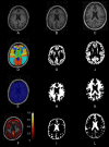
Figure 1
Figure 1
Slices of one example of the anatomical pipeline on one sample image. (A) The raw MPRAGE image, (B) the bias-field corrected MPRAGE, (C) the bias-field corrected MPRAGE in MNI space, (D) the Hammers atlas in anatomical space, (E) a brain mask, and (F) the result from the cortical thickness pipeline, (G–I) gray matter, white matter and cerebro-spinal fluid (tissue segmentations), and (J–L) tissue segmentations in MNI space.
Figure 2
Figure 2
Slices of partial results from one sample processed by the PET/MR pipeline. (A) The raw FDG-PET, (B) the MPRAGE, (C) the partial volume corrected PET, (D) a brain mask, (E) the Hammers' atlas, and (F) the PVC PET in MNI space.
Figure 3
Figure 3
Slices of one example from the DTI processing pipeline: (A) the raw DTI (b-value = 0) image, (B) the Eddy-currents corrected and nl-means denoized image, (C) the FA image, and (D) the atlas in DTI space. (E) Shows the structural connectivity matrix calculated with Camino.

Similar articles

Cited by

References

    1. Abraham A., Pedregosa F., Eickenberg M., Gervais P., Mueller A., Kossaifi J., et al. . (2014). Machine learning for neuroimaging with scikit-learn. Front. Neuroinform. 8:14. 10.3389/fninf.2014.0001400053 - DOI - PMC - PubMed
    1. Aiello M., Salvatore E., Cachia A., Pappatà S., Cavaliere C., Prinster A., et al. . (2015). Relationship between simultaneously acquired resting-state regional cerebral glucose metabolism and functional MRI: a PET/MR hybrid scanner study. NeuroImage 113, 111–121. 10.1016/j.neuroimage.2015.03.01700022 - DOI - PubMed
    1. Andersson J. L. R., Sotiropoulos S. N. (2016). An integrated approach to correction for off-resonance effects and subject movement in diffusion MR imaging. NeuroImage 125, 1063–1078. 10.1016/j.neuroimage.2015.10.019 - DOI - PMC - PubMed
    1. Ashburner J. (2012). SPM: a history. NeuroImage 62, 791–800. 10.1016/j.neuroimage.2011.10.02500107 - DOI - PMC - PubMed
    1. Avants B. B., Tustison N. J., Stauffer M., Song G., Wu B., Gee J. C. (2014). The Insight ToolKit image registration framework. Front. Neuroinform. 8:44. 10.3389/fninf.2014.00044 - DOI - PMC - PubMed

LinkOut - more resources