. 2017 Apr 11:8:193.
doi: 10.3389/fphar.2017.00193. eCollection 2017.

Inhibition of VEGF and Angiopoietin-2 to Reduce Brain Metastases of Breast Cancer Burden

Affiliations

Inhibition of VEGF and Angiopoietin-2 to Reduce Brain Metastases of Breast Cancer Burden

Kaci A Bohn et al. Front Pharmacol. .

Abstract

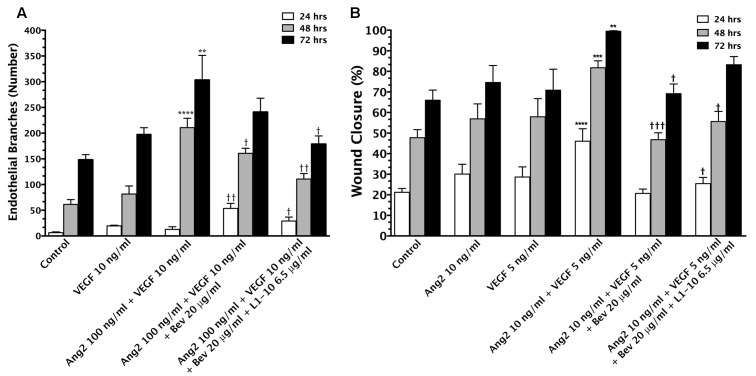
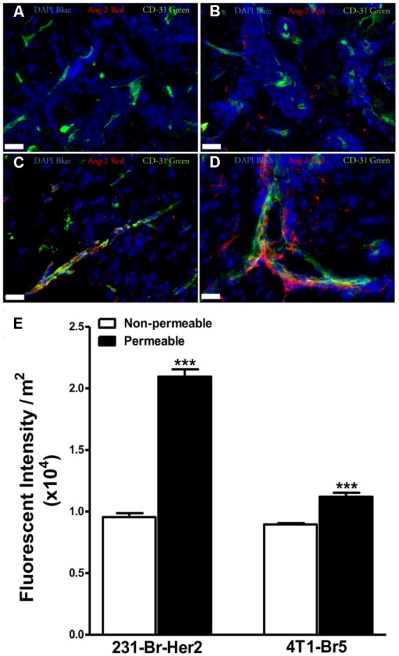
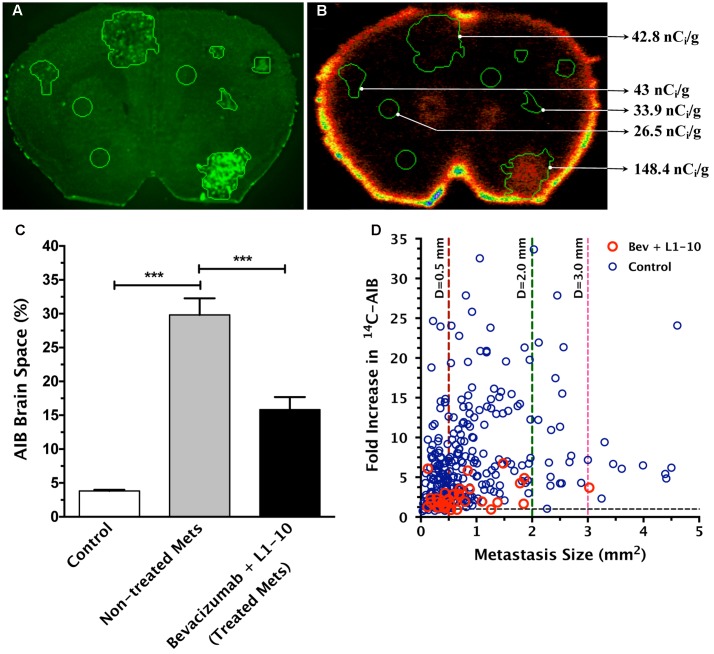
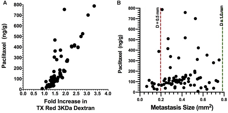
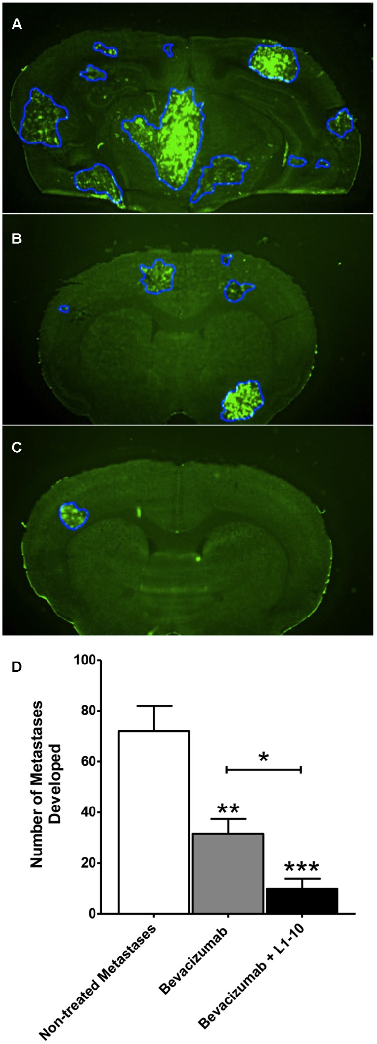
For metastases in the central nervous system, angiogenesis enhances metastatic potential and promotes progression. Primary factors which drive vessel growth are vascular endothelial growth factor (VEGF) and angiopoietin-2. Preclinical models show inhibition of either factor reduces metastases spread and inhibits growth. This work sets out to answer two questions in a preclinical mouse model. First, whether the combined inhibition of VEGF and angiopoietin-2, reduces passive permeability and limits drug uptake into brain metastases; and second, whether this inhibition reduces metastases burden in brain. We observed combinatorial inhibition of VEGF and angiopoietin-2, decreased (p < 0.05) angiogenesis and vascular branching in an aortic ring assay and decreased (p < 0.05) endothelial wound closure times. Using a brain metastases of breast cancer model (induced by intracardiac injections of brain seeking MDA-MB-231Br cells or 4T1Br cells), we observed, similar to VEGF, angiopoetin-2 expression correlates to increased angiogenesis (p < 0.05) and increased lesion permeability. To determine efficacy, animals were administered bevacizumab plus L1-10 (angiopoietin inhibitor) twice per week until neurological symptoms developed. Lesion permeability significantly decreased by ∼50% (p < 0.05) compared to untreated lesions, but remained ∼25% greater (p < 0.0%) than brain. In subsequent experiments, animals were administered similar regimens but sacrificed on day 32. The number of metastatic lesions developed was significantly (p < 0.001) reduced in the bevacizumab group (56%) and combination group (86%). Lesions' size was reduced in bevacizumab treated lesions (∼67%) and bevacizumab and L1-10 treated lesions (∼78%) developing area < 0.5 mm2. In summary, combinatorial inhibition of VEGF and angiopoietin reduces lesion permeability and brain metastatic burden.

Keywords: L1-10; angiogenesis; bevacizumab; brain metastases; permeability; prevention.


Figures

FIGURE 1
FIGURE 1
Vascular endothelial growth factor (VEGF) and Ang-2 promote angiogenesis and can be modulated by inhibition. The endothelial branching observed from in vitro aortic ring angiogenesis assays in the presence of VEGF (10 ng/mL) + Ang-2 (100 ng/mL) and the addition of bevacizumab (20 μg/mL) and L1-10 (6.5 μg/mL) for 24, 48, and 72 h duration of treatment (A). An increase in endothelial branching compared to control is denoted by (). A reduction in endothelial branching compared to VEGF + Ang-2 is denoted by (). Wound closure of bEND5 cells in the presence of Ang-2 (10 ng/mL) + VEGF (5 ng/mL) and the addition of bevacizumab (20 μg/mL) and L1-10 (6.5 μg/mL) in comparison to Ang-2 (10 ng/mL) and VEGF (5 ng/mL) over 24, 48, and 72 h (B). An increase in percent wound closure relative to control is denoted by () and a decrease in wound closure relative to Ang-2 + VEGF is denoted by (). Statistical evaluation was determined by student’s t-tests; and for p < 0.05, ∗∗ and †† for p < 0.01, ∗∗∗ and ††† for p < 0.001 and ∗∗∗∗ and †††† for p < 0.0001.
FIGURE 2
FIGURE 2
Expression of angiopoietin-2 is greater in lesions with higher permeability. Brain slices from un-treated metastases bearing animals were stained for the hypoxia induced vascular destabilizing protein Ang-2. Ang-2 expression in MDA-MB-231-BR-Her2 metastases is shown in non-permeable (A) and permeable (B) metastatic lesions. Representative images in the 4T1-BR5 model are shown in a lesion with low permeability (C) and a lesion with high permeability (D). Ang-2 (red), CD31 (green), DAPI (blue). Ang-2 intensity in permeable and non-permeable lesion in both preclinical models of brain metastases of breast cancer (E). Permeable lesions demonstrated greater Ang-2 expression than non-permeable in both models, with a >2-fold increase in the MDA-MB-231-BR-Her2 model, indicating the presence of vascular destabilization. Scale bar = 25 μm. Statistical significance was determined using Student’s t-test to determine difference in Ang-2 expression between the two permeability groups within each model (∗∗∗ for p < 0.001).
FIGURE 3
FIGURE 3
Treatment of bevacizumab and L1-10AIB during metastasis development decreases permeability of 14C-AIB, which correlates with metastasis size. Representative brain slice image of enhanced green fluorescent protein (eGFP) labeled lesions (A) and corresponding quantitative autoradiography (QAR) image (B) with 14C-AIB distribution. The percent brain space of 14C-AIB in normal brain and metastases (C). Fold-increase in permeability with metastasis size reveals a weak correlation in bevacizumab plus L1-10 treated lesions (n = 65, r2 = 0.162, p < 0.05) (D). Statistical significance was determined using ANOVA followed by Bonferroni’s multiple comparisons test for AIB brain space percentage, and linear regression analysis for fold increase in permeability versus lesion size (∗∗∗ for p < 0.001).
FIGURE 4
FIGURE 4
Brain uptake of 14C-paclitaxel in brain metastases after bevacizumab treatment. Bevacizumab reduces (p < 0.05) metastasis paclitaxel uptake to a mean of 163 ng/g, ranging from 7.76 to 788 ng/g (n = 81) as compared to our previously reported value of 276 ng/g ranging from 2.56 to 1768 (Lockman et al., 2010). In addition, there was a strong correlation with Texas Red 3 kDa dextran permeability (r2 = 0.72, p < 0.001) (A). In addition, paclitaxel concentrations in metastases from the bevacizumab group showed a weak correlation with lesion size (r2 = 0.071, p < 0.05) (B).
FIGURE 5
FIGURE 5
Bevacizumab and L1-10 treatment reduces metastatic burden in brain. Representative images of MDA-MB-231-BR-Her2 non-treated metastatic brain (A), bevacizumab treated brain (B), and bevacizumab + L1-10 treated brain (C). Bevacizumab (10 mg/kg) or bevacizumab + L1-10 (4 mg/kg) was administered on day 10 after inoculation (with cancer cells and the number of metastatic lesions developed was significantly (p < 0.001) reduced in the bevacizumab alone group (56%) and in the combination group (86%) (D). Treatment also reduced lesion size with 67% ± 1 of bevacizumab treated lesions and 78% ± 10 of bevacizumab + L1-10 treated lesions developing area < 0.5 mm2. Data illustrate a significant reduction in metastatic burden with simultaneous inhibition of the VEGF/angiopoietin-2 pathway. Significance was determined using ANOVA analysis followed by Bonferroni’s multiple comparisons test; for p < 0.05, ∗∗ for p < 0.01 and ∗∗∗ for p < 0.001.)

References

    1. Abbott N. J., Patabendige A. A., Dolman D. E., Yusof S. R., Begley D. J. (2009). Structure and function of the blood-brain barrier. Neurobiol. Dis. 37 13–25. 10.1016/j.nbd.2009.07.030 - DOI - PubMed
    1. Ahmad S. A., Liu W., Jung Y. D., Fan F., Wilson M., Reinmuth N., et al. (2001). The effects of angiopoietin-1 and -2 on tumor growth and angiogenesis in human colon cancer. Cancer Res. 61 1255–1259. - PubMed
    1. Asahara T., Chen D., Takahashi T., Fujikawa K., Kearney M., Magner M., et al. (1998). Tie2 receptor ligands, angiopoietin-1 and angiopoietin-2, modulate VEGF-induced postnatal neovascularization. Circ. Res. 83 233–240. 10.1161/01.RES.83.3.233 - DOI - PubMed
    1. Barresi V. D. G. C., Regiani-Bonetti L., Ponz-De Leon M., Barresi G., Vitarelli E. (2010). Stage I colorectal carcinoma: VEGF immunohistochemical expression, microvessel density, and their correlation with clinical outcome. Virchows Arch. 457 11–19. 10.1007/s00428-010-0933-5 - DOI - PubMed
    1. Bergers G., Song S., Meyer-Morse N., Bergsland E., Hanahan D. (2003). Benefits of targeting both pericytes and endothelial cells in the tumor vasculature with kinase inhibitors. J. Clin. Invest. 111 1287–1295. 10.1172/JCI17929 - DOI - PMC - PubMed

LinkOut - more resources