. 2017 Apr 10:8:58.
doi: 10.3389/fendo.2017.00058. eCollection 2017.

On the Molecular Evolution of Leptin, Leptin Receptor, and Endospanin

Affiliations

On the Molecular Evolution of Leptin, Leptin Receptor, and Endospanin

Richard Lyle Londraville et al. Front Endocrinol (Lausanne). .

Abstract

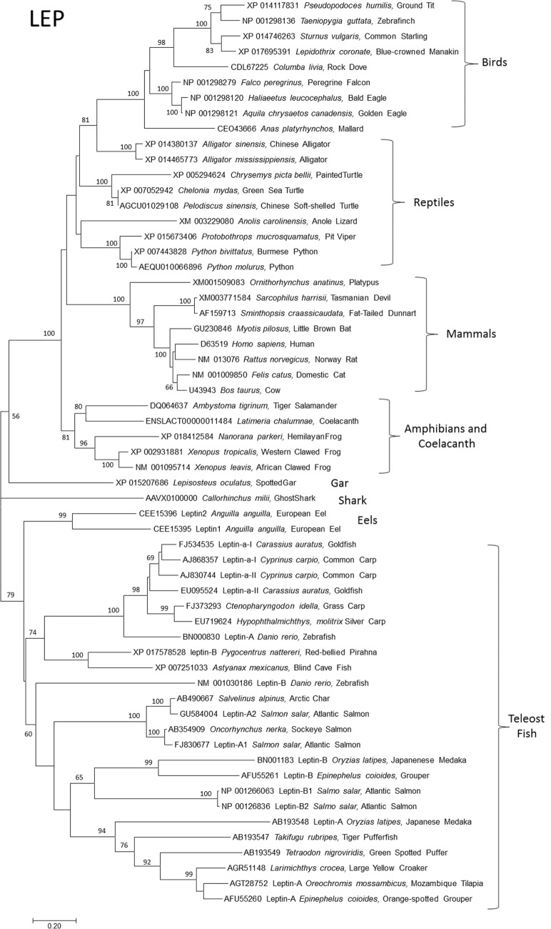
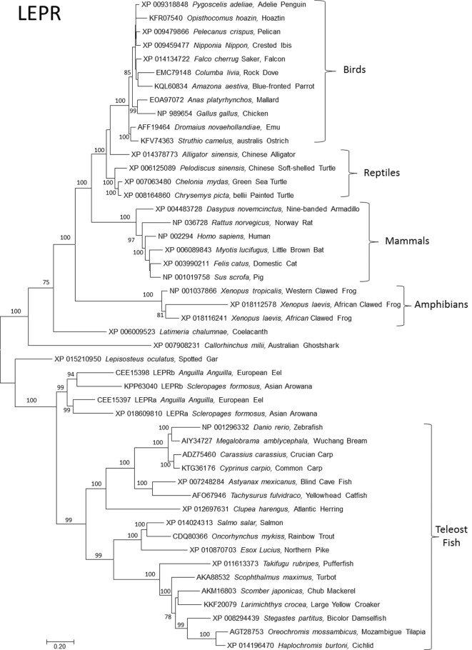
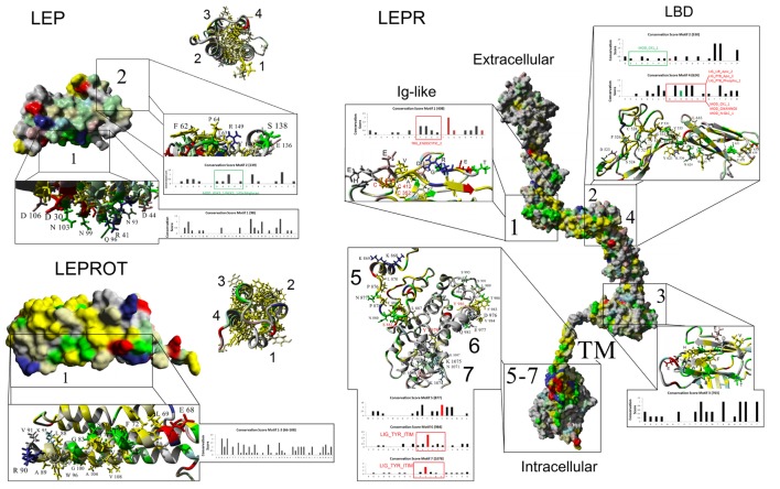
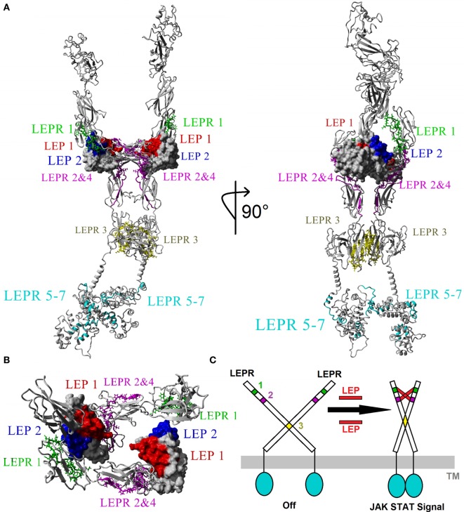
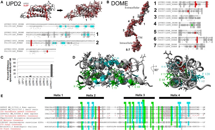
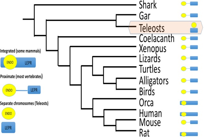
Over a decade passed between Friedman's discovery of the mammalian leptin gene (1) and its cloning in fish (2) and amphibians (3). Since 2005, the concept of gene synteny conservation (vs. gene sequence homology) was instrumental in identifying leptin genes in dozens of species, and we now have leptin genes from all major classes of vertebrates. This database of LEP (leptin), LEPR (leptin receptor), and LEPROT (endospanin) genes has allowed protein structure modeling, stoichiometry predictions, and even functional predictions of leptin function for most vertebrate classes. Here, we apply functional genomics to model hundreds of LEP, LEPR, and LEPROT proteins from both vertebrates and invertebrates. We identify conserved structural motifs in each of the three leptin signaling proteins and demonstrate Drosophila Dome protein's conservation with vertebrate leptin receptors. We model endospanin structure for the first time and identify endospanin paralogs in invertebrate genomes. Finally, we argue that leptin is not an adipostat in fishes and discuss emerging knockout models in fishes.

Keywords: adipostat; endospanin; fish models; in silico modeling; leptin; leptin receptor; molecular evolution; obesity.


Figures

Figure 1
Figure 1
Evolutionary relationships of vertebrate leptins (LEPs). Relationships of 59 amino acid sequences using the neighbor-joining method conducted in MEGA7. Numbers at nodes represent percentage of 500 bootstrap replicates. Nodes with no number indicate bootstrap support of less than 50%. Leptin amino acid sequences were manually aligned in MEGA7 informed by protein structural homologies. GenBank accession numbers represent protein accession.
Figure 2
Figure 2
Evolutionary relationships of vertebrate leptin receptor (LEPR). Relationships of 48 amino acid sequences using the neighbor-joining method conducted in MEGA7. Numbers at nodes represent percentage of 500 bootstrap replicates. Nodes with no number indicate bootstrap support of less than 50%. GenBank accession numbers represent protein accessions.
Figure 3
Figure 3
Mapping protein conservation of leptin (LEP), leptin receptor (LEPR), and LEPROT/endospanin. Consurf analysis of LEP (top left), LEPR (right), and LEPROT/endospanin (bottom left) are shown as molecular surface plots of each structure. For LEP and LEPR, a picture of the four-helix bundle with conserved hydrophobic amino acids is shown as a ribbon diagram beside the surface plots of conservation. Top conserved motifs are magnified, identifying conserved amino acids that contribute to each motif. Amino acids are colored as followed: yellow, conserved hydrophobic; red, conserved polar acidic; blue, conserved polar basic; green, conserved hydrophilic; gray, not conserved. Amino acids with known posttranslational modifications are red (disulfide bonds of Cys-C or phosphorylation of Ser-S/Thr-T/Tyr-Y) and green (glycosylation of Asn-N) on the bar graphs of conservation. Predicted eukaryotic linear motifs are boxed and labeled on the bar graphs.
Figure 4
Figure 4
Modeling the 2xLEP–2xLEPR interaction. (A) Each of the top motifs for leptin (LEP) and leptin receptor (LEPR) are colored in respective color coding. LEP: motif 1, red; motif 2, blue. LEPR: motif 1, green; motif 2 and 4, magenta; motif 3, yellow; motif 5–7, cyan. (B) Magnified view of the Ig-like and LBD of LEPR showing the 2–2 interaction model based on vertebrate evolution. (C) Model of endogenous dimerized LEPR being activated by LEP binding.
Figure 5
Figure 5
Defining the metazoan leptin system. (A) UPD2 model (gray) aligned with human LEP (hLEP, red). To the right of the structure overlay are identified amino acids in red that are conserved in the two proteins. Below the models are sequence alignments showing amino acids conserved in the top two motifs (gray), conserved posttranslational modifications (PTMs) (red), and known sites to interact with the receptor (cyan). (B) Dome model with amino acids in red conserved with human leptin receptor (LEPR). To the right of the model are sequence alignments showing amino acids conserved in the top seven motifs (gray) and conserved PTMs (red). (C) Using the top motifs of LEP, LEPR, and LEPROT, BLAST data for each in invertebrate genomes. (D,E) Models of LEPROT, now known as endospanin 1 (D), with conserved amino acids identified (E). Amino acids in red are conserved in all sequences, those in green conserved in at least four of the five groups, those in cyan conserved in at least three groups, and those in gray conserved in at least two groups.
Figure 6
Figure 6
Schematic of gene order for endospanin and leptin receptor (LEPR) among vertebrates. For most vertebrate classes, endospanin (LEPROT) is either embedded within the LEPR gene, or within 150,000 bp, and without any gene between LEPROT and LEPR. For teleost fishes only, LEPROT and LEPR are on separate chromosomes. Data mined from Genbank queries. For example, LEPR search term returns chromosome 1, acc# NC_000001.11 for human LEPR, which also maps LEPROT within the human LEPR sequence, and chromosome 6, acc# NC_07117.6 for zebrafish LEPR but chromosome 2, acc# NC_007113.6 for zebrafish LEPROT.

References

    1. Zhang Y, Proenca R, Maffei M, Barone M, Leopold L, Friedman JM. Positional cloning of the mouse obese gene and its human homologue. Nature (1994) 372:425–32. 10.1038/372425a0 - DOI - PubMed
    1. Kurokawa T, Uji S, Suzuki T. Identification of cDNA coding for a homologue to mammalian leptin from pufferfish, Takifugu rubripes. Peptides (2005) 26:745–50. 10.1016/j.peptides.2004.12.017 - DOI - PubMed
    1. Crespi EJ, Denver RJ. Leptin (ob gene) of the South African clawed frog Xenopus laevis. Proc Natl Acad Sci U S A (2006) 103:10092–7. 10.1073/pnas.0507519103 - DOI - PMC - PubMed
    1. Pelleymounter MA, Cullen MJ, Baker MB, Hecht R, Winters D, Boone T, et al. Effects of the obese gene product on body weight regulation in ob/ob mice. Science (1995) 269:540–3. 10.1126/science.7624776 - DOI - PubMed
    1. Halaas JL, Gajiwala KS, Maffei M, Cohen SL, Chait BT, Rabinowitz D, et al. Weight-reducing effects of the plasma protein encoded by the obese gene. Science (1995) 269:543–6. 10.1126/science.7624777 - DOI - PubMed

LinkOut - more resources