. 2017 Apr 11:8:522.
doi: 10.3389/fpls.2017.00522. eCollection 2017.

Cytosolic Glyceraldehyde-3-Phosphate Dehydrogenase Is Phosphorylated during Seed Development

Affiliations

Cytosolic Glyceraldehyde-3-Phosphate Dehydrogenase Is Phosphorylated during Seed Development

Claudia V Piattoni et al. Front Plant Sci. .

Abstract

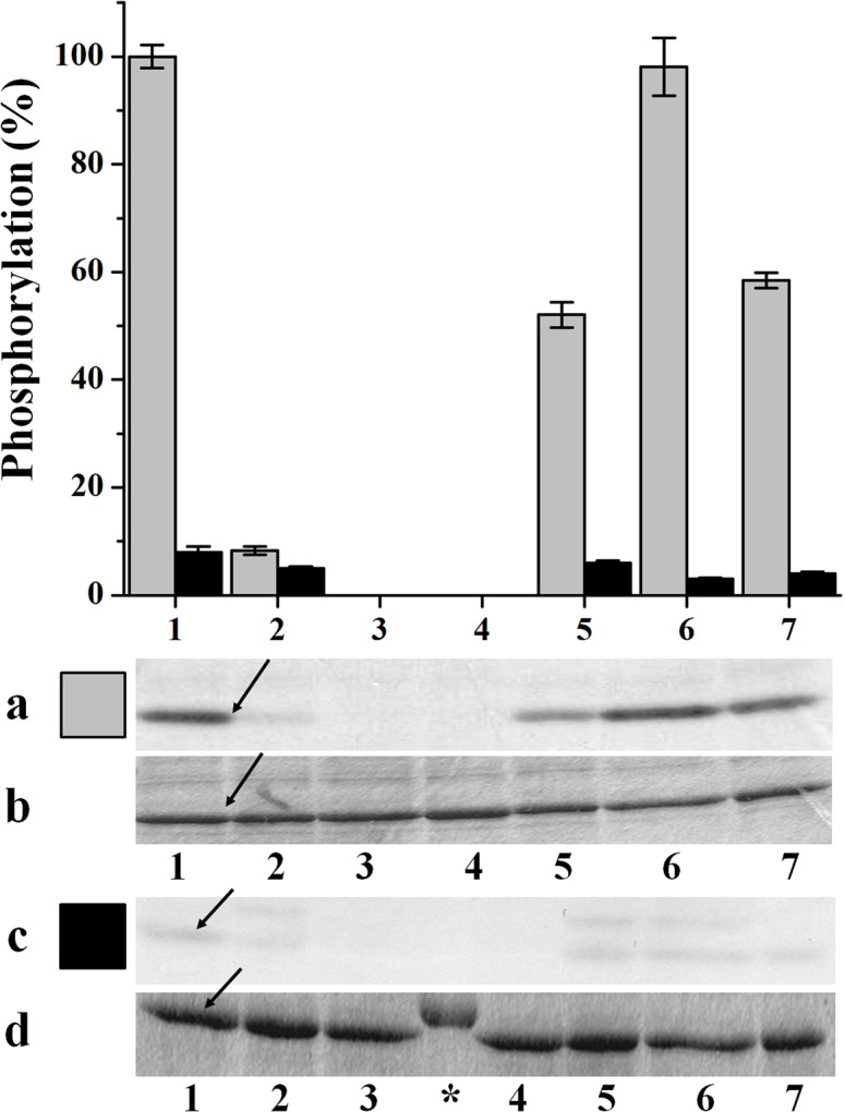
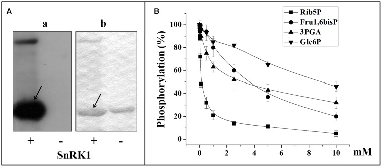
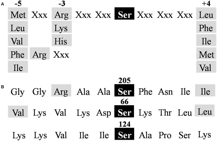
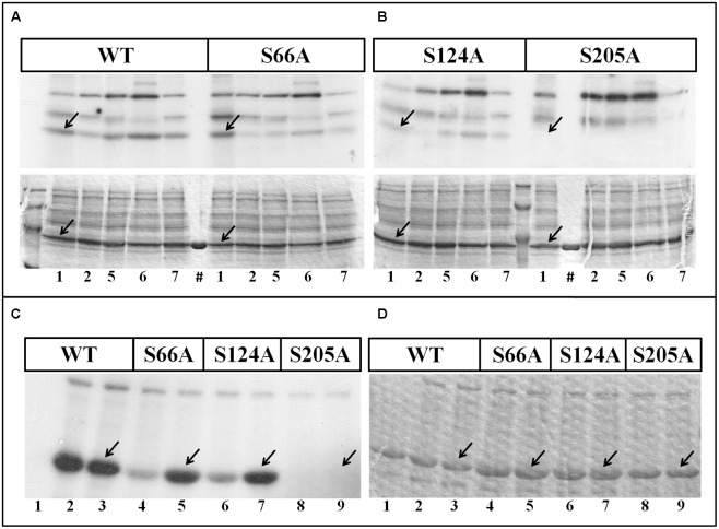
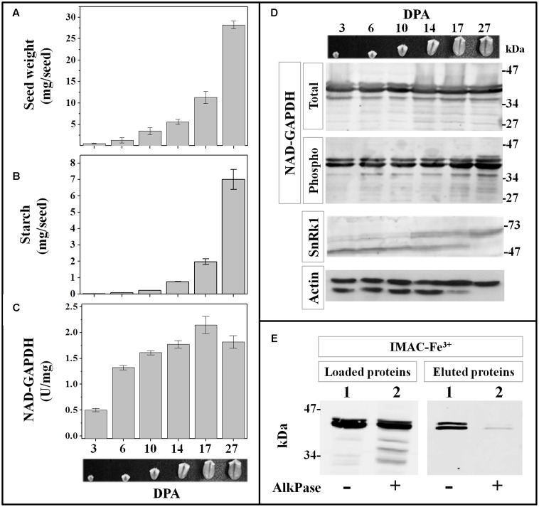
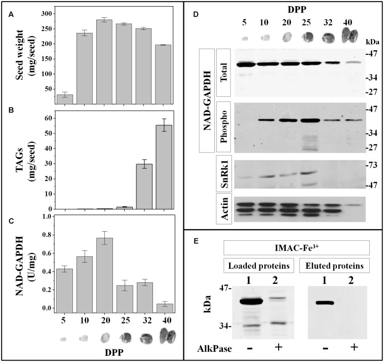
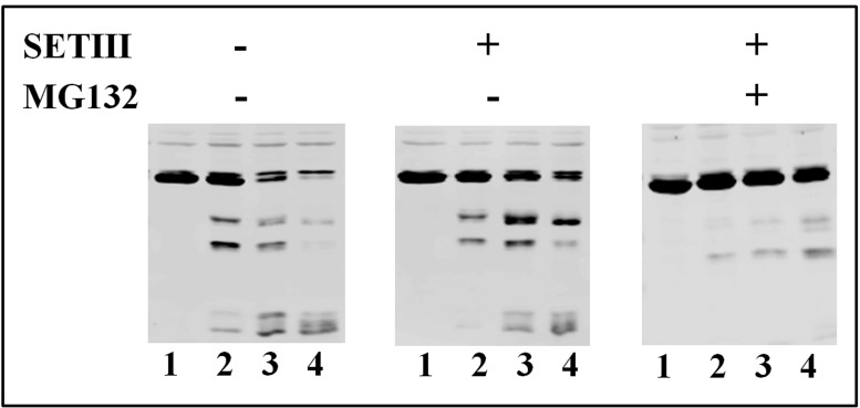
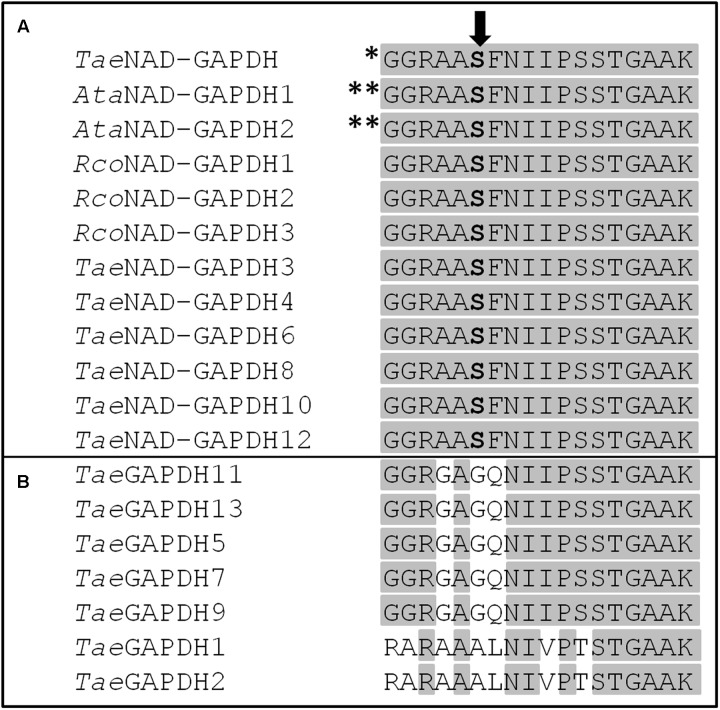
Cytosolic glyceraldehyde-3-phosphate dehydrogenase (NAD-GAPDH) is involved in a critical energetic step of glycolysis and also has many important functions besides its enzymatic activity. The recombinant wheat NAD-GAPDH was phosphorylated in vitro at Ser205 by a SNF1-Related protein kinase 1 (SnRK1) from wheat heterotrophic (but not from photosynthetic) tissues. The S205D mutant enzyme (mimicking the phosphorylated form) exhibited a significant decrease in activity but similar affinity toward substrates. Immunodetection and activity assays showed that NAD-GAPDH is phosphorylated in vivo, the enzyme depicting different activity, abundance and phosphorylation profiles during development of seeds that mainly accumulate starch (wheat) or lipids (castor oil seed). NAD-GAPDH activity gradually increases along wheat seed development, but protein levels and phosphorylation status exhibited slight changes. Conversely, in castor oil seed, the activity slightly increased and total protein levels do not significantly change in the first half of seed development but both abruptly decreased in the second part of development, when triacylglycerol synthesis and storage begin. Interestingly, phospho-NAD-GAPDH levels reached a maximum when the seed switch their metabolism to mainly support synthesis and accumulation of carbon reserves. After this point the castor oil seed NAD-GAPDH protein levels and activity highly decreased, and the protein stability assays showed that the protein would be degraded by the proteasome. The results presented herein suggest that phosphorylation of NAD-GAPDH during seed development would have impact on the partitioning of triose-phosphate between different metabolic pathways and cell compartments to support the specific carbon, energy and reducing equivalent demands during synthesis of storage products.

Keywords: castor oil seed; glyceraldehyde-3-phosphate; glycolysis; phosphorylation; seeds; wheat.


Figures

FIGURE 1
FIGURE 1
In vitro phosphorylation of recombinant TaeNAD-GAPDH by wheat seed and leaf extracts. Schematic representation of NAD-GAPDH phosphorylation by different plant protein kinases extracted from seeds (gray) and leaves (black). NAD-GAPDH phosphorylation reactions resolved by SDS-PAGE and revealed by autoradiography (a,c) to visualize the NAD-GAPDH radioactivity incorporation from the [32P]ATP or stained with Coomassie Blue (b,d) to visualize all the proteins after phosphorylation by the plant kinases extracted from seeds and leaves, respectively. The NAD-GAPDH phosphorylation (%) was calculated from autoradiography using LabImage Version 2.7.0 (free edition) and all data are means of three independent experiments and reproducible with differences below ± 10%. Numbers indicate phosphorylation conditions for (1) WPK4, (2) SOS2, (3) GSK3, (4) MAPK, (5), CKII, (6) Tsl, and (7) CDPK. Arrows indicate the migration of NAD-GAPDH, of molecular mass in the range of 37 kDa. () Molecular mass marker.
FIGURE 2
FIGURE 2
In vitro phosphorylation of recombinant TaeNAD-GAPDH by a SnRK1 purified from wheat seed. (A) NAD-GAPDH radioactivity incorporation from the [32P]ATP after phosphorylation by the purified SnRK1 resolved by SDS-PAGE and revealed by storing phospho-screen exposure and scanning with the TyphoonTM system (a) or stained with Coomassie Blue (b). Arrows indicate the migration of NAD-GAPDH. (B) Schematic representation of NAD-GAPDH phosphorylation by SnRK1 under the presence of different concentrations of Rib5P, Fru1,6bisP, 3PGA, or Glc6P. For schematic representations NAD-GAPDH phosphorylation (%) values were calculated from autoradiography using LabImage Version 2.7.0 (free edition) and all data are means of three independent experiments and reproducible with differences below ± 10%.
FIGURE 3
FIGURE 3
Identification of TaeNAD-GAPDH phosphorylation motifs. (A) Consensus sequence for SnRK1 targets (Halford et al., 2003). (B) NAD-GAPDH sequences identified as putative phosphorylation sites for SnRK1 in TaeNAD-GAPDH (NCBI N° ABQ81648.1). Residues required for recognition are highlighted in gray.
FIGURE 4
FIGURE 4
Phosphorylation of TaeNAD-GAPDH versions. (A,B) In vitro phosphorylation of NAD-GAPDH wild type and mutant S66A (A), and NAD-GAPDH mutant S124A and S205A (B), with plant extracts under kinases phosphorylation conditions for (1) WPK4, (2) SOS2, (5) CKII, (6) Tsl, and (7) CDPK. Incorporation of [32P]ATP was detected by autoradiography (upper image) of SDS-PAGE gel stained with Coomassie Blue (bottom image). (#) Recombinant proteins in the absence of the plant extract. (C,D) In vitro phosphorylation of all the NAD-GAPDH versions by the purified SnRK1 resolved by SDS-PAGE and revealed by storing phospho-screen exposure and scanning with the TyphoonTM system (C) or stained with Coomassie Blue (D). Numbers show the NAD-GAPDH versions: WT in the absence of SnRK1 (1); WT oxidized (2) or reduced (3) in the presence of SnRK1; S66A oxidized (4) or reduced (5) in the presence of SnRK1; S124A oxidized (6) or reduced (7) in the presence of SnRK1; S205A oxidized (8) or reduced (9) in the presence of SnRK1. Arrows indicate the migration of NAD-GAPDH. All the assays were also perform in the absence of any recombinant protein to identify the background phosphorylation.
FIGURE 5
FIGURE 5
NAD-GAPDH during wheat seed development. Wheat seeds were collected at different DPA along development and the seed fresh weight (A) and starch content (B) determined immediately after sample collection. (C) NAD-GAPDH activity determined in total proteins extracted from each seed sample. (D) Immunoidentification of total NAD-GAPDH in whole protein extracts, phosphorylated NAD-GAPDH after phosphoprotein enrichment by IMAC-Fe3+ chromatography, SnRK1 and actin (used as a control) in whole protein extracts. (E) Immunoidentification of NAD-GAPDH in total wheat seed protein extracts at 27 DPA non-treated (lane 1) or treated (lane 2) with alkaline phosphatase before loading and after the IMAC-Fe3+ purification. In A–C, values are means of three independent assays determined by triplicate in two different biological samples. All immunoidentifications were performed in two different biological replicates and repeated independently twice in each replicate.
FIGURE 6
FIGURE 6
NAD-GAPDH during castor oil seed development. Castor seeds were collected at different DPP along development and the seed fresh weight (A) and TAG content (B) determined immediately after sample collection. (C) NAD-GAPDH activity determined in total proteins extracted from each seed sample. (D) Immunoidentification of: total NAD-GAPDH in whole protein extracts, phosphorylated NAD-GAPDH after phosphoprotein enrichment by IMAC-Fe3+ chromatography, SnRK1 and actin (used as a control) in whole protein extracts. (E) Immunoidentification of NAD-GAPDH in total castor seed protein extracts at 25 DPP non-treated (lane 1) or treated (lane 2) with alkaline phosphatase before loading and after the IMAC-Fe3+ purification. In A–C, values are means of three independent assays determined by triplicate in two different biological samples. All immunoidentifications were performed in two different biological replicates and repeated independently twice in each replicate.
FIGURE 7
FIGURE 7
Stability of NAD-GAPDH in extracts from castor seed at 25 DPP. NAD-GAPDH was immunodetected in the respective extracts incubated at 37°C during 0 (lane 1), 60 (lane 2), 120 (lane 3), or 240 (lane 4) min; without further addition, in the presence of SETIII protease inhibitor cocktail alone and plus 10 μM of MG132 proteasome inhibitor.
FIGURE 8
FIGURE 8
Alignment of the phosphorylation motif studied in this work between different plant GAPDHs. (A) Cytosolic NAD-GAPDHs and (B) plastidic TaeGAPDHs. AtaNAD-GAPDH 1 and AtaNAD-GAPDH 2 sequences are from TAIR corresponding to AT3g04120.1 and AT1g13440.1, respectively. TaeNAD-GAPDH and RcoNAD-GAPDH 1-3 sequences are from NCBI NABQ81648.1, XP_002509572.1, XP_002511235.1, and XP_002535536.1, respectively. TaeGAPDH 1-13 sequences were from Zeng et al. (2016). TaeGAPDH 1, 2, 5, 7, 9, 11, and 13 are isoforms found in different plastids. The Ser residue studied in this work is highlighted in bold. The amino acids conserved in all proteins are highlighted in gray. Phosphorylation site identified in vitro in this work. ∗∗Phosphorylation sites identified in vivo for NAD-GAPDH in Arabidopsis with high number of items according to PhosPhAt database.

References

    1. Baud S., Graham I. A. (2006). A spatiotemporal analysis of enzymatic activities associated with carbon metabolism in wild-type and mutant embryos of Arabidopsis using in situ histochemistry. Plant J. 46 155–169. 10.1111/j.1365-313X.2006.02682.x - DOI - PubMed
    1. Baud S., Lepiniec L. (2010). Physiological and developmental regulation of seed oil production. Prog. Lipid Res. 49 235–249. 10.1016/j.plipres.2010.01.001 - DOI - PubMed
    1. Boyer P. D., Krebs E. G. (1986). The Enzymes: Control by Phosphorylation. Orlando, FL: Academic Press, Inc.
    1. Bradford M. M. (1976). A rapid and sensitive method for the quantitation of microgram quantities of protein utilizing the principle of protein-dye binding. Anal. Biochem. 72 248–254. 10.1016/0003-2697(76)90527-3 - DOI - PubMed
    1. Briarty L. G., Hughes C. E., Evers A. D. (1979). The developing endosperm of wheat-A stereological analysis. Ann. Bot. 44 641–658. 10.1093/oxfordjournals.aob.a085779 - DOI