

Retrograde Cerebral Air Embolism in a Patient with Intestinal Necrosis: A Case Report
- PMID: 28443583
- PMCID: PMC5615971
- DOI: 10.4274/balkanmedj.2016.0292
Retrograde Cerebral Air Embolism in a Patient with Intestinal Necrosis: A Case Report
Abstract
Background: Cerebral venous air embolism is a severe clinical condition related to an unfavourable outcome in patients with neurological impairment. Cerebral venous air embolism may occur secondarily to arterial or venous interventions. A rare mechanism of cerebral venous air embolism is retrograde embolism, which is characterized by gas flow in a direction that is opposite to that of the normal blood flow.
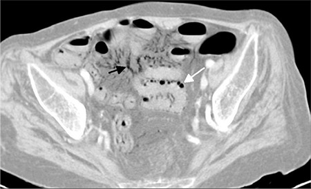
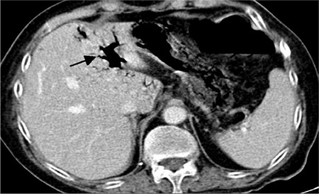
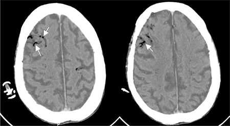
Case report: A 69-year-old female was admitted to our hospital with shortness of breath and abdominal pain. Abdominal computed tomography revealed intramural gas in the bowel and free gas in the mesenteric veins and portal vein. Cranial computed tomography, which was performed due to impaired consciousness, demonstrated cerebral air embolism with the appearance of a gyriform pattern. A bedside echocardiography and chest computed tomography revealed no evidence of right-to-left shunt.
Conclusion: Cerebral venous air embolism may occur after pneumatosis intestinalis by a retrograde flow of air from the mesenteric veins and portal vein. Low cardiac output and supine position are contributing factors for a retrograde flow of air bubbles into the venous circulation of the brain.
Keywords: Mesenteric ischaemia; air embolism; necrosis multidetector computed tomography..
Conflict of interest statement
Figures
References
-
- Bothma PA, Schlimp CJ. II. Retrograde cerebral venous gas embolism: are we missing too many cases? Br J Anaesth. 2014;112:401–4. - PubMed
-
- Ho LM, Paulson EK, Thompson WM. Pneumatosis intestinalis in the adult: benign to life-threatening causes. AJR Am J Roentgenol. 2007;188:1604–13. - PubMed
-
- Heckmann JG, Lang CJ, Kindler K, Huk W, Erbguth FJ, Neundörfer B. Neurologic manifestations of cerebral air embolism as a complication of central venous catheterization. Crit Care Med. 2000;28:1621–5. - PubMed
-
- Laurent PE, Coulange M, Bartoli C, Louis G, Souteyrand P, Gorincour G. Retrograde cerebral venous air embolism: a rare cause of intracranial gas. Diagn Interv Imaging. 2014;95:1113–5. - PubMed
Publication types
MeSH terms
LinkOut - more resources
Full Text Sources
Other Literature Sources