Case Reports
. 2017 Feb;22(1):110-125.
doi: 10.1590/2177-6709.22.1.110-125.sar.

Non-surgical treatment of transverse deficiency in adults using Microimplant-assisted Rapid Palatal Expansion (MARPE)

Affiliations
Case Reports

Non-surgical treatment of transverse deficiency in adults using Microimplant-assisted Rapid Palatal Expansion (MARPE)

Daniel Paludo Brunetto et al. Dental Press J Orthod. 2017 Feb.

Abstract

Introduction: Maxillary transverse deficiency is a highly prevalent malocclusion present in all age groups, from primary to permanent dentition. If not treated on time, it can aggravate and evolve to a more complex malocclusion, hindering facial growth and development. Aside from the occlusal consequences, the deficiency can bring about serious respiratory problems as well, due to the consequent nasal constriction usually associated. In growing patients, this condition can be easily handled with a conventional rapid palatal expansion. However, mature patients are frequently subjected to a more invasive procedure, the surgically-assisted rapid palatal expansion (SARPE). More recently, researches have demonstrated that it is possible to expand the maxilla in grown patients without performing osteotomies, but using microimplants anchorage instead. This novel technique is called microimplant-assisted rapid palatal expansion (MARPE).

Objective: The aim of the present article was to demonstrate and discuss a MARPE technique developed by Dr. Won Moon and colleagues at University of California - Los Angeles (UCLA).

Methods: All laboratory and clinical steps needed for its correct execution are thoroughly described. For better comprehension, a mature patient case is reported, detailing all the treatment progress and results obtained.

Conclusion: It was concluded that the demonstrated technique could be an interesting alternative to SARPE in the majority of non-growing patients with maxillary transverse deficiency. The present patient showed important occlusal and respiratory benefits following the procedure, without requiring any surgical intervention.

Introdução:: a deficiência transversa da maxila é uma má oclusão com alta prevalência em todas as faixas etárias, da dentição decídua à permanente. Se não for corrigida, pode agravar-se com o passar do tempo, prejudicando o crescimento e desenvolvimento facial. Além dos prejuízos oclusais, essa deficiência pode trazer problemas respiratórios também severos, devido à consequente constrição da cavidade nasal. Em pacientes em crescimento, a sua resolução é relativamente simples, por meio da expansão rápida convencional da maxila. Porém, os pacientes já maduros geralmente são encaminhados para um procedimento mais invasivo, a expansão rápida de maxila assistida cirurgicamente (SARPE). Mais recentemente, pesquisadores têm demonstrado que é possível executar a expansão palatal esquelética em pacientes adultos sem auxílio de osteotomias, mas sim com auxílio de mini-implantes. Essa técnica é denominada Microimplant-Assisted Rapid Palatal Expansion, ou MARPE.

Objetivo:: o objetivo do presente artigo é demonstrar e discutir uma das técnicas disponíveis de MARPE, desenvolvida por Won Moon e colaboradores, na University of California, Los Angeles (UCLA).

Métodos:: a técnica encontra-se detalhadamente descrita, com as etapas laboratoriais e clínicas que devem ser seguidas para sua correta execução. Para descrevê-la, é apresentado o caso clínico de uma paciente adulta, detalhando toda a sequência do tratamento e os resultados obtidos.

Conclusão:: a técnica apresentada pode ser uma alternativa não invasiva à SARPE na resolução da deficiência transversa de maxila, podendo ser empregada na maioria dos pacientes com crescimento facial finalizado. A paciente apresentada demonstrou benefícios significativos nos aspectos oclusal e respiratório, sem a necessidade de intervenção cirúrgica.


Conflict of interest statement

The authors report no commercial, proprietary or financial interest in the products or companies described in this article.

Patients displayed in this article previously approved the use of their facial and intraoral photographs.

Figures

Figure 1
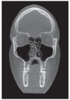
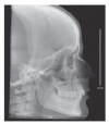
Figure 1. Lateral radiograph and coronal CBCT slice of a patient with true mandibular prognathism and excessive vertical growth; images show bilateral skeletal posterior crossbite due to mandibular anterior position and lower tongue posture.
Figure 2
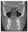
Figure 2. Coronal slice shows maxillary transverse deficiency and, consequently, nasal cavity narrowing in adult mouth-breather with moderate OSAS (AHI = 15.9). Are also noticeable the high-arched palate, low tongue position and anatomic disorders of nasal cavity (turbinate hypertrophy and septal deviation).
Figure 3
Figure 3. Laboratory procedures: midline (palatal raphe) and limit between soft and hard palate (clinically determined) traced using lead pencil on model; selection of MSE with greatest expansion capacity (8, 10 or 12 mm) that can be placed flush to palatal mucosa; appliance wire segments bended to outline palate curvature, holding at least a 2 mm gap from the mucosa; expander should be centralized to palatal raphe and placed at the most posterior position possible, slightly before limit between soft and hard palate; soldering of wire segments onto the bands, followed by polishing; posterior view shows that expander is flush to palatal mucosa, but should not touch it.
Figure 4
Figure 4. Clinical visit: Expander clinical proof, topical anesthesia applied, and expander cemented; after expander is cemented (as shown on plaster model, for teaching purposes), infiltrative anesthesia is applied close to orifices of MI; after region is anesthetized, MI are placed paying special attention to anteroposterior and lateral inclination. Most cases do not need previous perforation. Index finger of one hand should hold the digital key, and index and thumb of other hand firmly moves key counterclockwise. During posterior MI placement, patient mouth should be wide open to ensure correct anteroposterior inclination. MI should touch expander gently and not push it toward the mucosa. After the four MI are placed, initial stability is tested using clinical tweezers. After confirmation, expander is activated two to three times.
Figure 5
Figure 5. Force application too far from bone/microimplant interface, resulting in MI deformation.
Figure 6
Figure 6. Dry skull shows relation between the pterygoid plates of sphenoid bone and maxilla. These structures provide great resistance to lateral forces applied by the expander, and connection between them has to be split apart for real skeletal expansion.
Figure 7
Figure 7. When expander is placed at a more posterior position, forces concentrate closer to the pterygoid plates, structures that offer great resistance to palatal expansion. Therefore, occurs a parallel opening of the palatine suture anteroposteriorly and vertically, differently from conventional expansion, in which opening takes the form of a "V" (broader in anterior region).
Figure 8
Figure 8. A) In conventional palatal expansion, forces are applied to teeth, below the center of resistance of the maxilla. This system of forces generates buccal dentoalveolar tipping and an inverted-V opening (coronal view), indicated by the red dotted lines. The amount of momentum generated is directly associated with palatal depth. B) in MARPE, forces are applied directly into the maxillary center of resistance by means of the MI, which practically eliminates inclination forces of posterior teeth and promotes more parallel suture opening in a coronal view (indicated by red dotted lines).
Figure 9
Figure 9. Maxillary occlusal photograph showing removal of anterior wire segments of MSE, to improve vertical fit in a very narrow and high-arched palate.
Figure 10
Figure 10. Initial facial photographs: harmonic profile and proportional face thirds. Smiling photo shows excessive buccal corridors, particularly on left side.
Figure 11
Figure 11. Initial occlusal photographs; moderate crowding in mandibular arch due to constriction caused by the maxilla. Note the lingual inclination of left posterior teeth in maxillary arch.
Figure 12
Figure 12. Initial intraoral photographs: good occlusal relationship on the right side and edge-to-edge relationship of left canines (Class II); left superior buccal segment in crossbite.
Figure 13
Figure 13. Lateral radiograph obtained from CBCT shows harmonic maxillomandibular skeletal relationship and satisfactory position of maxillary and mandibular incisors: note that mandibular ramus heights are asymmetric.
Figure 14
Figure 14. Coronal CBCT slice at the level of maxillary first molars shows excessive palatal inclination of these teeth; tongue is at a low position; measurement indicated maxillary constriction and, consequently, nasal cavity constriction.
Figure 15
Figure 15. Sagittal slice shows incorrect position of condyles into articular fossa, especially on the right side (contralateral to the posterior crossbite).
Figure 16
Figure 16. Baseline in-home polysomnography shows an AHI of 7.9, defined as mild OSAS; despite that, patient has good oxygen saturation along the night.
Figure 17
Figure 17. Photograph taken after 20 activations (4 mm); interincisal diastema confirms suture opening. Discrete anterior open bite appears due to an overjet reduction of the posterior teeth in crossbite, which generates premature occlusal contacts.
Figure 18
Figure 18. Maxillary occlusal photograph taken after 34 activations (6.8 mm). Absence of tooth or alveolar bone tipping as left posterior teeth still show palatal inclination.
Figure 19
Figure 19. Photograph taken during MSE removal, after 44 activations (8.8 mm); high mechanical resistance bent expander built-in support wires; MI with no deformation when removed are signs of ideal progression of the expansion.
Figure 20
Figure 20. Intraoral photographs after expansion. Spaces created by the expansion were distributed along maxillary arch. When manipulated to centric occlusion, a substantial transverse increase was evident and the posterior crossbite corrected. Following the expander removal after the recommended retention time, right posterior teeth will return to their adequate inclination. A broader maxillary arch allows for the expansion of mandibular arch, which was also constricted.
Figure 21
Figure 21. Occlusal photo taken at Hyrax tie-lace; smaller expansion on left side, limited by crossbite occluding forces. Left posterior teeth aligned to canine and second molar, which reduces time of treatment with fixed appliance.
Figure 22
Figure 22. Facial photographs after expansion. Improved mandibular position and buccal corridors as a result of skeletal and dental expansion promoted by the two expanders.
Figure 23
Figure 23. CBCT slices show homogenous suture opening along anterior and posterior regions and uniform separation of the hemimaxillae.
Figure 24
Figure 24. Coronal slice after expansion shows more favorable buccolingual inclination (torque) of posterior maxillary teeth. Also, nasal cavity floor is 23.2 mm wide, larger than at baseline (15 mm). Wax-bite registration was sent to the radiologic laboratory, but the CBCT scan was obtained at maximal intercuspation, suggesting that posterior left crossbite is still present. There was no reason to irradiate the patient again.
Figure 25
Figure 25. Post-expansion in-home polysomnography depicted an AHI of 1.5, a substantial reduction from baseline value of 7.9. Patient presented a more homogeneous breathing pattern during sleep. Clinical symptoms have also improved significantly.
Figure 26
Figure 26. Photographs three months after inferior fixed appliance delivery. Note anterior overbite improvement, matching of the dental midlines and almost complete crowding dissolution.
Figure 27
Figure 27. The overjet created on the left cuspids will allow for an increase on the intercanine distance in the mandibular arch, completing its ideal alignment. On the right side, the upper cuspid will also present with some overjet when its torque is corrected.

Similar articles

Cited by

References

    1. Kurol J, Berglund L. Longitudinal study and cost-benefit analysis of the effect of early treatment of posterior cross-bites in the primary dentition. Eur J Orthod. 1992;14(3):173–179. - PubMed
    1. Silva OG, Filho, Santamaria M, Jr, Capelozza L., Filho Epidemiology of posterior crossbite in the primary dentition. J Clin Pediatr Dent. 2007;32(1):73–78. - PubMed
    1. Modeer T, Odenrick L, Lindner A. Sucking habits and their relation to posterior cross-bite in 4-year-old children. Scand J Dent Res. 1982;90(4):323–328. - PubMed
    1. Lione R, Franchi L, Huanca Ghislanzoni LT, Primozic J, Buongiorno M, Cozza P. Palatal surface and volume in mouth-breathing subjects evaluated with three-dimensional analysis of digital dental casts-a controlled study. Eur J Orthod. 2015;37(1):101–104. - PubMed
    1. Moss ML. The functional matrix hypothesis revisited. 1. The role of mechanotransduction. Am J Orthod Dentofacial Orthop. 1997 Jul;112(1):8–11. - PubMed

Publication types