. 2017 Jul 1;26(13):2480-2492.
doi: 10.1093/hmg/ddx143.

Arl3 and RP2 regulate the trafficking of ciliary tip kinesins

Affiliations

Arl3 and RP2 regulate the trafficking of ciliary tip kinesins

Nele Schwarz et al. Hum Mol Genet. .

Erratum in

  • Arl3 and RP2 regulate the trafficking of ciliary tip kinesins.
    Schwarz N, Lane A, Jovanovic K, Parfitt DA, Aguila M, Thompson CL, da Cruz L, Coffey PJ, Chapple JP, Hardcastle AJ, Cheetham ME. Schwarz N, et al. Hum Mol Genet. 2017 Sep 1;26(17):3451. doi: 10.1093/hmg/ddx245. Hum Mol Genet. 2017. PMID: 28854704 Free PMC article. No abstract available.

Abstract

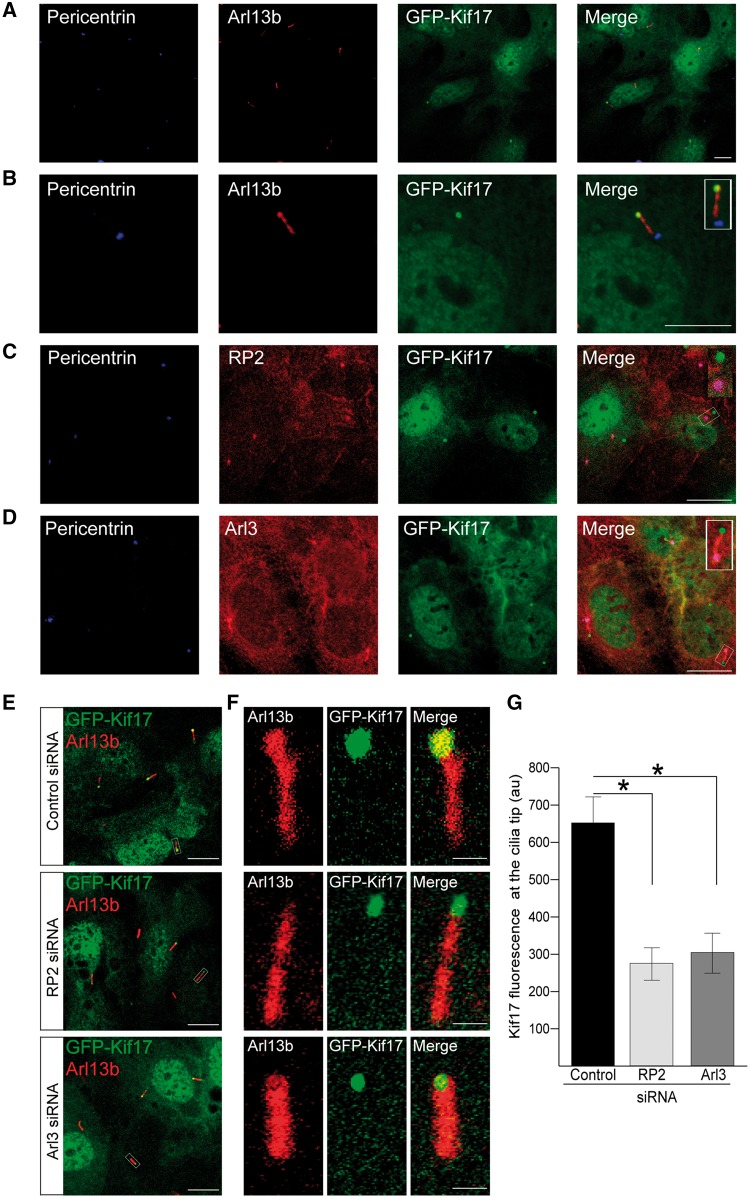
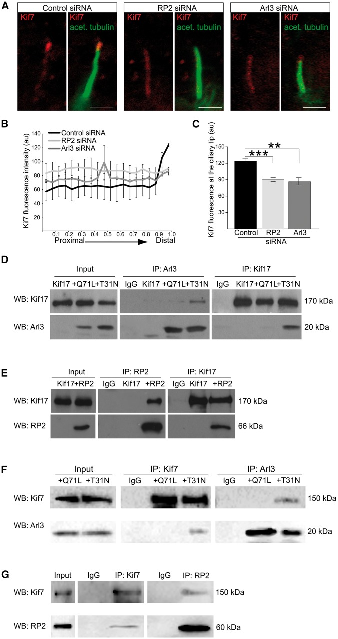
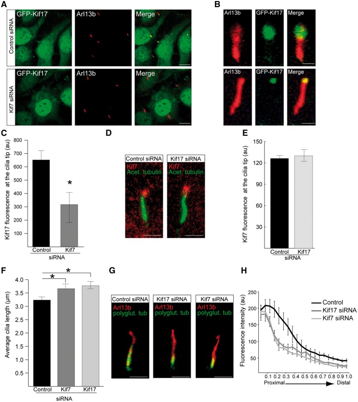
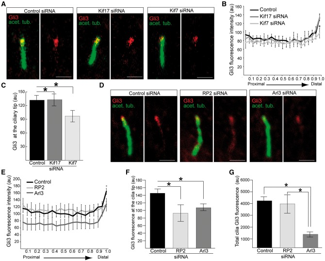
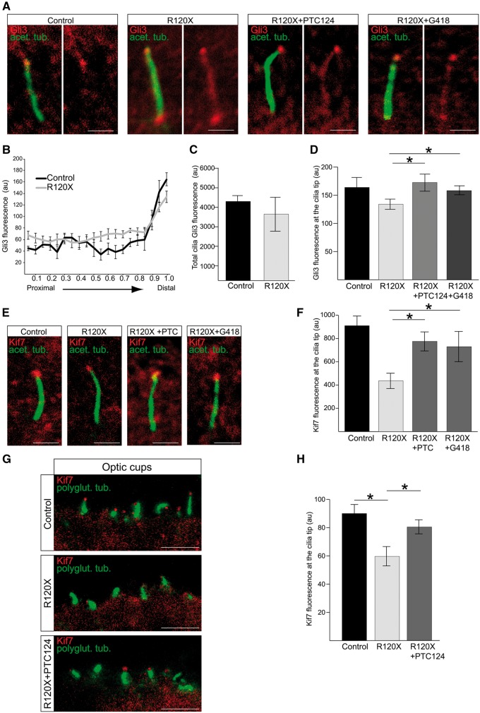
Ciliary trafficking defects are the underlying cause of many ciliopathies, including Retinitis Pigmentosa (RP). Anterograde intraflagellar transport (IFT) is mediated by kinesin motor proteins; however, the function of the homodimeric Kif17 motor in cilia is poorly understood, whereas Kif7 is known to play an important role in stabilizing cilia tips. Here we identified the ciliary tip kinesins Kif7 and Kif17 as novel interaction partners of the small GTPase Arl3 and its regulatory GTPase activating protein (GAP) Retinitis Pigmentosa 2 (RP2). We show that Arl3 and RP2 mediate the localization of GFP-Kif17 to the cilia tip and competitive binding of RP2 and Arl3 with Kif17 complexes. RP2 and Arl3 also interact with another ciliary tip kinesin, Kif7, which is a conserved regulator of Hedgehog (Hh) signaling. siRNA-mediated loss of RP2 or Arl3 reduced the level of Kif7 at the cilia tip. This was further validated by reduced levels of Kif7 at cilia tips detected in fibroblasts and induced pluripotent stem cell (iPSC) 3D optic cups derived from a patient carrying an RP2 nonsense mutation c.519C > T (p.R120X), which lack detectable RP2 protein. Translational read-through inducing drugs (TRIDs), such as PTC124, were able to restore Kif7 levels at the ciliary tip of RP2 null cells. Collectively, our findings suggest that RP2 and Arl3 regulate the trafficking of specific kinesins to cilia tips and provide additional evidence that TRIDs could be clinically beneficial for patients with this retinal degeneration.


Figures

Figure 1
Figure 1
RP2 and Arl3 mediate GFP-Kif17 cilia tip localisation. (A) In stable GFP-Kif17 hTERT-RPE cells Kif17 (green) localises to the nucleus, the cytoplasm and to cilia tips, as indicated by the cilia marker Arl13b (red) and the basal body marker pericentrin (blue). Scale bar 10 μm. (B) Zoomed image of GFP-Kif17 hTERT-RPE cell line. Inset shows a zoomed in image of the cilia tip. Scale bar 10 μm. (C) RP2 (red) localises to the basal body (pericentrin, blue) of cilia in GFP-Kif17 hTERT-RPE cells. Scale bar 10 μm. (D) Arl3 (red) localises to the basal body (pericentrin, blue) and the along the ciliary axoneme in GFP-Kif17 hTERT-RPE cells. Scale bar 10 μm. (E) siRNA-mediated knockdown of RP2 or Arl3 decreases GFP-Kif17 at cilia tips. Cilia marker Arl13b (red). Scale bar 10 μm. (F) Zoomed image of GFP-Kif17 at cilia tips following control, RP2 or Arl3 siRNA transfection. Cilia marker Arl13b (red). Scale bar 1 μm. (G) Quantification of GFP-Kif17 fluorescence at cilia tips after control, RP2 or Arl3 siRNA transfection. n = 3 independent experiments. *P ≤ 0.05, values are mean ± SEM.
Figure 2
Figure 2
RP2 and Arl3 mediate the localisation of Kif7 to cilia tips. (A) siRNA-mediated knockdown of RP2 or Arl3 in hTERT-RPE cells decreases Kif7 (red) at cilia tips. Cilia marker acetylated α-tubulin (green). Scale bar 1 μm. (B) Fluorescence intensity of Kif7 along the axoneme, normalised for cilium length. n = 3 independent experiments, with a total of 90 cilia measured for each siRNA treatment. For RP2 siRNA, P ≤ 0.0001 for the last measurement of the cilia tip. For Arl3 siRNA, P ≤ 0.05 and P ≤ 0.001, for the distal part of cilia. Values are mean ± SEM. (C) Quantification of Kif7 fluorescence at cilia tips. n = 3 independent experiments, with a total of 90 cilia measured for each siRNA treatment. ***P ≤ 0.0001, **P ≤ 0.001, values are mean ± SEM. (D-G) Reciprocal co-IPs of different combinations; (D) GFP-Kif17 with the myc-tagged Arl3 conformational mimics Arl3-GTP (Q71L) and Arl3-GDP (T31N) shows that GFP-Kif17 preferentially binds to Arl3-T31N. (E) GFP-Kif17 binds to RP2. (F) V5-Kif7 with the myc-tagged Arl3 conformational mimics Arl3-GTP (Q71L) and Arl3-GDP (T31N) shows that Kif7 preferentially binds to Arl3-T31N. Kif7 and Arl3 were immunopurified with the V5 and myc antibody, respectively. (G) V5-Kif7 binds to RP2. Kif7 and RP2 were immunopurified with the V5 antibody and GFP-trap magnetic beads, respectively.
Figure 3
Figure 3
Kif7 functions upstream of Kif17 in cilia and both kinesins contribute to cilia stability. (A) siRNA-mediated knockdown of Kif7 in GFP-Kif17 hTERT-RPE cells reduces GFP-Kif17 at cilia tips. Scale bar 10 μm. (B) Zoomed in image of GFP-Kif17 cilia localisation following control or Kif7 siRNA treatment. Scale bar 1 μm. (C) Quantification of GFP-Kif17 fluorescence at the cilia tip following siRNA treatment. n = 3 independent experiments with 150 cilia measured for each siRNA. *P ≤ 0.05, values are mean ± SEM. (D) hTERT-RPE cells stained for Kif7 (red) and acetylated α-tubulin (green) following siRNA treatment. Scale bar 1 μm. (E) Quantification of Kif7 fluorescence at the cilia tip following siRNA treatment. n = 3 independent experiments with 150 cilia analysed for each siRNA. (F) Cilia length in hTERT-RPE cells treated with Kif7 and Kif17 siRNA is increased compared to controls. n = 3 independent experiments with 150 cilia measured for each siRNA. *P ≤ 0.05, values are mean ± SEM. (G) hTERT-RPE cilia treated with Kif17 or Kif7 siRNA show decreased levels of polyglutamylated tubulin (green) compared to controls. Cilia marker, Arl13b (red). Scale bar 1 μm. (H) Fluorescence intensity of polyglutamylated tubulin, normalised for cilium length. n = 3 independent experiments, with a total of 90 cilia measured for each siRNA treatment. For Kif7 and Kif17 siRNA, P ≤ 0.05 for the proximal part of cilia. Values are mean ± SEM.
Figure 4
Figure 4
Loss of RP2 and Arl3 mediated reduction of Kif7 affects the ciliary tip localisation of Gli3. (A) hTERT-RPE cells treated with siRNA against Kif7, but not Kif17, reduces Gli3 (red) at cilia tips. Cilia marker acetylated α-tubulin (green). Scale bar 1 μm. (B) Fluorescence intensity of Gli3, normalised for cilium length. n = 3 independent experiments, with a total of 90 cilia measured for each siRNA treatment. *P ≤ 0.05, values are mean ± SEM. (C) Quantification of Gli3 fluorescence at the cilia tip following Kif7 or Kif17 siRNA treatment. n = 3 independent experiments with 150 cilia analysed for each siRNA. *P ≤ 0.05, values are mean ± SEM. (D) hTERT-RPE cells treated with siRNA against RP2 or Arl3 reduces Gli3 (red) at cilia tips. Cilia marker acetylated α-tubulin (green). Scale bar 1 μm. (E) Fluorescence intensity of Gli3, normalised for cilium length. n = 3 independent experiments, with a total of 90 cilia measured for each siRNA treatment. For RP2 and Arl3 siRNA, *P ≤ 0.05 for the distal part of cilia. For Arl3 siRNA, P ≤ 0.05 for the proximal part of cilia. Values are mean ± SEM. (F) Gli3 fluorescence is reduced at the cilia tip following RP2 or Arl3 siRNA treatment. n = 3 independent experiments with 150 cilia analysed for each siRNA. *P ≤ 0.05, values are mean ± SEM. (G) Total Gli3 fluorescence is reduced in cilia in Arl3 knockdown cells, compared to RP2 knockouts and control. n = 3 independent experiments with 150 cilia analysed for each siRNA. *P ≤ 0.05, values are mean ± SEM.
Figure 5
Figure 5
Treatment with TRIDs rescues Kif7 cilia localisation in RP2 R120X fibroblasts and 3D optic cups. (A) Ciliary tip localisation of Gli3 is restored in RP2 R120X fibroblasts. Cells were treated with a single 24-h dose of either 750 μM G418 or 10 μg/ml PTC124 and stained for Gli3 (red) and acetylated α-tubulin (green). Scale bar 1 μm. (B) Fluorescence intensity of Gli3, normalised for cilium length in control and RP2 R120X fibroblasts shows an increase of Gli3 fluorescence along the ciliary axoneme and reduction of Gli3 at the cilia tips. For RP2 R120X, P ≤ 0.05 at the proximal, central and distal part of cilia. n = 3 independent experiments, with a total of 90 cilia measured. Values are mean ± SEM (C) Total cilia Gli3 fluorescence levels in cilia from RP2 R120X fibroblasts are comparable to control cilia. (D) Quantification of Gli3 fluorescence at cilia tips. n = 3 independent experiments, with a total of 90 cilia measured. *P ≤ 0.05, values are mean ± SEM. (E) Treatment of RP2 R120X fibroblasts with a single 24-h dose of either 750 μM G418 or 10 μg/ml PTC124 restores Kif7 (red) levels at cilia tips. Cilia marker acetylated α-tubulin (green). Scale bar 1 μm. (F) Quantification of Kif7 fluorescence intensity at cilia tips in untreated R120X fibroblasts compared to control cells. This RP2-null cellular phenotype was reversed with treatment of either G418 or PTC124. n = 3 independent experiments, with a total of 90 cilia measured. *P ≤ 0.05, values are mean ± SEM. (G) PTC124 treated and untreated RP2 R120X iPSC-derived 3D optic cups were stained for Kif7 (red) and polyglutamylated tubulin (green) and compared to control 3D optic cups. Scale bar 5 μm. (H) Kif7 levels are restored at cilia tips in 3D optic cups following treatment with three doses of 10 μg/ml PTC124 over the course of seven days. n ≥ 60 cilia analysed. *P ≤ 0.05, values are mean ± SEM.

Similar articles

Cited by

References

    1. Christensen S.T., Pedersen L.B., Schneider L., Satir P. (2007) Sensory cilia and integration of signal transduction in human health and disease. Traffic, 8, 97–109. - PubMed
    1. Fliegauf M., Benzing T., Omran H. (2007) When cilia go bad: cilia defects and ciliopathies. Nat. Rev. Mol. Cell. Biol., 8, 880–893. - PubMed
    1. Hildebrandt F., Benzing T., Katsanis N. (2011) Ciliopathies. N. Engl. J. Med., 364, 1533–1543. - PMC - PubMed
    1. Hildebrandt F., Otto E. (2005) Cilia and centrosomes: a unifying pathogenic concept for cystic kidney disease? Nat. Rev. Gen., 6, 928–940. - PubMed
    1. Wheway G., Parry D.A., Johnson C.A. (2014) The role of primary cilia in the development and disease of the retina. Organogenesis, 10, 69–85. - PMC - PubMed

MeSH terms

Supplementary concepts