. 2017 Aug;174(15):2427-2443.
doi: 10.1111/bph.13836. Epub 2017 Jun 18.

Dual cyclin-dependent kinase 4/6 inhibition by PD-0332991 induces apoptosis and senescence in oesophageal squamous cell carcinoma cells

Affiliations

Dual cyclin-dependent kinase 4/6 inhibition by PD-0332991 induces apoptosis and senescence in oesophageal squamous cell carcinoma cells

Liang Chen et al. Br J Pharmacol. 2017 Aug.

Abstract

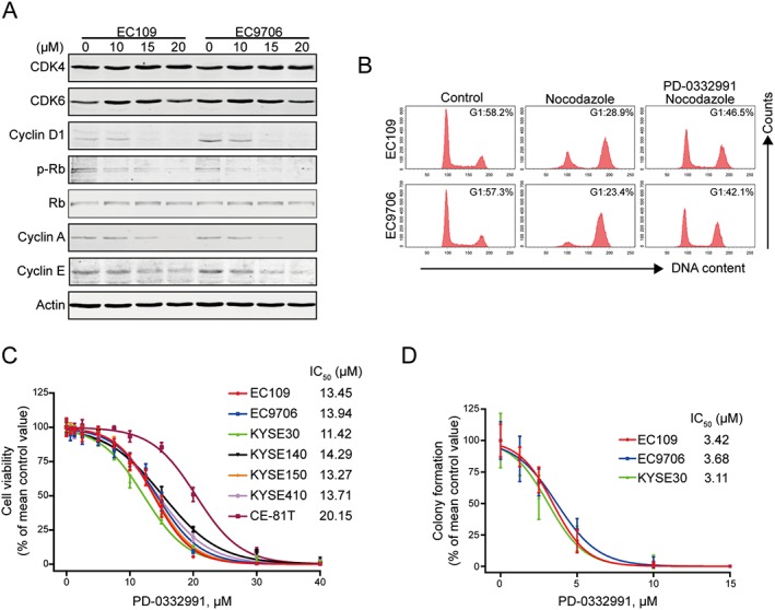
Background and purpose: Aberrant activation of the cyclin D1-cyclin-dependent kinase 4/6 (CDK4/6)-Rb signalling pathway is common in oesophageal squamous cell carcinoma (ESCC). PD-0332991, a highly specific inhibitor of CDK4/6, has potent antitumour activity against many types of cancer. The purpose of this study was to examine the in vitro and in vivo antineoplastic effect of PD-0332991 against the growth and metastasis of ESCC cells.

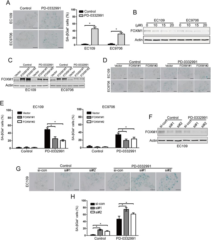
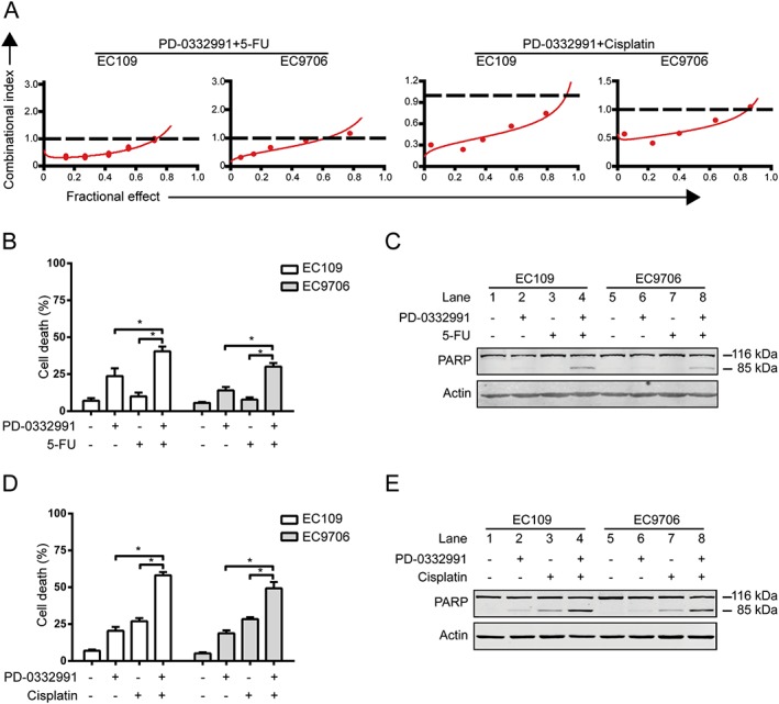
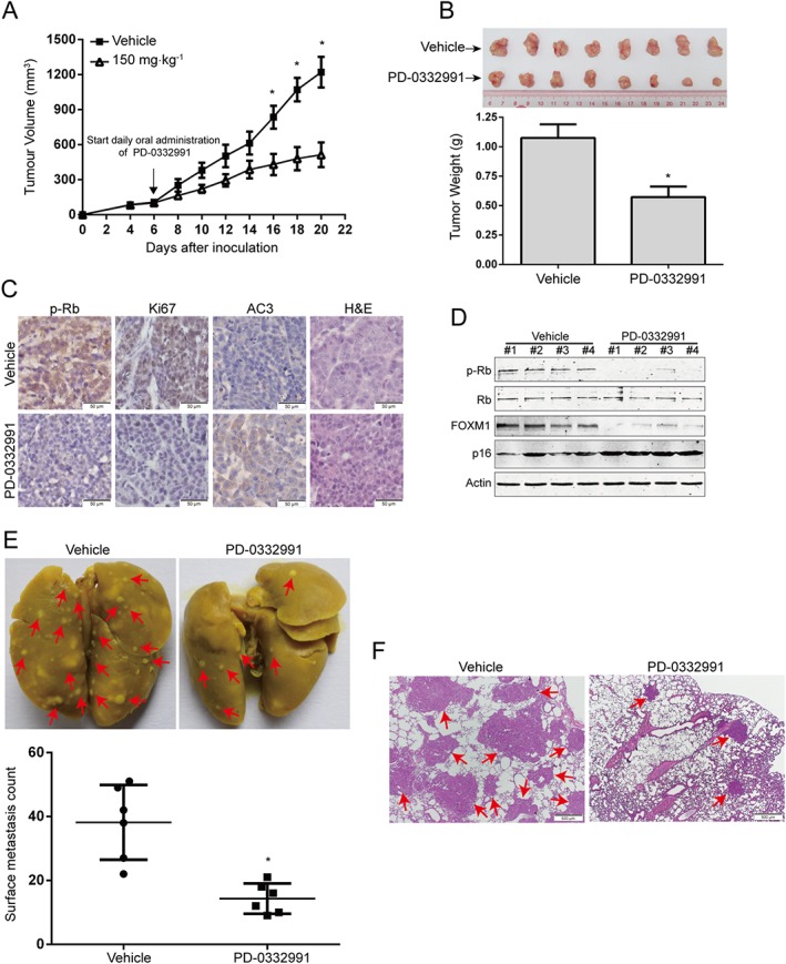
Experimental approach: Cell viability and any synergy between PD-0332991 and 5-fluorouracil or cisplatin were measured by MTS assay and CalcuSyn software respectively. Cell migration and invasion were detected by wound healing and transwell assays. Apoptosis was evaluated by flow cytometry after staining annexin V-FITC/PI. Cellular senescence was assessed by measuring SA-β-gal activity. Nude mouse xenograft models of ESCC were employed to determine the in vivo activity of PD-0332991 against tumour growth and lung metastasis.

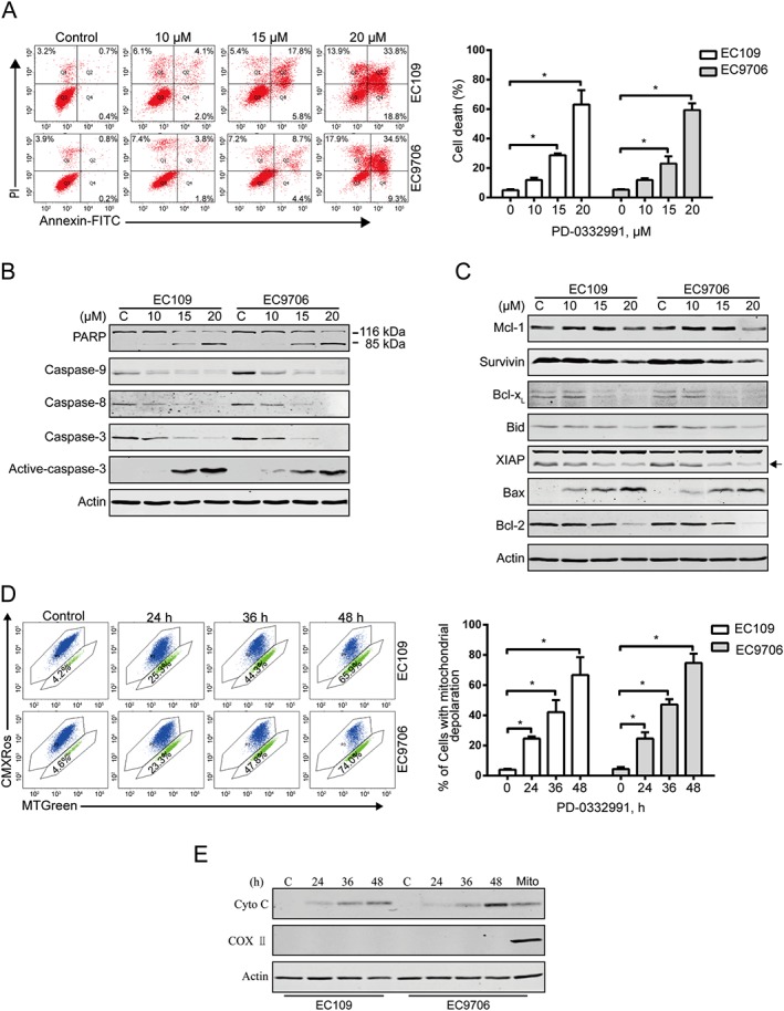
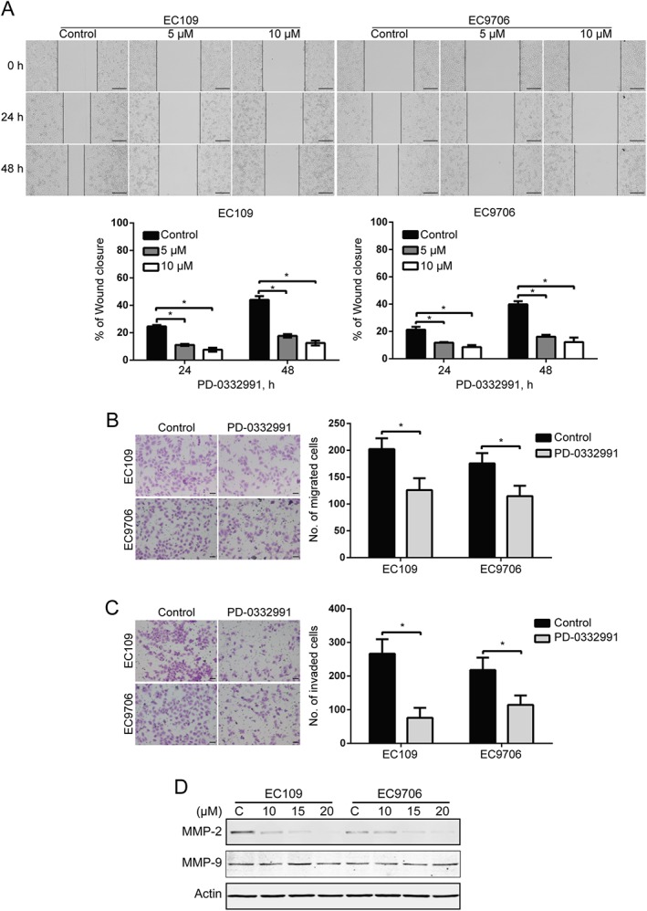
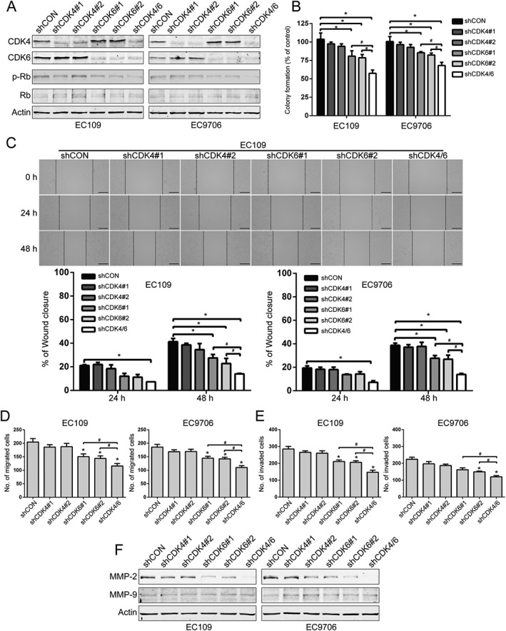
Key results: PD-0332991 inhibited cellular growth and induced mitochondrial-dependent apoptosis in ESCC cells. PD-0332991 also suppressed migration, invasion and the expression of MMP-2 in ESCC cells. Furthermore, PD-0332991 treatment caused cell senescence in a FOXM1-dependent manner. In addition, there was synergy between PD-0332991 and cisplatin or 5-fluorouracil. Importantly, the xenografted tumour experiments demonstrated that PD-0332991 potently inhibits ESCC cell growth and lung metastasis.

Conclusions and implications: PD-0332991 can elicit a strong antitumour activity against ESCC growth and metastasis and may be a promising candidate drug for the treatment of patients with ESCC. Our results warrant a clinical trial to further evaluate the efficacy of PD-0332991 in ESCC patients, even those with metastasis.


Figures

Figure 1
Figure 1
PD‐0332991 induces cellular Rb dephosphorylation and G1‐phase arrest in ESCC cells. (A) EC109 and EC9706 cells were incubated with increasing concentrations of PD‐0332991 for 48 h; immunoblotting analysis was performed with the indicated antibodies. (B) EC109 and EC9706 were treated with or without 2.5 μM PD‐0332991 for 8 h, followed by addition of 20 ng·mL−1 nocodazole for another 16 h. The cells were then fixed and stained with PI before flow cytometry analysis. (C) ESCC cells were incubated with increasing concentrations of PD‐0332991 for 72 h. Cell viability was determined with MTS assay (n = 8 per group). (D) EC109, EC9706 and KYSE30 cells were exposed to increasing concentrations of PD‐0332991 for 48 h. The cells were then washed and subjected to a drug‐free soft agar assay (n = 6 per group).
Figure 2
Figure 2
PD‐0332991 induces apoptosis in ESCC cells. (A) EC109 and EC9706 cells were treated with increasing concentrations of PD‐0332991 for 48 h and then underwent flow cytometry analysis after staining with annexin V‐FITC/PI. Right panel: the vertical axis presents the sum of the top left, top right and bottom right quadrants. Data are expressed as mean ± SD, from five independent experiments. *P < 0.05, one‐way ANOVA with post hoc intergroup comparison by the Tukey's test. (B, C) Immunoblotting analysis of apoptosis‐related proteins was performed in EC109 and EC9706 cells treated with increasing concentrations of PD‐0332991 for 48 h. Actin was used as loading control. (D) EC109 and EC9706 cells treated with or without 10 μM PD‐0332991 for 24 h, and then the mitochondrial potential was analysed by flow cytometry after staining with CMXRos and MTGreen. Right: results from five independent experiments. * P < 0.05, one‐way ANOVA with post hoc intergroup comparison by the Tukey's test. (E) PD‐0332991 led to release of cytochrome c into cytosol in ESCC cells. Levels of cytochrome c in the cytosolic extracts prepared with digitonin buffer were detected by immunoblotting. Actin was used as an internal control. The cytosolic fractionations were not contaminated as indicated by COX II.
Figure 3
Figure 3
PD‐0332991 suppresses the migration and invasion of ESCC cells. (A) EC109 and EC9706 cells were treated with 0, 5 or 10 μM PD‐0332991 for 0, 24 and 48 h after scratching. Scale bar: 200 μm. The average gap width was used to evaluate migration. Bottom: quantitative analysis of the relative breadth of the wound. The wound breadth was normalized to the initial time point (0 h). Columns and error bars represent mean ± SD (n = 6 per group). * P < 0.05, one‐way ANOVA with post hoc intergroup comparison with the Tukey's test. (B, C) EC109 and EC9706 cells were treated with 5 μM PD‐0332991 for 48 h and then underwent transwell migration (B) and invasion (C) assay. Left: representative images; Right: quantitative analysis from six random fields. Scale bar: 50 μm. Mean; error bar, SD. *P < 0.05 by Student's t‐test. (D) Western blotting analysis of whole cell lysates of EC109 and EC9706 cells that were treated with PD‐0332991 for 48 h.
Figure 4
Figure 4
Knockdown of CDK4/6 inhibits the proliferation and migration of ESCC cells. (A) Western blotting analysis of CDK4 and CDK6, as well as the phosphorylated and total levels of Rb in EC109 and EC9706 cells stably transfected with the indicated shRNA. (B) Tumourigenicity was detected by colony formation in ESCC cells that was stably knocked down for CDK4/6 by shRNA (n = 6 per group). shCON: control shRNA; shCDK4#1 and shCDK4#2: two different shRNAs targeting CDK4; shCDK6#1 and shCDK6#2, two different shRNAs targeting CDK6; shCDK4/6, both CDK4 and CDK6 shRNA. * P < 0.05 versus shCON; # P < 0.05 versus shCDK4/6; P values were obtained by one‐way ANOVA with post hoc intergroup comparison by the Tukey's test. (C–E) Wound healing (C), transwell migration (D) and invasion (E) assays of EC109 and EC9706 cell stable clones with knockdown of CDK4, CDK6 alone or both (n = 6 per group). Scale bar for wound healing: 200 μm. *, P < 0.05 versus shCON; #, P < 0.05 versus shCDK4/6; P values were obtained by one‐way ANOVA with post hoc intergroup comparison with Tukey's test. (F) Western blotting analysis of MMP‐2 and MMP‐9 expression in CDK4/6‐knockdown ESCC cells by shRNA. Actin served as a loading control.
Figure 5
Figure 5
PD‐0332991 induces FOXM1‐dependent senescence in ESCC cells. (A) Senescence phenotype analysis was conducted in ESCC cells after treatment with 2.5 μM PD‐0332991 for 6 days. Left: representative images of SA‐β‐galactosidase assay. Right: quantitative analysis of SA‐β‐galactosidase positive cells. The percentages of cells with senescence morphology are from six random fields. Scale bar: 100 μm. Mean; error bar, SD. * P < 0.05 by Student's t‐test. (B) Immunoblotting of FOXM1 in EC109 and EC9706 cells was analysed after treatment with increasing concentrations of PD‐0332991 for 48 h. (C–E) ESCC cells ectopically overexpressing FOXM1 were exposed to 2.5 μM PD‐0332991 for 6 days, Western blotting of FOXM1 (C) and SA‐β‐galactosidase positive cells (D, E) were analysed. (D) Representative images of SA‐β‐galactosidase assay. Scale bar: 100 μm. (E) Quantitative analysis of SA‐β‐galactosidase positive (n = 6 per group). * P < 0.05, one‐way ANOVA with post hoc intergroup comparison with Tukey's test. (F–H) EC109 cells was transiently transfected with the siRNA duplexes against Control or FOXM1 for 48 h and then treated with 2.5 μM PD‐0332991 for 6 days. si‐CON, control siRNA; si#1 and si#2, two different siRNA dulplexes against FOXM1. Western blotting of FOXM1 (F) and SA‐β‐galactosidase positive cells (G‐H) were analysed. (G) Representative images of SA‐β‐galactosidase assay. Scale bar: 100 μm. (H) Quantitative analysis of SA‐β‐galactosidase positive cells (n = 6 per group). *P < 0.05, one‐way ANOVA with post hoc intergroup comparison with Tukey's test.
Figure 6
Figure 6
PD‐0332991 is synergistic with cisplatin and 5‐FU. (A) EC109 and EC9706 cells were incubated in a serially diluted mixture (a fixed ratio) of PD‐0332991 and cisplatin or 5‐FU for 72 h; the cell viability was determined by MTS assay. The synergistic effect was estimated using the median‐effect method of Chou and Talalay. The CI was the ratio of the combined dose to the sum of the single‐agent doses at an isoeffective level. CI < 1 indicates synergy; CI > 1, antagonism; and CI = 1, additive. (B, D) EC109 and EC9706 cells were exposed to PD‐0332991 (10 μM) combined with 5‐FU (200 μM) or cisplatin (12.5 μM) for 48 h and then examined with a haemocytometer by trypan blue exclusion assay (n = 6 per group). Column, mean; error bar, SD. * P < 0.05, one‐way ANOVA with post hoc intergroup comparison with Tukey's test. (C, E) The cleavage of PARP was detected by Western blotting analysis. + indicates the presence, and – indicates the absence of the drugs.
Figure 7
Figure 7
PD‐0332991 inhibits ESCC cell growth and metastasis in nude mice. (A) The growth curves of s.c. xenografts of EC109 cells are shown. Nude mice bearing EC109 xenograft tumours were treated with vehicle or PD‐0332991 (150 mg·kg−1, administered by oral gavage daily) from day 6 to 20 after inoculation of EC109 cells. Point, mean; error bar, SD. n = 8 per group. * P < 0.05 by Student's t‐test. (B) After 14 days of PD‐0332991 treatment, the mice were killed, and tumours were dissected, weighed and photographed. Top: representative tumours from the control and experiment group are shown; bottom: comparison of tumour weights in control and experimental group (n = 8 per group). * P < 0.05 by Student's t‐test. (C) Immunohistochemical analysis of phospho‐Rb, Ki67 and active caspase‐3 (AC3) in xenograft tissues from mice. Haematoxylin and eosin (H&E)‐stained serial sections of the same xenografts are presented. Scale bar: 50 μm. (D) Immunoblotting of the phospho‐Rb, total Rb, FOXM1 and p16 proteins in xenograft tissues is shown. (E) KYSE150 cells were injected i.v. into nude mice via the tail vein. Top: representative images of lungs harvested 6 weeks post‐injection (arrows indicate the metastatic nodules). Bottom: surface metastatic nodules in the lungs were counted. Error bars represent mean ± SD (n = 6 per group). * P < 0.05 by Student's t‐test. (F) Representative images of lung sections stained with H&E (arrows indicate the metastatic colonization of tumour cells in the lung tissues). Scale bar, 500 μm.

Similar articles

Cited by

References

    1. Alexander SP, Fabbro D, Kelly E, Marrion N, Peters JA, Benson HE et al. (2015a). The Concise Guide to PHARMACOLOGY 2015/16: Enzymes. Br J Pharmacol 172: 6024–6109. - PMC - PubMed
    1. Alexander SP, Kelly E, Marrion N, Peters JA, Benson HE, Faccenda E et al. (2015b). The Concise Guide to PHARMACOLOGY 2015/16: Overview. Br J Pharmacol 172: 5729–5743. - PMC - PubMed
    1. Anders L, Ke N, Hydbring P, Choi YJ, Widlund HR, Chick JM et al. (2011). A systematic screen for CDK4/6 substrates links FOXM1 phosphorylation to senescence suppression in cancer cells. Cancer Cell 20: 620–634. - PMC - PubMed
    1. Baughn LB, Di Liberto M, Wu K, Toogood PL, Louie T, Gottschalk R et al. (2006). A novel orally active small molecule potently induces G1 arrest in primary myeloma cells and prevents tumor growth by specific inhibition of cyclin‐dependent kinase 4/6. Cancer Res 66: 7661–7667. - PubMed
    1. Chen L, Li M, Li Q, Wang CJ, Xie SQ (2013). DKK1 promotes hepatocellular carcinoma cell migration and invasion through β‐catenin/MMP7 signaling pathway. Mol Cancer 12: 157. - PMC - PubMed

MeSH terms