. 2018 Feb;79(2):968-973.
doi: 10.1002/mrm.26724. Epub 2017 Apr 25.

2D multi-spectral imaging for fast MRI near metal

Affiliations

2D multi-spectral imaging for fast MRI near metal

Brian A Hargreaves et al. Magn Reson Med. 2018 Feb.

Abstract

Purpose: To develop a fast 2D method for MRI near metal with reduced B0 in-plane and through-slice artifacts.

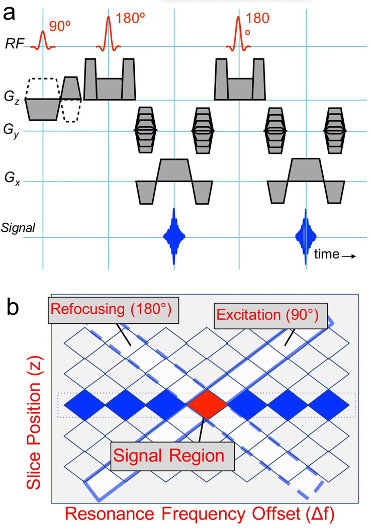
Methods: Multi-spectral imaging (MSI) approaches reduce artifacts in MR images near metal, but require 3D imaging of multiple excited volumes regardless of imaging geometry or artifact severity. The proposed 2D MSI method rapidly excites a limited slice and spectral region using gradient reversal between excitation and refocusing pulses, then uses standard 2D imaging, with the process repeating to cover multiple spectral offsets that are combined as in other MSI techniques. 2D MSI was implemented in a spin-echo-train sequence and validated in phantoms and in vivo by comparing it with standard spin-echo imaging and existing MSI techniques.

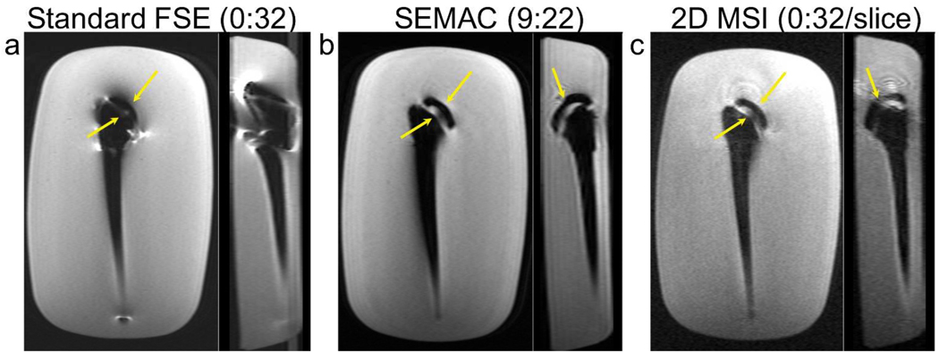
Results: 2D MSI images for each spatial-spectral region follow isocontours of the dipole-like B0 field variation, and thus frequency variation, near metal devices. Artifact correction in phantoms and human subjects with metal is comparable to 3D MSI methods, and superior to standard spin-echo techniques. Scan times are reduced compared with 3D MSI methods in cases where a limited number of slices are needed, though signal-to-noise ratio is also reduced as expected.

Conclusion: 2D MSI offers a fast and flexible alternative to 3D MSI for artifact reduction near metal. Magn Reson Med 79:968-973, 2018. © 2017 International Society for Magnetic Resonance in Medicine.

Keywords: MAVRIC; SEMAC; artifact; metal; multi-spectral imaging.


Figures

Figure 1:
Figure 1:
Excited regions and imaging strategies with purely-spectral MAVRIC (a), selective approaches such as SEMAC or MAVRIC-SL (b), and the proposed 2D MSI (c). Existing methods excite frequency bands (a) or slice/slab regions in the presence of a gradient (b), and use 3D encoding to resolve the image. 2D MSI aims to excite a finite spatial and spectral extent so that only 2D encoding is necessary, to reduce the time needed when the volume is limited. [1.5 column]
Figure 2:
Figure 2:
(a) The 2D MSI pulse sequence is a standard 2D spin-echo-train sequence, but the selection and refocusing gradients for the excitation (90°) pulse are negated. In practice, slice-refocusing gradients are typically combined with crusher pulses, and the refocusing RF pulse flip angles are usually reduced from 180°, and modulated over the echo train. (b) Gradient reversal between excitation and refocusing RF pulses results in an “inner-volume” excitation in frequency-slice space (on-resonance band at center slice position is highlighted in red). For a given slice location on-resonance, the modulation frequency for excitation (fi,j) and refocusing (fi,j) pulses has opposite sign, since the gradients are opposite. Additional frequency bands centered at frequencies Δfj are excited by adding Δfj (equal sign) to excitation and refocusing pulse frequencies to include all frequencies at a given slice location (blue regions). [1 column wide]
Figure 3:
Figure 3:
Comparison of FSE (a), SEMAC (b) and 2D MSI (c) images in a total-shoulder-replacement phantom. The acquired plane, corresponding to coronal when the device is oriented in a patient (left), and a “sagittal” reformat (right) are shown for each scan. FSE images show severe distortion artifacts near the device. These are substantially reduced, both in-plane and through-slice in both SEMAC and 2D MSI, with similar depiction of the device structure between the latter methods. SEMAC images have higher SNR than 2D MSI due to averaging, and reduced ripple. [1.5 columns wide]
Figure 4:
Figure 4:
Demonstration of 2D MSI component images in a subject with titanium screw in his knee. (a) Images acquired for frequency offsets of −2 to 2 kHz. (b) Field map obtained using a center-of-mass combination of component images. (c) Single-shot 2D fast spin echo (d) Single-shot 2D MSI images without deblurring and (e) Single-shot 2D MSI images with deblurring, showing sharper resolution in the frequency-encode (S/I) direction (solid arrows). Note that there is some minor through-slice artifactual signal (dotted arrows) and signal loss (dashed arrow) in (c) that is avoided in (d) and (e). [1.5 columns wide]
Figure 5:
Figure 5:
Comparison of SSFSE (a), 2D MSI (b) and MAVRIC-SL (c) in a subject with spinal fixation hardware includig pedicle screws. The L5 nerve root and surrounding area is obscured in standard SSFSE images by signal loss (solid white arrow) and through-slice distortion (dotted white arrow), but comparably visible in 2D MSI and MAVRIC-SL (yellow arrows). [1.5 columns wide]

References

    1. Schenck JF. The role of magnetic susceptibility in magnetic resonance imaging: MRI magnetic compatibility of the first and second kinds. Med Phys 1996; 23:815–850. - PubMed
    1. Ladd ME, Erhart P, Debatin JF, Romanowski BJ, Boesiger P, McKinnon GC. Biopsy needle susceptibility artifacts. ”Magn Reson Med.” 1996; 36:646–651. - PubMed
    1. Koch K, Hargreaves B, Pauly K, Chen W, Gold G, King K. Magnetic resonance imaging near metal implants. Journal of Magnetic Resonance Imaging 2010; 32:773–787. - PubMed
    1. Koch KM, Lorbiecki JE, Hinks RS, King KF. A multispectral three-dimensional acquisition technique for imaging near metal implants. Magn Reson Med 2009; 61:381–390. - PubMed
    1. Lu W, Pauly KB, Gold GE, Pauly JM, Hargreaves BA. SEMAC: Slice encoding for metal artifact correction in MRI. Magn Reson Med 2009; 62:66–76. - PMC - PubMed

Publication types