. 2017 Jul;242(3):358-370.
doi: 10.1002/path.4911. Epub 2017 Jun 1.

Focal Adhesion Kinase (FAK) tyrosine 397E mutation restores the vascular leakage defect in endothelium-specific FAK-kinase dead mice

Affiliations

Focal Adhesion Kinase (FAK) tyrosine 397E mutation restores the vascular leakage defect in endothelium-specific FAK-kinase dead mice

Annika N Alexopoulou et al. J Pathol. 2017 Jul.

Abstract

Focal adhesion kinase (FAK) inhibitors have been developed as potential anticancer agents and are undergoing clinical trials. In vitro activation of the FAK kinase domain triggers autophosphorylation of Y397, Src activation, and subsequent phosphorylation of other FAK tyrosine residues. However, how FAK Y397 mutations affect FAK kinase-dead (KD) phenotypes in tumour angiogenesis in vivo is unknown. We developed three Pdgfb-iCreert -driven endothelial cell (EC)-specific, tamoxifen-inducible homozygous mutant mouse lines: FAK wild-type (WT), FAK KD, and FAK double mutant (DM), i.e. KD with a putatively phosphomimetic Y397E mutation. These ECCre+;FAKWT/WT , ECCre+;FAKKD/KD and ECCre+;FAKDM/DM mice were injected subcutaneously with syngeneic B16F0 melanoma cells. Tumour growth and tumour blood vessel functions were unchanged between ECCre+;FAKWT/WT and ECCre-;FAKWT/WT control mice. In contrast, tumour growth and vessel density were decreased in ECCre+;FAKKD/KD and ECCre+;FAKDM/DM mice, as compared with Cre - littermates. Despite no change in the percentage of perfused vessels or pericyte coverage in either genotype, tumour hypoxia was elevated in ECCre+;FAKKD/KD and ECCre+;FAKDM/DM mice. Furthermore, although ECCre+;FAKKD/KD mice showed reduced blood vessel leakage, ECCre+;FAKDM/DM and ECCre-;FAKDM/DM mice showed no difference in leakage. Mechanistically, fibronectin-stimulated Y397 autophosphorylation was reduced in Cre+;FAKKD/KD ECs as compared with Cre+;FAKWT/WT cells, with no change in phosphorylation of the known Src targets FAK-Y577, FAK-Y861, FAK-Y925, paxillin-Y118, p130Cas-Y410. Cre+;FAKDM/DM ECs showed decreased Src target phosphorylation levels, suggesting that the Y397E substitution actually disrupted Src activation. Reduced VE-cadherin-pY658 levels in Cre+;FAKKD/KD ECs were rescued in Cre+FAKDM/DM ECs, corresponding with the rescue in vessel leakage in the ECCre+;FAKDM/DM mice. We show that EC-specific FAK kinase activity is required for tumour growth, angiogenesis, and vascular permeability. The ECCre+;FAKDM/DM mice restored the KD-dependent tumour vascular leakage observed in ECCre+;FAKKD/KD mice in vivo. This study opens new fields in in vivo FAK signalling. © 2017 The Authors. The Journal of Pathology published by John Wiley & Sons Ltd on behalf of Pathological Society of Great Britain and Ireland.

Keywords: focal adhesion kinase; tumour angiogenesis.


Figures

Figure 1
Figure 1
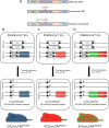
Generation of Pdgfb‐iCreert;FAKfl/fl;R26FAKWT/WT, Pdgfb‐iCreert;FAKfl/fl;R26FAKKD/KD and Pdgfb‐iCreert;FAKfl/fl;R26FAKDM/DM mice. (A) Schematic representation of WT FAK, the K454R KD mutant FAK and the DM harbouring both the K454R and Y397E mutations. (B–D) Schematic representations of Pdgfb‐iCreert;FAKfl/fl;R26FAKWT/WT, Pdgfb‐iCreert;FAKfl/fl;R26FAKKD/KD and Pdgfb‐iCreert;FAKfl/fl;R26FAKDM/DM mouse genotypes. Endogenous FAK is floxed, and knockin chicken FAK mutants were produced as homozygotes. Administration of tamoxifen induces endogenous mouse FAK deletion and WT, KD or DM knockin chicken FAK expression, to generate (B) ECCre + FAKWT/WT, (C) ECCre + FAKKD/KD, and (D) ECCre + FAKDM/DM mice, respectively.
Figure 2
Figure 2
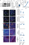
ECCre+;FAKWT/WT mice and ECCre−;FAKWT/WT mice show similar tumour growth and angiogenesis. (A) Immunoprecipitation (IP) for FAK and Western blotting (WB) for the myc‐tag demonstrate increased myc expression in Cre+;FAKWT/WT EC lysates as compared with Cre−;FAKWT/WT control lysates, indicating WT chicken FAK knockin expression in the Cre + ECs. Knockin was also verified in vivo, by similar analysis of fresh heart lysates made from tamoxifen‐treated PdgfbCre+;FAKfl/fl;R26FAKWT/WT (ECCre + FAKWT/WT) mice, but not PgdfbCre−;FAKfl/fl;R26FAKWT/WT (ECCre−;FAKWT/WT) mice. WB for FAK indicated that the knockin was expressed at a similar level to endogenous FAK. The images shown are representative of five experiments. (B) qPCR analysis of mouse FAK shows reduced endogenous mouse FAK mRNA in Cre+;FAKWT/WT EC lysates and ECCre+;FAKWT/WT fresh heart lysates, as compared with Cre − controls. The graphs represent mean values ± standard errors of the mean (SEMs) relative to the glyceraldehyde‐3‐phosphate dehydrogenase gene; n = 5 replicates. (C) Tumour growth was not statistically different between ECCre−;FAKWT/WT and ECCre+;FAKWT/WT mice injected subcutaneously with 1 million B16F0 cells. The graph represents means ± SEMs; n = 13 (ECCre−;FAKWT/WT) and n = 15 (ECCre+;FAKWT/WT). (D–H) Sections of end‐stage tumours grown in ECCre−;FAKWT/WT and ECCre+;FAKWT/WT mice were analysed by immunohistochemistry for: (D) blood vessel density (arrows, endomucin‐positive blood vessels); (E) blood vessel perfusion (arrows, PE‐PECAM/endomucin‐positive blood vessels); (F) pericyte coverage of blood vessels (arrowheads, NG2/endomucin‐positive blood vessels); (G) tumour hypoxia (dotted lines delineate pimonidazole‐positive tumour areas); and (H) blood vessel leakage (dotted lines delineate Hoechst‐positive areas around PE‐PECAM‐positive blood vessels). No significant differences in any of these features was observed between tumours grown in ECCre−;FAKWT/WT and ECCre+;FAKWT/WT mice. Representative immunofluorescence images are shown. Scale bars: 100 µm in (D), (E), (F) and (H); 250 µm in (G). Quantification is given as scatter plots: points represent mean values for each mouse, with means ± SEMs for each genotype. n = 4–6 mice per genotype. BV, blood vessels; DAPI, 4′,6‐diamidino‐2‐phenylindole; nsd, no statistically significant difference.
Figure 3
Figure 3
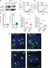
ECCre+;FAKKD/KD mice show reduced tumour growth and associated angiogenesis, and this is not rescued in ECCre+;FAKDM/DM mice. (A) Immunoprecipitation (IP) for FAK and Western blotting (WB) for the myc‐tag demonstrates increased myc‐tag expression in Cre+;FAKKD/KD EC lysates as compared with Cre−;FAKKD/KD control lysates, and in Cre+;FAKDM/DM EC lysates as compared with Cre−;FAKDM/DM control lysates, indicating mutant FAK knockin expression in Cre + ECs. FAK WB shows similar expression levels of endogenous and knockin FAK. Blots are representative of three experimental repeats. (B) qPCR analysis of mouse FAK shows reduced endogenous mouse FAK mRNA levels in Cre+;FAKKD/KD EC lysates as compared with Cre−;FAKKD/KD control lysates, and in Cre+;FAKDM/DM EC lysates as compared with Cre−;FAKDM/DM control lysates. Graphs represent mean values ± standard errors of the mean, relative to the glyceraldehyde‐3‐phosphate dehydrogenase gene; n = 2 experimental repeats. (C, D) B16F0 tumour cells were injected subcutaneously into (C) ECCre−;FAKKD/KD (n = 14) and ECCre+;FAKKD/KD (n = 15) mice, or (D) ECCre−;FAKDM/DM (n = 13) and ECCre+;FAKDM/DM (n = 16), mice and tumour growth was monitored over time. (E, F) Endomucin‐positive blood vessel density was quantified in midline tumour sections from tumours grown in (E) ECCre−;FAKKD/KD and ECCre+;FAKKD/KD mice, or (F) ECCre−;FAKDM/DM and ECCre+;FAKDM/DM mice. n = 6 tumours per genotype. (G) Immunofluorescence detection of endomucin‐positive blood vessels in tumours grown in ECCre−;FAKKD/KD and ECCre+;FAKKD/KD mice, or ECCre−;FAKDM/DM and ECCre+;FAKDM/DM mice. Arrows: endomucin‐positive blood vessels. Scale bar: 50 µm. Student's t‐test: *p < 0.05, ***p < 0.001. DAPI, 4′,6‐diamidino‐2‐phenylindole.
Figure 4
Figure 4
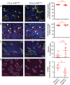
Reduced tumour vasculature leakage and enhanced tumour hypoxia in ECCre+;FAKKD/KD mice. Sections of end‐stage tumours grown in ECCre−;FAKKD/KD and ECCre+;FAKKD/KD mice were analysed by immunohistochemistry for: (A) blood vessel perfusion (arrows, PE‐PECAM/endomucin‐positive blood vessels); (B) pericyte coverage of blood vessels (arrowheads, NG2/endomucin‐positive blood vessels); (C) tumour hypoxia (dotted lines delineate pimonidazole‐positive tumour areas); and (D) blood vessel leakage (dotted lines delineate Hoechst‐positive areas around PE‐PECAM‐positive blood vessels). Representative immunofluorescence images are shown. Quantification is given in scatter plots: points represent mean values for individual mice, with means ± standard errors of the mean for each genotype. Scale bars: 100 µm in (A), (B), and (D); 250 µm in (C). Student's t‐test: *p < 0.05. n = 4–6 mice per genotype. BV, blood vessels; DAPI, 4′,6‐diamidino‐2‐phenylindole; nsd, no statistically significant difference.
Figure 5
Figure 5
Tumour vascular leakage is not affected in ECCre+;FAKDM/DM mice. Sections of end‐stage tumours grown in ECCre−;FAKDM/DM and ECCre+;FAKDM/DM mice were analysed by immunohistochemistry for: (A) blood vessel perfusion (arrows, PE‐PECAM/endomucin‐positive blood vessels); (B) pericyte coverage of blood vessels (arrowheads, NG2/endomucin‐positive blood vessels); (C) tumour hypoxia (dotted lines delineate pimonidazole‐positive tumour areas); and (D) blood vessel leakage (dotted lines delineate Hoechst‐positive areas around PE‐PECAM‐positive blood vessels). Representative immunofluorescence images are shown. Quantification is given in scatter plots: points represent mean values for individual mice, with means ± standard errors of the mean for each genotype. Scale bars: 100 µm in (A), (B), and (D); 250 µm in (C). Student's t‐test: **p < 0.01. n = 3–6 mice per genotype. BV, blood vessels; DAPI, 4′,6‐diamidino‐2‐phenylindole; nsd, no statistically significant difference.
Figure 6
Figure 6
The EC FAK Y397E KD double mutation results in dual FAK and Src kinase inhibition and restores VECAD‐pY658 levels and permeability‐related signalling as compared with Cre+;FAKKD/KD ECs. (A) Cre+;FAKWT/WT, Cre+;FAKKD/KD and Cre+;FAKDM/DM ECs were allowed to adhere to fibronectin (FN) for 0, 5, 10 and 30 min, lysed, and analysed by western blotting for levels of myc‐tag, FAK, pY397‐FAK, pY577‐FAK, pY861‐FAK, pY925‐FAK, pY118‐paxillin, paxillin, pY410‐p130Cas, p130Cas, pY416‐Src, and Src. Glyceraldehyde‐3‐phosphate dehydrogenase was used as a loading control. Blots are representative of three experimental repeats. (B) Western blot analysis of Cre+;FAKWT/WT, Cre+;FAKKD/KD and Cre+;FAKDM/DM EC lysates for VECAD‐pY658, total VECAD, and GAPDH as a loading control. The bar chart represents mean densitometric readings for duplicate samples. (C) Cre+;FAKWT/WT, Cre+;FAKKD/KD and Cre+;FAKDM/DM EC lysates were subjected to RPPA analysis, and expression levels of β‐actin, total IRS‐1, IRS‐1‐pS636/639, XIAP, Tyk2‐pY1054/1055 and PLCγ1 are shown. Bars represent mean values from two experimental repeats.

Similar articles

Cited by

References

    1. Sulzmaier FJ, Jean C, Schlaepfer DD. FAK in cancer: mechanistic findings and clinical applications. Nat Rev Cancer 2014; 14 : 598–610. - PMC - PubMed
    1. Tavora B, Batista S, Reynolds LE, et al Endothelial FAK is required for tumour angiogenesis. EMBO Mol Med 2010; 2 : 516–528. - PMC - PubMed
    1. Lechertier T, Hodivala‐Dilke K. Focal adhesion kinase and tumour angiogenesis. J Pathol 2012; 226 : 404–412. - PubMed
    1. Luo M, Guan JL. Focal adhesion kinase: a prominent determinant in breast cancer initiation, progression and metastasis. Cancer Lett 2010; 289 : 127–139. - PMC - PubMed
    1. Arold ST. How focal adhesion kinase achieves regulation by linking ligand binding, localization and action. Curr Opin Struct Biol 2011; 21 : 808–813. - PMC - PubMed

Publication types

MeSH terms