. 2017 Jul;242(3):371-383.
doi: 10.1002/path.4912. Epub 2017 Jun 2.

The histone methyltransferase EZH2 is a therapeutic target in small cell carcinoma of the ovary, hypercalcaemic type

Affiliations

The histone methyltransferase EZH2 is a therapeutic target in small cell carcinoma of the ovary, hypercalcaemic type

Yemin Wang et al. J Pathol. 2017 Jul.

Abstract

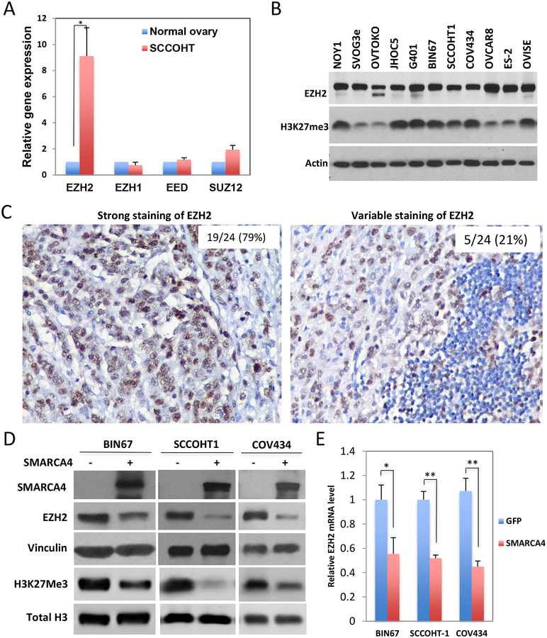
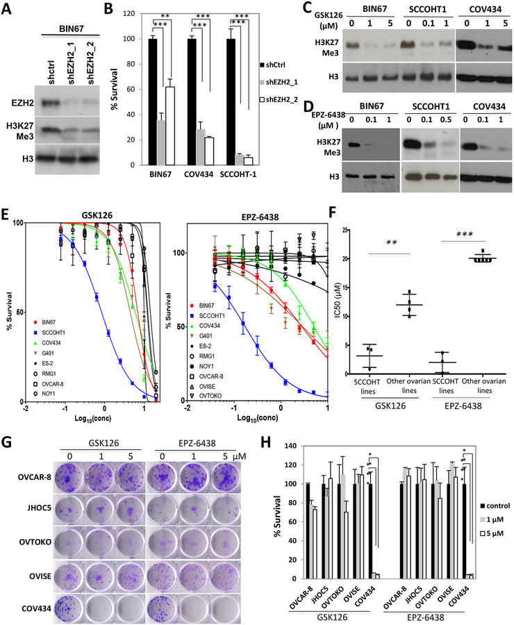
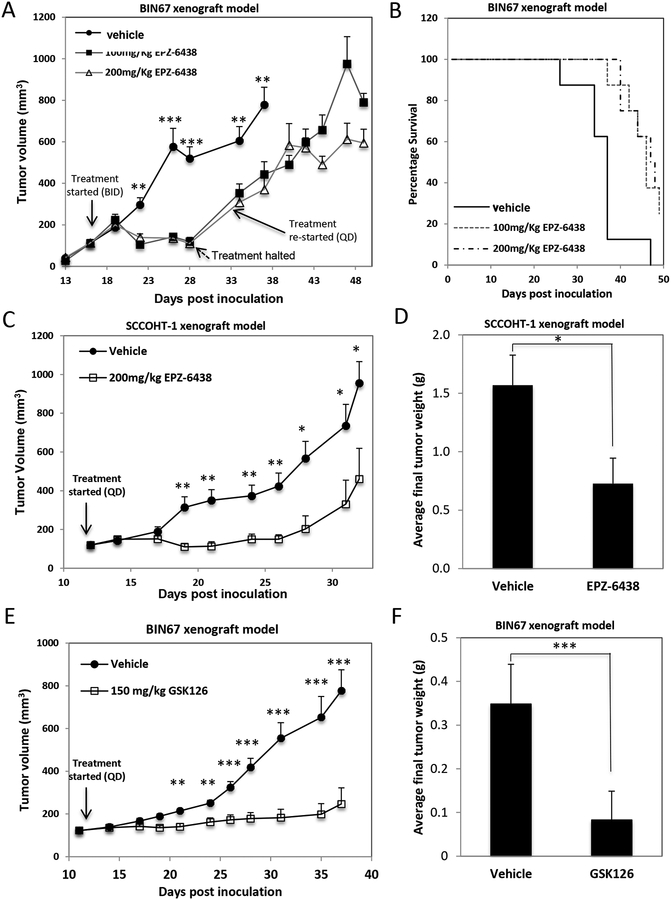
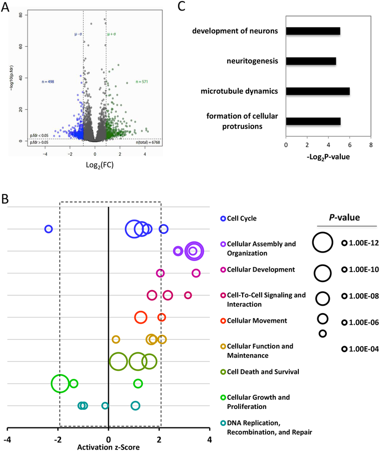
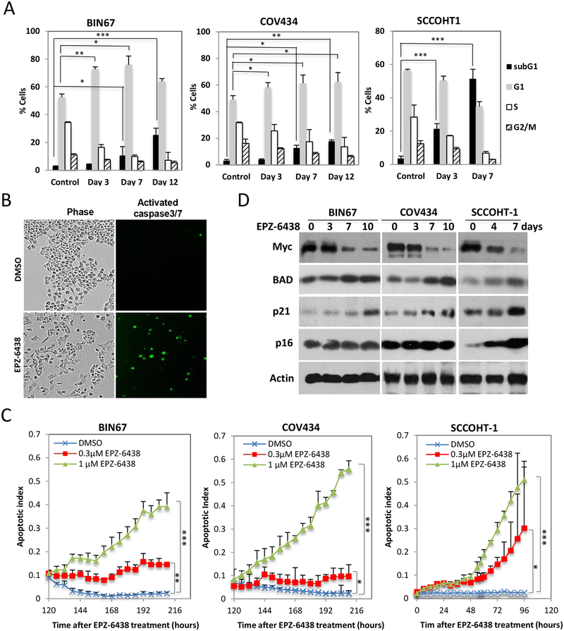
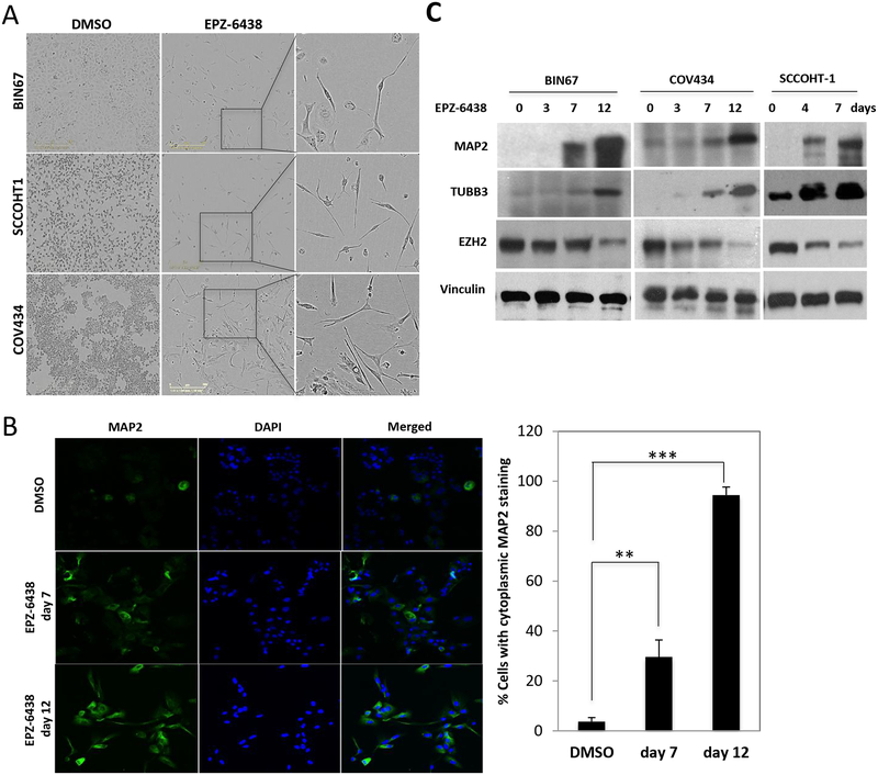
Small cell carcinoma of the ovary, hypercalcaemic type (SCCOHT) is a rare but aggressive and untreatable malignancy affecting young women. We and others recently discovered that SMARCA4, a gene encoding the ATPase of the SWI/SNF chromatin-remodelling complex, is the only gene recurrently mutated in the majority of SCCOHT. The low somatic complexity of SCCOHT genomes and the prominent role of the SWI/SNF chromatin-remodelling complex in transcriptional control of genes suggest that SCCOHT cells may rely on epigenetic rewiring for oncogenic transformation. Herein, we report that approximately 80% (19/24) of SCCOHT tumour samples have strong expression of the histone methyltransferase EZH2 by immunohistochemistry, with the rest expressing variable amounts of EZH2. Re-expression of SMARCA4 suppressed the expression of EZH2 in SCCOHT cells. In comparison to other ovarian cell lines, SCCOHT cells displayed hypersensitivity to EZH2 shRNAs and two selective EZH2 inhibitors, GSK126 and EPZ-6438. EZH2 inhibitors induced cell cycle arrest, apoptosis, and cell differentiation in SCCOHT cells, along with the induction of genes involved in cell cycle regulation, apoptosis, and neuron-like differentiation. EZH2 inhibitors suppressed tumour growth and improved the survival of mice bearing SCCOHT xenografts. Therefore, our data suggest that loss of SMARCA4 creates a dependency on the catalytic activity of EZH2 in SCCOHT cells and that pharmacological inhibition of EZH2 is a promising therapeutic strategy for treating this disease. Copyright © 2017 Pathological Society of Great Britain and Ireland. Published by John Wiley & Sons, Ltd.

Keywords: EZH2; SCCOHT; SMARCA4; SWI/SNF; chromatin remodelling complex; differentiation; ovarian cancer.


Conflict of interest statement

Conflict of interest: None of the authors report any conflicts of interest.

Figures

Figure 1.
Figure 1.. EZH2 is expressed abundantly in SCCOHT.
(A) The expression of PRC2 complex proteins in three SCCOHT versus two normal ovaries were extracted out from our previous Agilent microarray analyses (GE access No.: GSE49887 and GSE66434). (B) The expression levels of EZH2 and H3K27Me3 in multiple cell lines were analyzed by Western blotting analysis. (C) The expression of EZH2 protein in primary SCCOHT samples was determined using immunohistochemistry on TMA. Representative images of strong or variable staining are shown. (D, E) SCCOHT cell lines were infected with lentivirus expressing either GFP or SMARCA4. Cells were harvested 72 h post infection for determining the effect of SMARCA4 re-expression on EZH2 and histone H3 K27me3 by Western blotting (D) or the mRNA level of EZH2 by RT-qPCR (E). * P<0.05, ** P<0.001.
Figure 2.
Figure 2.. SCCOHT cells are sensitive to EZH2 depletion and suppression.
(A, B) Cells were infected with lentivirus expressing control scramble shRNA or EZH2 shRNA followed by puromycin selection for 48 h. Cells were then reseeded in 24-well plates for 6 d before being fixed and quantitated by crystal violet staining assay. (C, D) Cells were treated as indicated for 3 d for Western blot analysis of histone H3K27me3 level. (E) Cells were seeded in 96-well plates, treated with EZH2 inhibitors GSK126 or EPZ-6438 at indicated doses and incubated for 6 d and measured for cell survival by crystal violet assay. (F) The IC50s of cell lines to EZH2 inhibitors in (E) were compared between SCCOHT lines and other ovarian lines. Note: 20 μM was assigned to the cell lines that were not responsive to EZH2 inhibitors at 10 μM. (G, H) Cells were seeded in 12-well plates, treated with EZH2 inhibitors GSK126 or EPZ-6438 at indicated doses for 14 d and measured for clonogenic abilities by crystal violet assay. ** P<0.01, *** P<0.001
Figure 3.
Figure 3.. In vivo efficacy of EPZ-6438 in BIN67 mouse xenograft model.
(A-B) The efficacy of EPZ-6438 was evaluated in SCCOHT-1-derived mouse subcutaneous xenograft model. Tumor volume of either vehicle or 200 mg/kg EPZ-6438-treated group was plotted against the days post cell inoculation (A). Final tumor weight was determined and compared between two treated groups (B). (C, D) The efficacy of GSK126 was evaluated in BIN67 mouse subcutaneous xenograft model. Tumor volume of either vehicle or 150 mg/kg GSK126-treated group was plotted against the days post cell inoculation (C) with final tumor weights being compared between two treated groups (D). * P<0.05, ** P<0.01, *** P<0.001
Figure 4.
Figure 4.. The effect of EPZ-6438 on the proteome of BIN67 cells.
(A) Volcano plot of the proteome of BIN67 cells exposed to EPZ-6438 or vehicle. Cells were treated with either DMSO or 1 μM EPZ-6438 for 7 d and then processed for proteomic profiling. Peptide data were subjected to PECA analysis for identification of significantly altered proteins (p.fdr<0.05 and Log2FC>mean+SD or <mean-SD). (B) IPA analysis of significantly altered proteins caused by EPZ-6438 identified top affected biological functions by EPZ-6438 treatment. Any biological function with an activation z-score greater than 2 or less than −2 was predicted to be significantly increased or decreased by IPA analysis. (C) IPA analysis identified significantly increased cellular activities related to neuronal development. (D) Clustering analysis of proteins in significantly altered biological functions.
Figure 5.
Figure 5.. EPZ-6438 induces SCCOHT cell cycle arrest and apoptosis.
(A) FACS analysis of cell cycle profiles in SCCOHT cells after DMSO or 1 μM EPZ-6438 treatment for the times shown. (B, C) SCCOHT cell lines were treated with 1 μM EPZ-6438 and cultured in the presence of a cell-permeable fluorescent dye for monitoring activated caspase-3/7 activity with IncuCyte live cell imaging system (see Materials and Methods). Apoptotic index was calculated by dividing the overall fluorescent object counts to cell numbers under each condition and plotted over incubation time. (D) The effects of EPZ-6438 (BIN67/COV434: 1 μM; SCCOHT-1: 0.25 μM) on the expression of Myc, BAD, p16 and p21 proteins was determined by western blotting. * P<0.05, ** P<0.01, *** P<0.001
Figure 6.
Figure 6.. EPZ-6438 induces neuronal differentiation in SCCOHT cells.
(A) Morphology of SCCOHT cells after prolonged exposure to EPZ-6438. Cells were treated with EPZ-6438 at either 1 μM (BIN67/COV434) or 0.25 μM (SCCOHT-1) for 12 days and characterized by phase contrast microscopy. (B) BIN67 cells were fixed and immunostained for MAP2, a selective neuronal marker. Percentage of cells with cytoplasmic staining of MAP2 was quantitated. (C) Western blotting for neuronal proteins from SCCOHT cells treated with EPZ-6438 at either 1 μM (BIN67/COV434) or 0.25 μM (SCCOHT-1) for the days shown in the panel. Vinculin served as a loading control. ** P<0.01, *** P<0.001

References

    1. Longy M, Toulouse C, Mage P, et al. Familial cluster of ovarian small cell carcinoma: a new mendelian entity? J Med Genet 1996; 33: 333–335. - PMC - PubMed
    1. Martinez-Borges AR, Petty JK, Hurt G, et al. Familial small cell carcinoma of the ovary. Pediatr Blood Cancer 2009; 53: 1334–1336. - PubMed
    1. Florell SR, Bruggers CS, Matlak M, et al. Ovarian small cell carcinoma of the hypercalcemic type in a 14 month old: the youngest reported case. Med Pediatr Oncol 1999; 32: 304–307. - PubMed
    1. Estel R, Hackethal A, Kalder M, et al. Small cell carcinoma of the ovary of the hypercalcaemic type: an analysis of clinical and prognostic aspects of a rare disease on the basis of cases published in the literature. Arch Gynecol Obstet 2011; 284: 1277–1282. - PubMed
    1. Young RH, Oliva E, Scully RE. Small cell carcinoma of the ovary, hypercalcemic type. A clinicopathological analysis of 150 cases. Am J Surg Pathol 1994; 18: 1102–1116. - PubMed

MeSH terms