. 2017 Jun 13;8(24):39309-39322.
doi: 10.18632/oncotarget.16947.

PKC and CaMK-II inhibitions coordinately rescue ischemia-induced GABAergic neuron dysfunction

Affiliations

PKC and CaMK-II inhibitions coordinately rescue ischemia-induced GABAergic neuron dysfunction

Li Huang et al. Oncotarget. .

Abstract

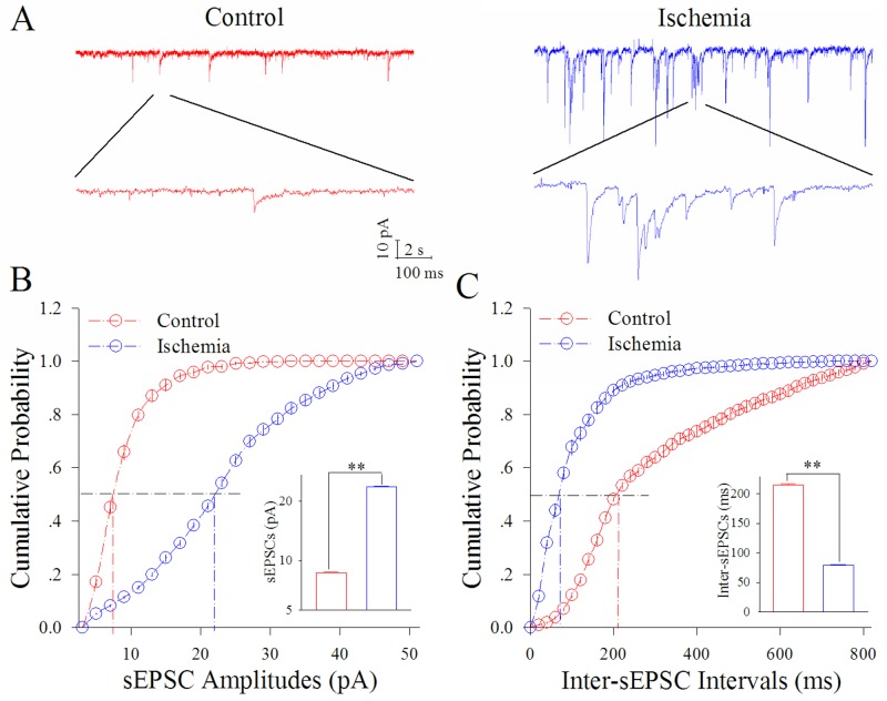
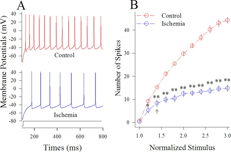
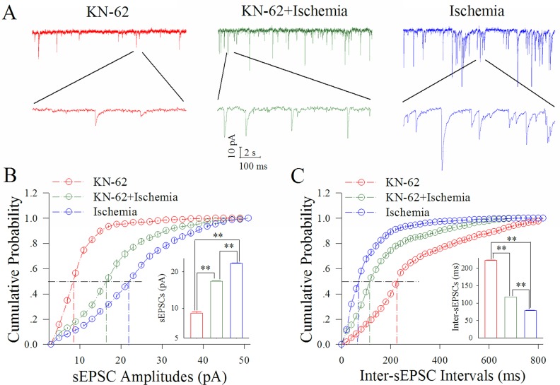
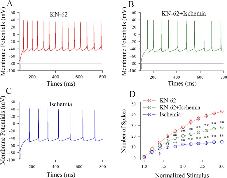
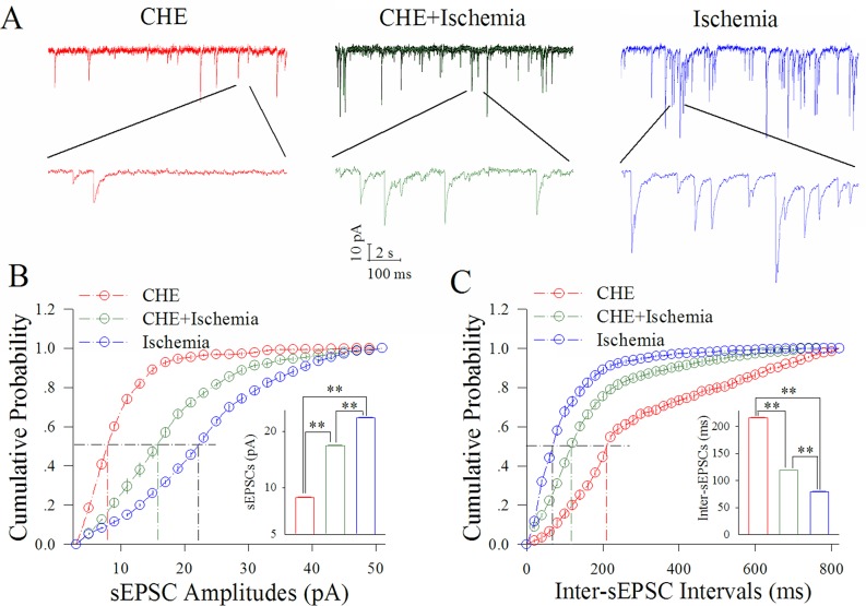
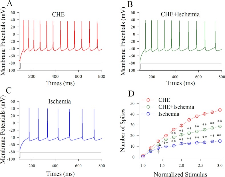
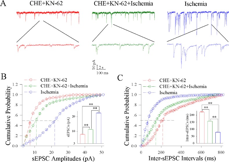
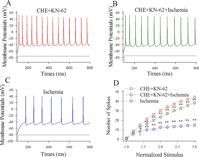
Cerebral ischemia leads to neuronal death for stroke, in which the imbalance between glutamatergic neurons and GABAergic neurons toward neural excitotoxicity is presumably involved. GABAergic neurons are vulnerable to pathological factors and impaired in an early stage of ischemia. The rescue of GABAergic neurons is expected to be the strategy to reserve ischemic neuronal impairment. As protein kinase C (PKC) and calmodulin-dependent protein kinase II (CaMK-II) are activated during ischemia, we have investigated whether the inhibitions of these kinases rescue the ischemic impairment of cortical GABAergic neurons. The functions of GABAergic neurons were analyzed by whole-cell recording in the cortical slices during ischemia and in presence of 1-[N,O-bis(5-isoquinolinesulfonyl)-N-methyl-L-tyrosyl]-4-phenylpiperazine (CaMK-II inhibitor) and chelerythrine chloride (PKC inhibitor). Our results indicate that PKC inhibitor or CaMK-II inhibitor partially prevents ischemia-induced functional deficits of cortical GABAergic neurons. Moreover, the combination of PKC and CaMK-II inhibitors synergistically reverses this ischemia-induced deficit of GABAergic neurons. One of potential therapeutic strategies for ischemic stroke may be to rescue the ischemia-induced deficit of cortical GABAergic neurons by inhibiting PKC and CaMK-II.

Keywords: GABA; PKC and CaMK-II; ischemia; neuron; synapse.


Conflict of interest statement

CONFLICTS OF INTEREST

The authors declare no conflicts of interest.

COMPETING INTERESTS

All authors declare no competing interest. All authors have read and approved the final version of the manuscript.

Figures

Figure 1
Figure 1. Ischemia upregulates excitatory synaptic transmission at cortical GABAergic neurons
sEPSCs were recorded by whole-cell voltage-clamp in cortical GABAergic neurons and analyzed in terms of their amplitudes and frequencies. (A) shows sEPSCs recorded in GABAergic neurons during control (red trace) and subsequent ischemia (blue). Calibration bars are 10 pA, 2 seconds (top traces) and 100 ms (bottom trace). (B) shows cumulative probability versus sEPSC amplitudes in the control (red symbols) and ischemia (blue). The insert shows sEPSC amplitudes at 50% cumulative probability under the control (red bar) and after ischemia (blue bar; two asterisks, p<0.01; n=13). (C) shows cumulative probability versus inter-sEPSC intervals in the control (red symbols) and ischemia (blue). The insert shows that sEPSC intervals at 50% cumulative probability under the control (red bar) and after ischemia (blue bar; two asterisks, p<0.01).
Figure 2
Figure 2. Ischemia impairs spiking ability at cortical GABAergic neurons
Sequential spikes were recorded by whole-cell current-clamp in cortical GABAergic neurons and analyzed in terms of spikes per second. (A) shows sequential spikes of GABAergic neurons in response to depolarization pulse with the same intensity under the conditions of control (red trace on top panel) and control (blue trace on bottom) at a GABAergic neuron. (B) shows that spike frequencies versus normalized stimuli at GABAergic neurons (n=13) under the control (red symbols) and subsequent ischemia (blue; two asterisks, p<0.01).
Figure 3
Figure 3. KN-62 partially blocks the ischemic upregulation of excitatory synaptic transmission at cortical GABAergic neurons
KN-62 was added into the ACSF with a final concentration at 0.9 μM. (A) illustrates sEPSC recorded on GABAergic neurons under the conditions of KN-62 application (red traces), KN-62 plus ischemia (green) and ischemia (blue). (B) illustrates cumulative probability versus sEPSC amplitudes under the conditions of KN-62 application (red symbols), KN-62 plus ischemia (green) as well as ischemia (blue). Insert figure shows sEPSC amplitudes at 50% cumulative probability in KN-62 application (red bar), KN-62 plus ischemia (green) and during ischemia only (blue; two asterisks, p<0.01; n=13). (C) illustrates cumulative probability versus inter-sEPSC intervals under the conditions of KN-62 application (red symbols), KN-62 plus ischemia (green) and ischemia (blue). Insert figure demonstrates sEPSC intervals at 50% cumulative probability in KN-62 application (red bar), KN-62 plus ischemia (green) and during ischemia only (blue; two asterisks, p<0.01; n=13).
Figure 4
Figure 4. KN-62 prevents the ischemic impairment of spiking capability at cortical GABAergic neurons
KN-62 was added into the ACSF with a final concentration at 0.9 μM. (A) shows sequential spikes in presence of KN-62 application. (B) shows sequential spikes under the condition of KN-62 plus ischemia. (C) shows sequential spikes during ischemia. Spikes at the GABAergic neuron in A~C respond to the same stimulus intensity of depolarization pulse. (D) shows that spike frequencies versus normalized stimuli at GABAergic neurons (n=13) under the conditions of KN-62 application (red symbols), KN-62 plus ischemia (green) and ischemia (blue; two asterisks, p<0.01).
Figure 5
Figure 5. CHE partially blocks the ischemic upregulation of excitatory synaptic transmission at cortical GABAergic neurons
CHE was added into the ACSF with a final concentration at 0.6 μM. (A) illustrates sEPSC recorded on GABAergic neurons under the conditions of CHE application (red traces), CHE plus ischemia (green) and ischemia (blue). (B) illustrates cumulative probability versus sEPSC amplitudes under the conditions of CHE application (red symbols), CHE plus ischemia (green) as well as ischemia (blue). Insert figure shows sEPSC amplitudes at 50% cumulative probability in CHE application (red bar), CHE plus ischemia (green) and during ischemia only (blue; two asterisks, p<0.01; n=13). (C) shows cumulative probability versus inter-sEPSC intervals under the conditions of CHE application (red symbols), CHE plus ischemia (green) and ischemia (blue). Insert figure demonstrates sEPSC intervals at 50% cumulative probability in CHE application (red bar), CHE plus ischemia (green) and during ischemia only (blue; two asterisks, p<0.01; n=13).
Figure 6
Figure 6. CHE prevents the ischemic impairment of spiking capability at cortical GABAergic neurons
CHE was added into the ACSF with a final concentration at 0.6 μM. (A) shows sequential spikes in presence of CHE application. (B) shows sequential spikes under the condition of CHE plus ischemia. (C) shows sequential spikes during ischemia. Spikes at the GABAergic neuron in A~C respond to the same stimulus intensity of depolarization pulse. (D) shows that spike frequencies versus normalized stimuli at GABAergic neurons (n=13) under the conditions of CHE application (red symbols), CHE plus ischemia (green) and ischemia (blue; two asterisks, p<0.01).
Figure 7
Figure 7. CHE and KN-62 synergistically block ischemic upregulation of excitatory synaptic transmission at cortical GABAergic neurons
Final concentrations of CHE and KN-62 in the ACSF are 0.6 μM and 0.9 μM, respectively. (A) shows sEPSC recorded on GABAergic neurons under the conditions of CHE/KN-62 application (red traces), CHE/KN-62 plus ischemia (green) and ischemia (blue). (B) illustrates cumulative probability versus sEPSC amplitudes under the conditions of CHE/KN-62 application (red symbols), CHE/KN-62 plus ischemia (green) and ischemia (blue). Insert figure illustrates sEPSC amplitudes at 50% cumulative probability in CHE/KN-62 application (red bar), CHE/KN-62 plus ischemia (green) and during ischemia (blue; two asterisks, p<0.01; n=13). (C) shows cumulative probability versus inter-sEPSC intervals under the conditions of CHE/KN-62 application (red symbols), CHE/KN-62 plus ischemia (green) and ischemia (blue). Insert figure shows sEPSC intervals at 50% cumulative probability in CHE/KN-62 application (red bar), CHE/KN-62 plus ischemia (green) and during ischemia (blue; two asterisks, p<0.01; n=13).
Figure 8
Figure 8. CHE and KN-62 synergistically prevent the ischemic impairment of spiking capability at cortical GABAergic neurons
Final concentrations of CHE and KN-62 in the ACSF are 0.6 μM and 0.9 μM, respectively. (A) illustrates sequential spikes in presence of CHE/KN-62 application. (B) illustrates sequential spikes under the condition of CHE/KN-62 plus ischemia. (C) illustrates sequential spikes during ischemia. Spikes at the GABAergic neuron in A~C respond to the same stimulus intensity of depolarization pulse. (D) shows that spike frequencies versus normalized stimuli at GABAergic neurons (n=13) under the conditions of CHE/KN-62 application (red symbols), CHE/KN-62 plus ischemia (green) and ischemia only (blue; two asterisks, p<0.01).

Similar articles

Cited by

References

    1. Candelario-Jalil E. Injury and repair mechanisms in ischemic stroke: considerations for the development of novel neurotherapeutics. Curr Opin Investig Drugs. 2009;10:644–54. - PubMed
    1. Metha SL, Manhas N, Raghubir R. Molecular targets in cerebral ischemia for developing novel therapeutics. Brain Research Review. 2007;54:34–66. - PubMed
    1. Schwartz-Bloom RD, Sah R. r-aminobutyric acid A neurotransmission and cerebral ischemia. Journal of neurochemistry. 2001;77:353–71. - PubMed
    1. Taoufik E, Probert L. Ischemic neuronal damage. Current Pharm Des. 2008;14:3565–73. - PubMed
    1. Welsh JP, Yuen G, Placantonkis DG, Yu TQ, Haiss F, O'Heaen E, Molliver ME, Aicher SA. Why do Purkinje cells die so easily after global brain ischemia? Aldolase C, EAAT4, and the cerebellar contribution to posthypoxic myoclonus. Advanced Neurology. 2002;89:331–59. - PubMed

MeSH terms