. 2017 Jun 20;8(25):40115-40131.
doi: 10.18632/oncotarget.16969.

Role of Delta-like 4 in Jagged1-induced tumour angiogenesis and tumour growth

Affiliations

Role of Delta-like 4 in Jagged1-induced tumour angiogenesis and tumour growth

Chern Ein Oon et al. Oncotarget. .

Abstract

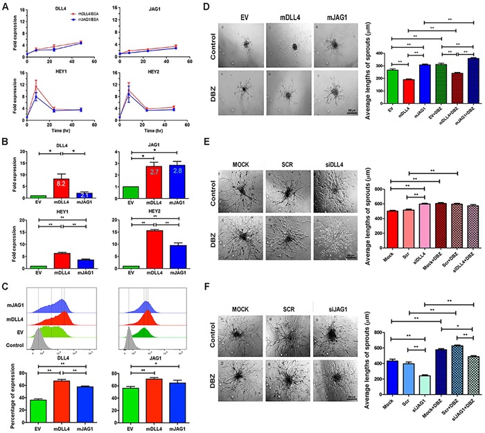
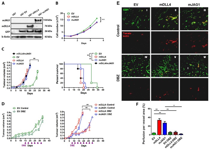
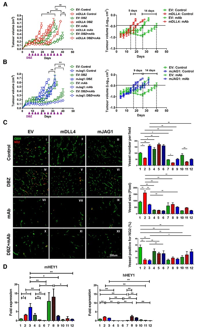
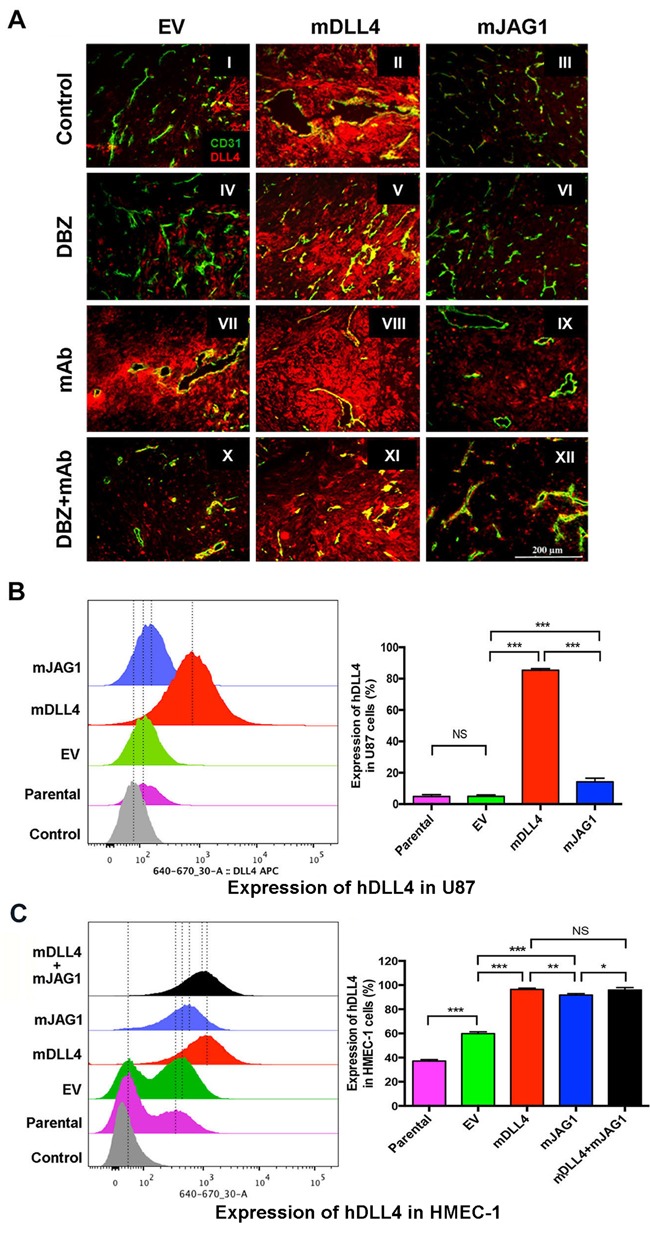
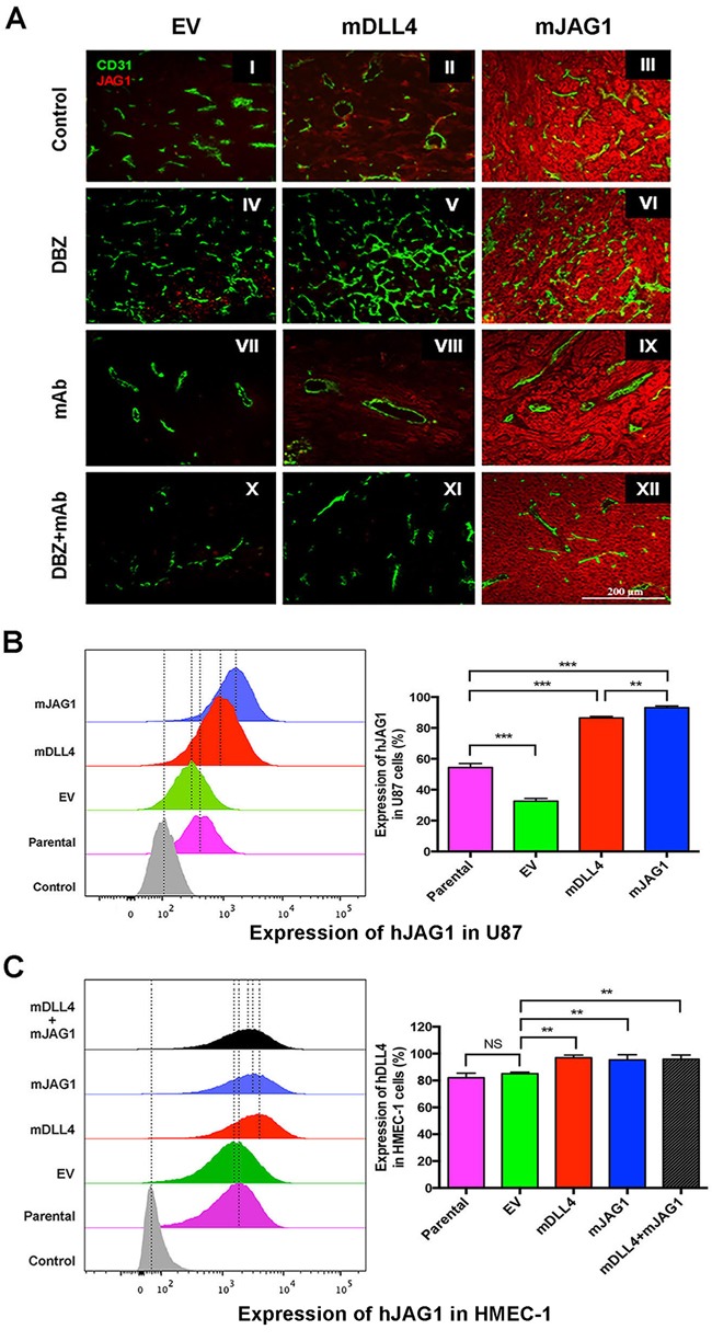
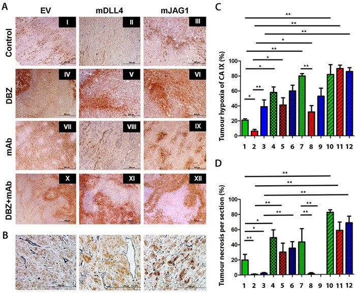
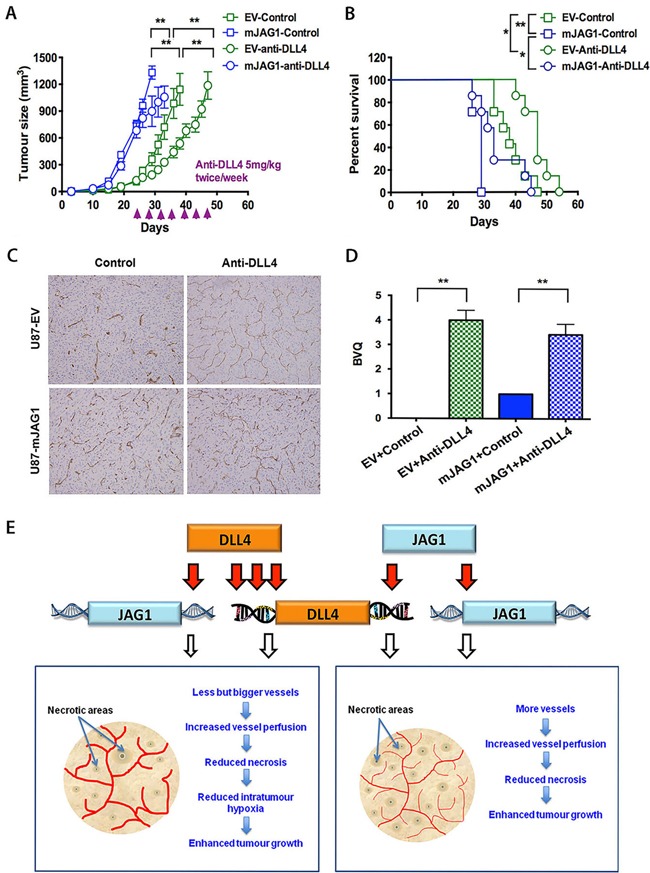
Delta-like 4 (DLL4) and Jagged1 (JAG1) are two key Notch ligands implicated in tumour angiogenesis. They were shown to have opposite effects on mouse retinal and adult regenerative angiogenesis. In tumours, both ligands are upregulated but their relative effects and interactions in tumour biology, particularly in tumour response to therapeutic intervention are unclear. Here we demonstrate that DLL4 and JAG1 displayed equal potency in stimulating Notch target genes in HMEC-1 endothelial cells but had opposing effects on sprouting angiogenesis in vitro. Mouse DLL4 or JAG1 expressed in glioblastoma cells decreased tumour cell proliferation in vitro but promoted tumour growth in vivo. mDLL4-expressing tumours showed fewer but larger vessels whereas mJAG1-tumours produced more vessels. In both tumour types pericyte coverage was decreased but the vessels were more perfused. Both ligands increased tumour resistance towards anti-VEGF therapy but the resistance was higher in mDLL4-tumours versus mJAG1-tumours. However, their sensitivity to the therapy was restored by blocking Notch signalling with dibenzazepine. Importantly, anti-DLL4 antibody blocked the effect of JAG1 on tumour growth and increased vessel branching in vivo. The mechanism behind the differential responsiveness was due to a positive feedback loop for DLL4-Notch signalling, rendering DLL4 more dominant in activating Notch signalling in the tumour microenvironment. We concluded that DLL4 and JAG1 promote tumour growth by modulating tumour angiogenesis via different mechanisms. JAG1 is not antagonistic but utilises DLL4 in tumour angiogenesis. The results suggest that anti-JAG1 therapy should be explored in conjunction with anti-DLL4 treatment in developing anti-Notch therapies in clinics.

Keywords: DLL4; JAG1; Notch signalling; angiogenesis; bevacizumab.


Conflict of interest statement

CONFLICTS OF INTEREST

The authors declare no potential conflicts of interests.

Figures

Figure 1
Figure 1. DLL4 and JAG1 activated Notch signalling and affected sprouting angiogenesis
in vitro. A. Expression profile of Notch target genes in HMEC-1 stimulated with rhDLL4 or rrJAG1 over a time course. QPCR was used to determine mRNA levels of DLL4, JAG1, HEY1 and HEY2. Fold changes were obtained by normalizing against the EV control.B. Expression profile of Notch target genes in HMEC-1 over-expressing mDLL4 or mJAG1 by retrovirus transductions. QPCR was used to determine mRNA levels of DLL4, JAG1, HEY1 and HEY2 (ANOVA with Bonferroni's post-test). C. Expression of endogenous hDLL4 and hJAG1 proteins in parental HMEC-1 cells (GFP-negative) sorted from co-culture of EV-, mDLL4- or mJAG1-overexpressing HMEC-1 with an equal amount of parental HMEC-1 cells by FACS analysis (ANOVA with Bonferroni's post-test). D. Effect of mDLL4 and mJAG1 expressed in HMEC-1 cells on sprouting in HMEC-1 spheroids and treated with DBZ. Average lengths of three longest sprouts were calculated for statistical test (ANOVA with Bonferroni's post-test). Graphs are means of 3 independent experiments. E. Effect of knockdown of DLL4 in HMEC-1 cells by specific siDLL4 on sprouting in HMEC-1 spheroids. Average lengths of three longest sprouts were calculated for statistical test (ANOVA with Bonferroni's post-test). Graphs are means of 3 independent experiments. F. Effect of knockdown of JAG1 in HMEC-1 cells by specific siJAG1 on sprouting in HMEC-1 spheroids. Average lengths of three longest sprouts were calculated for statistical test (ANOVA with Bonferroni's post-test). Graphs are means of 3 independent experiments. *P<0.05, ** P<0.01.
Figure 2
Figure 2. mDLL4 and mJAG1 reduced proliferation
in vitro but promoted tumour growth of U87 cells in vivovia different vasculature. A. Upregulation of mDLL4 and mJAG1 in U87 cells by retrovirus transductions. Western blotting confirmed the expression of mDLL4 and mJAG1. GFP encoded by the retrovirus vector was served as an internal control for the transduction efficacy. β-actin was served as a protein loading control. B. In vitro cell proliferation assays of U87 cells over a time course. N = 4. Error bars represent SD. C. Tumour growth and mouse survival of U87-EV, U87-mDLL4, U87-mJAG1 and U87-mDLL4+U87-mJAG1 (50%/50%) in mice. Parametric generalized linear model with random effects was used for tumour growth test and Kaplan-Meier analysis for overall survival test. Each group consisted of 5 mice. D. Tumour growth of U87-EV, U87-mDLL4 and U87-mJAG1 in mice treated with DBZ (8.1µmol/kg body weight, ip every 3 days). Each group consisted of 5 mice. Parametric generalized linear model with random effects was used for statistical test. E. Tumour vascular phenotype and vascular perfusion results. Immunofluorescence double staining for vessels (CD31, green) and tomato-lectin (red). Magnification 200X. F. Quantification of vascular perfusion. Graphs are means of perfusion per vessel area in 5 tumours. ANOVA with Bonferroni's post-test. *P<0.05, ** P<0.01, *** P<0.001.
Figure 3
Figure 3. DLL4 and JAG1 mediated tumour resistance to anti-VEGF therapy
A. Tumour growth curves (left panel) and linear plot of tumour volume (right panel) of U87-EV and U87-mDLL4 treated with PBS control, DBZ, bevacizumab (mAb), and combination of DBZ with bevacizumab. Each group consisted of 5 mice. A generalized linear model with random effects for tumour growth was used to analysis growth curves. ANOVA F-test was used to assess significance between curve fits. B. Tumour growth curves (left panel) and linear plot of tumour volume (right panel) of U87-EV and U87-mJAG1 treated with PBS control, DBZ, bevacizumab (mAb), and combination of DBZ with bevacizumab. Each group consisted of 5 mice. C. Tumour vascular phenotypes revealed by immunofluorescence double staining for vessels (CD31, green) and for pericytes (NG2, red) (left panel) as well as quantifications of vessel number, vessel size and vessel positive for pericytes (right panels). Immunofluorescent images (magnification 200X) were acquired for five randomly chosen fields per tumour section. Each group consisted of 5 tumours. ANOVA with Bonferroni's post-test.D. Quantification of Notch target expression of mHEY1 in tumour stroma (mouse) and hHEY in tumour cells (human) by QPCR. ANOVA with Bonferroni's post-test. Each group consisted of 5 tumours. *P<0.05, ** P<0.01. Each image in left panel or column in right panels represents: I/1, U87-EV control; II/2, U87-mDLL4 control; III/3, U87-mJAG1 control; IV/4, U87-EV DBZ; V/5, U87-mDLL4 DBZ; VI/6, U87-mJAG1 DBZ; VII/7, U87-EV mAb; VIII/8, U87-mDLL4 mAb; IX/9, U87-mJAG1 mAb; X/10, U87-EV DBZ+mAb; XI/11, U87-mDLL4 DBZ+mAb; XII/12, U87-mJAG1 DBZ+mAb.
Figure 4
Figure 4. DLL4 and JAG1 induced endogenous DLL4 expression in tumour vessels
A. Expression of DLL4 (red) and CD31 (green) as revealed by immunofluorescence double staining of tumour sections of U87. Magnification 200X.B. Endogenous hDLL4 expression in parental U87 cells (GFP-negative) sorted from co-culture of parental U87, U87-EV, U87-mDLL4 or U87-mJAG1 (GFP-positive) with an equal amount of parental U87 cells. hDLL4 expression in GFP-negative U87 cells was detected by FACS staining with anti-DLL4 antibody (recognised both human and mouse DLL4). ANOVA with Bonferroni's post-test. NS, no statistical difference. N = 3. Error bars represent SD. C. Endogenous hDLL4 expression in parental HMEC-1 cells (GFP-negative) sorted from co-culture of parental U87, U87-EV, U87-mDLL4 or U87-mJAG1 (GFP-positive) with an equal amount of parental HMEC-1 cells. hDLL4 expression in GFP-negative HMEC-1 cells was detected by FACS staining with anti-DLL4 antibody. ANOVA with Bonferroni's post-test. NS, no statistical difference. N = 3, Error bars represent SD. *P<0.05, ** P<0.01, *** P<0.001.
Figure 5
Figure 5. DLL4 and JAG1 induced endogenous JAG1 expression in tumour tissues
A. Expression of JAG1 (red) and CD31 (green) as revealed by immunofluorescence double staining of tumour sections of U87. Magnification 200X. B. Endogenous hJAG1 expression in parental U87 cells (GFP-negative) sorted from co-culture of parental U87, U87-EV, U87-mDLL4 or U87-mJAG1 (GFP-positive) with an equal amount of parental U87 cells. hJAG1 expression in GFP-negative U87 cells was detected by FACS staining with anti-human JAG1 mAb (64D). ANOVA with Bonferroni's post-test. N = 3, Error bars represent SD. C. Endogenous hJAG1 expression in parental HMEC-1 cells (GFP-negative) sorted from co-culture of parental U87, U87-EV, U87-mDLL4 or U87-mJAG1 (GFP-positive) with an equal amount of parental HMEC-1 cells. hJAG1 expression in GFP-negative HMEC-1 cells was detected by FACS staining with anti-human JAG1 mAb (64D). ANOVA with Bonferroni's post-test. NS, no statistical difference. N = 3, Error bars represent SD. *P<0.05, ** P<0.01, *** P<0.001.
Figure 6
Figure 6. Immunohistochemical staining for hypoxia and tumour vessels in xenograft tumours
A. Intra-tumour hypoxia staining of U87 tumour sections using carbonic anhydrase IX (CAIX) (brown). B. Double staining for CAIX (brown) and blood vessels (CD34) (blue) on tumour sections of U87. C. Tumour hypoxia quantification. ANOVA with Bonferroni's post-test. Each group contained 5 tumours. D. Tumour necrosis quantification. ANOVA with Bonferroni's post-test. Each group contained 5 tumours. *P<0.05, ** P<0.01. Each image in left panel or column in right panels represents: I/1, U87-EV control; II/2, U87-mDLL4 control; III/3, U87-mJAG1 control; IV/4, U87-EV DBZ; V/5, U87-mDLL4 DBZ; VI/6, U87-mJAG1 DBZ; VII/7, U87-EV mAb; VIII/8, U87-mDLL4 mAb; IX/9, U87-mJAG1 mAb; X/10, U87-EV DBZ+mAb; XI/11, U87-mDLL4 DBZ+mAb; XII/12, U87-mJAG1 DBZ+mAb.
Figure 7
Figure 7. Anti-DLL4 antibody inhibited
in vivo growth of U87-EV and U87-mJAG1 tumours and increased vessel branching. A. Tumour growth curves of U87-EV and U87-mJAG1 treated with anti-DLL4 blocking antibody (5mg/kg body weight) or PBS control. Each group consisted of 7 mice. Parametric generalized linear model with random effects was used for statistical test. B. Anti-DLL4 antibody increased tumour-bearing mouse survival. Kaplan-Meier analysis for overall survival test. Each group consisted of 7 mice. C. Visualisation of tumour xenograft vasculatures as revealed by CD34 staining. D. Quantification of vessel branching by branching vessel index (BVQ). Each group consisted of 7 tumours. Student's t-test. *P<0.05, ** P<0.01, *** P<0.001. E. A model demonstrating the effects of DLL4 or JAG1 on tumour angiogenesis and tumour growth. Angiogenic sprouting is aided by filopodia in response to VEGF gradient. DLL4-Notch signalling is promoted in vessels and tumours by increasing downstream gene expression. Signalling from tip cell to stalk cell specifies cell fate contributing to tumour growth. DLL4 and JAG1 amply Notch signalling by increasing their expressions. DLL4 is downstream of JAG1. JAG1 requires DLL4 for effect rather than antagonising it. The less induction of DLL4 by JAG1 than by DLL4 itself may produce less extreme phenotype with an increase in tip cells induced by JAG1 balanced by differentiation of stalk cells, implying a model of reciprocal regulation of the two cell populations. Both DLL4 and JAG1 promoted tumour growth but effectively reduced tumour necrosis through formation of different vasculature morphology.

References

    1. Hedlund EM, Yang X, Zhang Y, Yang Y, Shibuya M, Zhong W, Sun B, Liu Y, Hosaka K, Cao Y. Tumor cell-derived placental growth factor sensitizes antiangiogenic and antitumor effects of anti-VEGF drugs. Proc Natl Acad Sci U S A. 2013;110:654–659. - PMC - PubMed
    1. Li JL, Harris AL. Crosstalk of VEGF and Notch pathways in tumour angiogenesis: therapeutic implications. Front Biosci. 2009;14:3094–3110. - PubMed
    1. Gale NW, Dominguez MG, Noguera I, Pan L, Hughes V, Valenzuela DM, Murphy AJ, Adams NC, Lin HC, Holash J, Thurston G, Yancopoulos GD. Haploinsufficiency of delta-like 4 ligand results in embryonic lethality due to major defects in arterial and vascular development. Proc Natl Acad Sci U S A. 2004;101:15949–15954. - PMC - PubMed
    1. Jubb AM, Soilleux EJ, Turley H, Steers G, Parker A, Low I, Blades J, Li JL, Allen P, Leek R, Noguera-Troise I, Gatter KC, Thurston G, Harris AL. Expression of vascular notch ligand delta-like 4 and inflammatory markers in breast cancer. Am J Pathol. 2010;176:2019–2028. - PMC - PubMed
    1. Jubb AM, Turley H, Moeller HC, Steers G, Han C, Li JL, Leek R, Tan EY, Singh B, Mortensen NJ, Noguera-Troise I, Pezzella F, Gatter KC, et al. Expression of delta-like ligand 4 (Dll4) and markers of hypoxia in colon cancer. Br J Cancer. 2009;101:1749–1757. - PMC - PubMed

MeSH terms

LinkOut - more resources