

Cases of primary malignant melanoma and melanocytosis of the esophagus observed by magnifying endoscopy: Application to differential diagnosis: case series
- PMID: 28445275
- PMCID: PMC5413240
- DOI: 10.1097/MD.0000000000006701
Cases of primary malignant melanoma and melanocytosis of the esophagus observed by magnifying endoscopy: Application to differential diagnosis: case series
Abstract
Rationale: Primary malignant melanoma of the esophagus (PMME) is a rare disease with an extremely poor prognosis. In contrast, melanocytosis is a benign condition defined as melanocytic proliferation with melanin deposition. PMME is often accompanied by melanocytosis, but differentiating between them is difficult because of their similar appearance.
Patient concerns: Here, we reported 3 PMME cases, 2 with melanocytosis.
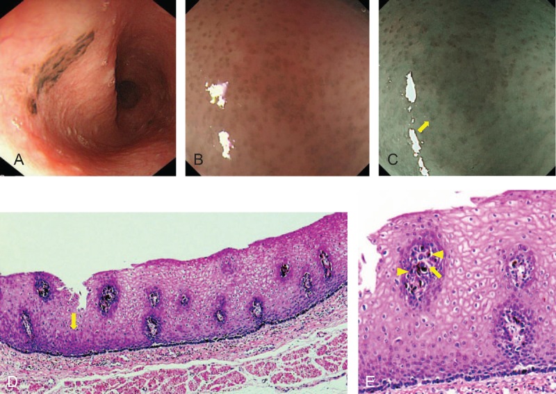
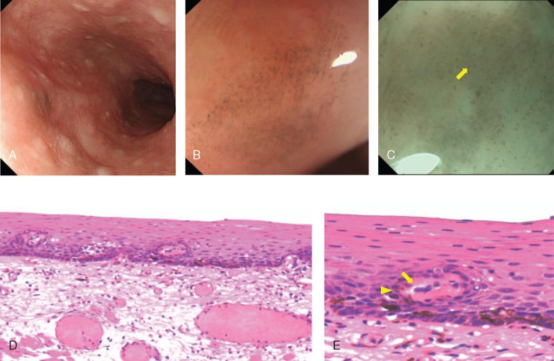
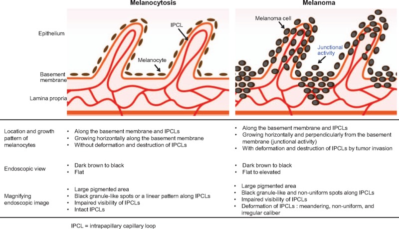
Diagnoses: Magnifying endoscopy revealed characteristic non-uniform pigmented spots along deformed intrapapillary capillary loops (IPCLs) in PMME, while melanocytosis showed fine granule-like or linearly arranged spots and intact IPCLs.
Interventions: The patients underwent endoscopic or surgical resection of each lesion.
Outcomes: Histologically, magnified images reflected melanocyte growth. For cases 1 and 2, the patients remained disease-free for 61 and 15 months after endoscopic resection, respectively. In case 3, liver metastases developed two months after surgery, and the patient died from liver failure after six months.
Lessons: This is the first report describing differences in magnified views of the 2 diseases, which aids a differential diagnosis.
Conflict of interest statement
The authors have no funding and conflicts of interest to disclose.
Figures
Similar articles
-
Primary Malignant Melanoma of the Esophagus With Unusual Endoscopic Findings: A Case Report and Literature Review.Medicine (Baltimore). 2016 Apr;95(17):e3479. doi: 10.1097/MD.0000000000003479. Medicine (Baltimore). 2016. PMID: 27124046 Free PMC article. Review.
-
Primary malignant melanoma of the esophagus with extensive intraepithelial extension.Ann Thorac Surg. 2012 Dec;94(6):2105-7. doi: 10.1016/j.athoracsur.2012.04.127. Ann Thorac Surg. 2012. PMID: 23176922
-
Primary malignant melanoma of the esophagus: a case report.World J Gastroenterol. 2014 Mar 14;20(10):2731-4. doi: 10.3748/wjg.v20.i10.2731. World J Gastroenterol. 2014. PMID: 24627611 Free PMC article.
-
Primary malignant melanoma of the esophagus treated with subtotal esophagectomy: a case report.BMC Surg. 2017 Dec 2;17(1):122. doi: 10.1186/s12893-017-0326-7. BMC Surg. 2017. PMID: 29197361 Free PMC article.
-
Current status of primary malignant melanoma of the esophagus: clinical features, pathology, management and prognosis.J Gastroenterol. 2012 Jan;47(1):21-8. doi: 10.1007/s00535-011-0490-y. Epub 2011 Nov 3. J Gastroenterol. 2012. PMID: 22048255 Review.
Cited by
-
The Thousand Faces of Malignant Melanoma: A Systematic Review of the Primary Malignant Melanoma of the Esophagus.Cancers (Basel). 2022 Jul 30;14(15):3725. doi: 10.3390/cancers14153725. Cancers (Basel). 2022. PMID: 35954389 Free PMC article. Review.
-
A case of primary malignant melanoma of the esophagus with a widely expanded surface area.Clin J Gastroenterol. 2019 Oct;12(5):424-428. doi: 10.1007/s12328-019-00961-3. Epub 2019 Mar 18. Clin J Gastroenterol. 2019. PMID: 30887427
-
Esophageal melanosis: Two case reports and review of literature.World J Gastroenterol. 2024 Nov 14;30(42):4557-4565. doi: 10.3748/wjg.v30.i42.4557. World J Gastroenterol. 2024. PMID: 39563752 Free PMC article. Review.
References
-
- Sabanathan S, Eng J, Pradhan GN. Primary malignant melanoma of the esophagus. Am J Gastroenterol 1989;84:1475–81. - PubMed
-
- Chalkiadakis G, Wihlm JM, Morand G, et al. Primary malignant melanoma of the esophagus. Ann Thorac Surg 1985;39:472–5. - PubMed
-
- Makuuchi H, Takubo K, Yanagisawa A, et al. Esophageal malignant melanoma: analysis of 134 cases collected by the Japan Esophageal Society. Esophagus 2015;12:158–69.
-
- Chang F. Esophageal melanocytosis. Arch Pathol Lab Med 2006;130:552–7. - PubMed
-
- Yamaguchi T, Shioaki Y, Koide K, et al. A case of primary malignant melanoma of the esophagus and analysis of 193 patients in Japan. J Jpn Gastroenterol 2004;101:1087–94. - PubMed
Publication types
MeSH terms
LinkOut - more resources
Full Text Sources
Other Literature Sources
Medical
