. 2017 Apr 26;12(4):e0176421.
doi: 10.1371/journal.pone.0176421. eCollection 2017.

Various mutations compensate for a deleterious lacZα insert in the replication enhancer of M13 bacteriophage

Affiliations

Various mutations compensate for a deleterious lacZα insert in the replication enhancer of M13 bacteriophage

Emily M Zygiel et al. PLoS One. .

Abstract

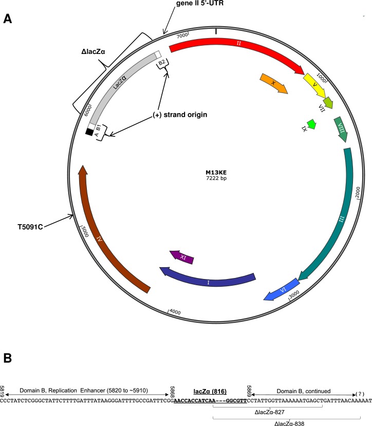
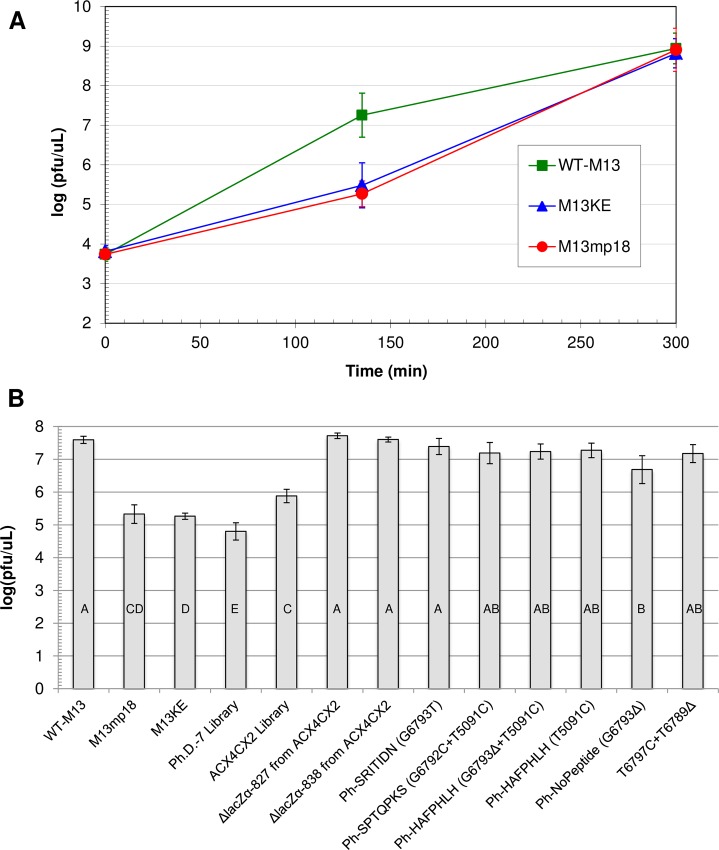
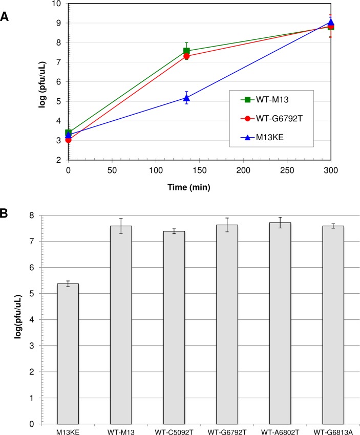
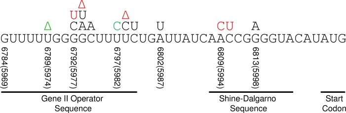
M13 and other members of the Ff class of filamentous bacteriophages have been extensively employed in myriad applications. The Ph.D. series of phage-displayed peptide libraries were constructed from the M13-based vector M13KE. As a direct descendent of M13mp19, M13KE contains the lacZα insert in the intergenic region between genes IV and II, where it interrupts the replication enhancer of the (+) strand origin. Phage carrying this 816-nucleotide insert are viable, but propagate in E. coli at a reduced rate compared to wild-type M13 phage, presumably due to a replication defect caused by the insert. We have previously reported thirteen compensatory mutations in the 5'-untranslated region of gene II, which encodes the replication initiator protein gIIp. Here we report several additional mutations in M13KE that restore a wild-type propagation rate. Several clones from constrained-loop variable peptide libraries were found to have ejected the majority of lacZα gene in order to reconstruct the replication enhancer, albeit with a small scar. In addition, new point mutations in the gene II 5'-untranslated region or the gene IV coding sequence have been spontaneously observed or synthetically engineered. Through phage propagation assays, we demonstrate that all these genetic modifications compensate for the replication defect in M13KE and restore the wild-type propagation rate. We discuss the mechanisms by which the insertion and ejection of the lacZα gene, as well as the mutations in the regulatory region of gene II, influence the efficiency of replication initiation at the (+) strand origin. We also examine the presence and relevance of fast-propagating mutants in phage-displayed peptide libraries.


Conflict of interest statement

Competing Interests: The authors have declared that no competing interests exist. Our affiliation with New England Biolabs, Inc. does not alter our adherence to PLOS ONE policies on sharing data and materials.

Figures

Fig 1
Fig 1. Schematics of the M13KE genome.
(A) The map of M13KE is shown. The (+) strand origin is divided into Domains A and B [20]. Domain A (nucleotides 5769–5819 in both WT-M13 and M13KE) is the “core origin” and is required for both (+) strand initiation and termination. Domain A is extremely sensitive to deletions and insertions, which reduce biological activity to ≤ 0.01% [20]. Domain B stretches from position 5820 to about 5910 in wild-type M13 (WT-M13), but it is interrupted in M13KE by the lacZα insert (the separated segments are indicated as B1 and B2). Dubbed the “replication enhancer,” Domain B is required for (+) strand initiation and is moderately sensitive to inserts and deletions, which reduce biological activity to ≥ 1% [20,31]. The locations of spontaneous mutations and ejections in M13KE are labeled as gene II 5’-UTR, ΔlacZα, and T5091C. The T5091C mutation is a reversion back to the WT-M13 nucleotide at position 5092 (the number is lower by 1 nt due a missing 1565T in M13mp18,19 and M13KE). The map of WT-M13 would be identical to M13KE with the exception of the lacZα insert (all downstream numbering is 815 nt lower in WT-M13). The (-) strand origin (not labeled) is upstream of the (+) strand origin in the intergenic region. The map was constructed using SnapGene®. (B) The exact locations of the lacZα insert and ejections are indicated. Nucleotide numbering corresponds to WT-M13. Domain A is not shown except for last downstream base, 5819C. In M13mp-based phage, an 816-nt insert containing the lacZα gene is placed in Domain B between nucleotides 5868 and 5869. Two different spontaneous ejections have arisen in M13KE, ΔlacZα-827 and ΔlacZα-838, both of which left behind eleven nucleotides of the lacZα insert. The vast majority of the lacZα insert was removed, in addition to a small section of Domain B: 22 nucleotides in the smaller (827-nt) ejection and 33 nt in the larger (838-nt) ejection.
Fig 2
Fig 2. Comparison of propagation rates for various M13-based phage.
(A) Time course for amplification of WT-M13, M13KE, and M13mp18. Each phage clone was amplified in three separate early log cultures of E. coli ER2738. From each culture, one aliquot was diluted and plated at the indicated incubation times, and the concentration of phage (pfu/μL) in each growing culture was determined based on plaque counts. Data points represent the mean log(pfu/μL) of the three separate cultures for each type of phage, and the error bars show the 95% confidence interval. Statistical analysis indicated significant differences among the phage concentrations for the three sets of data at 135 minutes (ANOVA; F2,8 = 89.2, P < 0.0001). Post-hoc analysis showed that M13KE and M13mp18 are not significantly different from each other (Tukey’s HSD; α = 0.05, P = 0.4471). Both M13KE and M13mp18 are significantly different from WT-M13 (P < 0.0001). (B) Phage concentrations of various M13-based clones at 135 minutes of incubation. Each phage clone was amplified separately in an ER2738 culture. At 135 minutes, three aliquots from each flask of growing culture were diluted and plated, and the concentration of phage (pfu/μL) was determined based on plaque counts. The M13KE control was run 21 times for a total of n = 63 platings. WT-M13 was run 6 times (n = 18 platings), M13mp18 and the Ph.D.-7 library were run 4 times each (n = 12 platings), and all other phage clones were run twice each (n = 6 platings). Each bar represents the mean log(pfu/μL) of all platings for a given clone, and the error bars show the 95% confidence interval. Statistical analysis indicated significant differences among the phage concentrations for all the data sets (ANOVA; F12,158 = 139.1, P < 0.0001). Post-hoc analysis showed that the 135-min concentrations of some clones are significantly different from one another, while others are not (Tukey’s HSD; α = 0.05, see the connecting letters at the bottom of the bars).
Fig 3
Fig 3. Comparison of propagation rates of wild-type M13 mutants.
(A) Time course for amplification of WT-M13, WT-G6792T, and M13KE. Each phage clone was amplified in three separate early log cultures of E. coli ER2738. From each culture, one aliquot was diluted and plated at the indicated incubation times, and the concentration of phage (pfu/μL) in each growing culture was determined based on plaque counts. Data points represent the mean log(pfu/μL) of the three separate cultures for each type of phage, and the error bars show the 95% confidence interval. Statistical analysis indicated significant differences among the phage concentrations for the three sets of data at 135 minutes (ANOVA; F2,8 = 318.2, P < 0.0001). Post-hoc analysis showed that WT-M13 and WT-G6792T were not significantly different from each other (Tukey’s HSD; α = 0.05, P = 0.0981). Both WT-M13 and WT-G6792T were significantly different from M13KE (P < 0.0001). (B) Phage concentrations of all WT-M13 mutant clones at 135 minutes of incubation. Each phage clone was amplified separately in an ER2738 culture. At 135 minutes, three aliquots from each flask of growing culture were diluted and plated, and the concentration of phage (pfu/μL) was determined based on plaque counts. The M13KE control was run 12 times for a total of n = 36 platings. WT-M13 was run 6 times (n = 18 platings) and all other phage clones were run twice each (n = 6 platings). Each bar represents the mean log(pfu/μL) of all platings for a given clone, and the error bars show the 95% confidence interval. Statistical analysis indicated significant differences among the phage concentrations for all the data sets (ANOVA; F5,77 = 239.3, P < 0.0001). Post-hoc analysis showed that M13KE is significantly different from all the other clones (Tukey’s HSD; α = 0.05, P < 0.0001) and that there is no significant difference between any pair among WT-M13 and the WT-mutant clones (range in P = 0.34–1.00).
Fig 4
Fig 4. Mutations in the 5’-UTR of gene II mRNA in M13KE.
The normal 5’-UTR region of the mRNA is shown, containing the gene II operator sequence, the Shine-Dalgarno sequence, and the start codon. The nucleotide numbering corresponds to M13KE. Thirteen different spontaneous 5’-UTR mutations are indicated above their respective nucleotides within the 5’-UTR (black and red). The majority of these mutations were previously reported [41], and were discovered in one of five ways: (i) in Ph.D.-7 or Ph.D.-12 phage display experiments that used Zn2+ as a target, (ii) during serial amplification of the Ph.D.-7 library, (iii) in a 135 minute screen of amplified Ph.D.-7 or Ph.D.-12 libraries, (iv) during amplification of M13KE, or (v) as a clone that contaminated a concurrent experiment in our lab. The mutations G6793T and G6792C are new to this publication, and were discovered via method (iii). The T6797C mutation was synthetically introduced using an insert containing the mutation, and was accompanied by a concomitant deletion, T6789Δ (both green). The mutations shown in red were made using a randomized synthetic insert, but had already arisen spontaneously in our experiments. Several mutations have been discovered repeatedly with different displayed peptides (or no peptide), including the two clones reported herein, Ph-VTAHGGR and Ph-SDLVLRP, which have new peptides but previously found mutations. These repeated mutations are listed here as: Mutation (Number of times observed): G6813A (4), C6810T (2), A6809C (3), T6798C (4), T6798Δ (3), G6793A (3), G6793Δ (2), G6792T (4). Two additional mutations arose through method (i) and are not shown here because they are upstream of the mRNA sequence: G6748Δ (in the gene II promoter) and C6589T (in the lacZα insert) [41].

Similar articles

Cited by

References

    1. Cao B, Yang M, Mao C. Phage as a Genetically Modifiable Supramacromolecule in Chemistry, Materials and Medicine. Acc Chem Res 2016. June 21;49(6):1111–1120. doi: 10.1021/acs.accounts.5b00557 - DOI - PMC - PubMed
    1. Henry KA, Arbabi-Ghahroudi M, Scott JK. Beyond phage display: non-traditional applications of the filamentous bacteriophage as a vaccine carrier, therapeutic biologic, and bioconjugation scaffold. Front Microbiol 2015. August 4;6:755 doi: 10.3389/fmicb.2015.00755 - DOI - PMC - PubMed
    1. Hamzeh-Mivehroud M, Alizadeh AA, Morris MB, Church WB, Dastmalchi S. Phage display as a technology delivering on the promise of peptide drug discovery. Drug Discov Today 2013. December;18(23–24):1144–1157. doi: 10.1016/j.drudis.2013.09.001 - DOI - PubMed
    1. Messing J. Phage M13 for the treatment of Alzheimer and Parkinson disease. Gene 2016. June 1;583(2):85–89. doi: 10.1016/j.gene.2016.02.005 - DOI - PubMed
    1. Sattar S, Bennett NJ, Wen WX, Guthrie JM, Blackwell LF, Conway JF, et al. Ff-nano, short functionalized nanorods derived from Ff (f1, fd, or M13) filamentous bacteriophage. Front Microbiol 2015. April 20;6:316 doi: 10.3389/fmicb.2015.00316 - DOI - PMC - PubMed