

Clinical Characteristics of Connective Tissue Nevi in Tuberous Sclerosis Complex With Special Emphasis on Shagreen Patches
- PMID: 28445558
- PMCID: PMC5817464
- DOI: 10.1001/jamadermatol.2017.0298
Clinical Characteristics of Connective Tissue Nevi in Tuberous Sclerosis Complex With Special Emphasis on Shagreen Patches
Abstract
Importance: Patients with tuberous sclerosis complex (TSC) frequently develop collagenous connective tissue nevi. The prototypical lesion is a large shagreen patch located on the lower back, but some patients only manifest small collagenomas or have lesions elsewhere on the body. The ability to recognize these variable presentations can be important for the diagnosis of TSC.
Objective: To describe the clinical characteristics of connective tissue nevi on the trunk and extremities of patients with tuberous sclerosis complex.
Design, setting, and participants: A retrospective analysis of patient medical records and skin photography was performed; 104 adult patients with TSC were enrolled in an observational cohort study that was enriched for those with pulmonary lymphangioleiomyomatosis, and was therefore composed mostly of women (99 women, 5 men). All patients included were examined at the National Institutes of Health (NIH) in Bethesda, Maryland, from 1998 to 2013. Connective tissue nevi were categorized per anatomic location and size. Lesions less than 1 cm in diameter were termed collagenomas. Shagreen patches were characterized as small (1 to <4 cm), medium (4 to <8 cm), and large (≥8 cm).
Main outcome and measures: Frequency, anatomic location, size, and histological appearance of connective tissue nevi in patients with TSC.
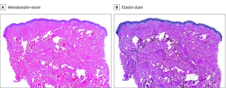
Results: Overall, 58 of 104 patients (median [range] age, 42 [19-70] years) with TSC (56%) had at least 1 connective tissue nevus on the trunk or thighs; of these, 28 of 58 patients (48%) had a solitary lesion, and 30 of 58 patients (52%) had 2 or more lesions. Overall, 120 lesions from 55 patients were classified by size; 46 lesions (38%) were collagenomas; 39 lesions (32%) were small shagreen patches; 21 lesions (18%), medium shagreen patches; and 14 lesions (12%), large shagreen patches. The distribution of lesions was 9% (n = 11), upper back; 29% (n = 35), middle back; 51% (n = 61), lower back; and 11% (n = 13), other locations. All 26 shagreen patches that were analyzed histopathologically had coarse collagen fibers and 24 of 26 stained with Miller elastic stain had decreased elastic fibers. On immunoblot analysis, fibroblasts grown from shagreen patches expressed higher levels of phosphorylated ribosomal protein S6 than paired fibroblasts from normal-appearing skin.
Conclusions and relevance: Tuberous sclerosis complex-related connective tissue nevi are not limited to the lower back, and occasionally present on the central or upper back, buttocks, or thighs. Elastic fibers are typically decreased. Recognition of these variable presentations can be important for TSC diagnosis.
Conflict of interest statement
Figures


Similar articles
-
Fibrous cephalic plaques in tuberous sclerosis complex.J Am Acad Dermatol. 2018 Apr;78(4):717-724. doi: 10.1016/j.jaad.2017.12.027. Epub 2017 Dec 16. J Am Acad Dermatol. 2018. PMID: 29258863 Free PMC article.
-
Miliary fibromas in tuberous sclerosis complex.J Eur Acad Dermatol Venereol. 2021 May;35(5):1226-1229. doi: 10.1111/jdv.17161. Epub 2021 Feb 23. J Eur Acad Dermatol Venereol. 2021. PMID: 33565654 Free PMC article.
-
Zosteriform Collagen Nevus in an Infant.Acta Dermatovenerol Croat. 2016 Jun;24(2):148-9. Acta Dermatovenerol Croat. 2016. PMID: 27477177
-
Histological Patterns of Skin Lesions in Tuberous Sclerosis Complex: A Panorama.Dermatopathology (Basel). 2021 Jul 4;8(3):236-252. doi: 10.3390/dermatopathology8030029. Dermatopathology (Basel). 2021. PMID: 34287284 Free PMC article. Review.
-
Dermatological manifestations of tuberous sclerosis complex (TSC).J Dtsch Dermatol Ges. 2017 Jul;15(7):695-700. doi: 10.1111/ddg.13264. Epub 2017 Jun 9. J Dtsch Dermatol Ges. 2017. PMID: 28598544 Review.
Cited by
-
Animals in Dermatology.Indian J Dermatol. 2024 Jul-Aug;69(4):333-337. doi: 10.4103/ijd.ijd_502_23. Epub 2024 Aug 19. Indian J Dermatol. 2024. PMID: 39296695 Free PMC article. No abstract available.
-
Segmental Collagenoma in Tuberous Sclerosis - Think Beyond the Skin: A Rare Case Report.Indian J Dermatol. 2023 Jan-Feb;68(1):123. doi: 10.4103/ijd.ijd_1038_20. Indian J Dermatol. 2023. PMID: 37151256 Free PMC article.
-
Fibrous cephalic plaques in tuberous sclerosis complex.J Am Acad Dermatol. 2018 Apr;78(4):717-724. doi: 10.1016/j.jaad.2017.12.027. Epub 2017 Dec 16. J Am Acad Dermatol. 2018. PMID: 29258863 Free PMC article.
-
Miliary fibromas in tuberous sclerosis complex.J Eur Acad Dermatol Venereol. 2021 May;35(5):1226-1229. doi: 10.1111/jdv.17161. Epub 2021 Feb 23. J Eur Acad Dermatol Venereol. 2021. PMID: 33565654 Free PMC article.
-
Collagenoma of the Eyelid.Ophthalmic Plast Reconstr Surg. 2019 Mar/Apr;35(2):e29-e30. doi: 10.1097/IOP.0000000000001298. Ophthalmic Plast Reconstr Surg. 2019. PMID: 30624411 Free PMC article.
References
-
- Roach ES. Applying the lessons of tuberous sclerosis: the 2015 Hower Award lecture. Pediatr Neurol. 2016;63:6-22. - PubMed
-
- Darling TN, Moss J, Mausner M Dermatologic Manifestations of Tuberous Sclerosis Complex. In: Kwiatkowski DJ, Whittemore VH, Thiele EA, eds. Tuberous Sclerosis Complex: Genes, Clinical Features, and Therapeutics. Weinheim, Germany: WILEY-VCH Verlag GmbH & Co. KGaA;2010:285-309.
-
- Hunt A. Tuberous sclerosis: a survey of 97 cases. II: physical findings. Dev Med Child Neurol. 1983;25(3):350-352. - PubMed
Publication types
MeSH terms
Substances
Supplementary concepts
Grants and funding
LinkOut - more resources
Full Text Sources
Other Literature Sources
Medical
