

Interferon alpha antagonizes the anti-hepatoma activity of the oncolytic virus M1 by stimulating anti-viral immunity
- PMID: 28445966
- PMCID: PMC5421880
- DOI: 10.18632/oncotarget.15788
Interferon alpha antagonizes the anti-hepatoma activity of the oncolytic virus M1 by stimulating anti-viral immunity
Abstract
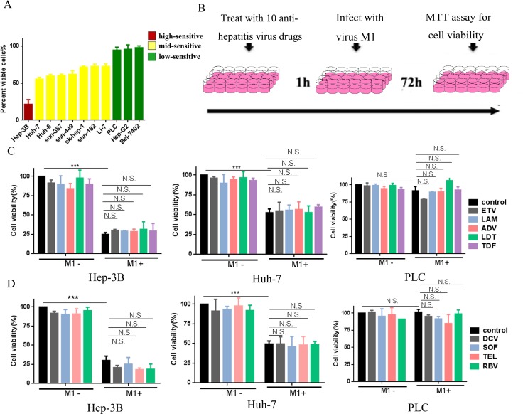
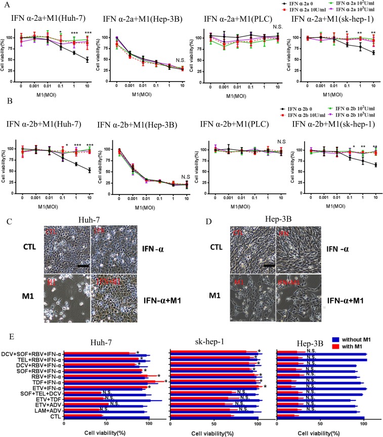
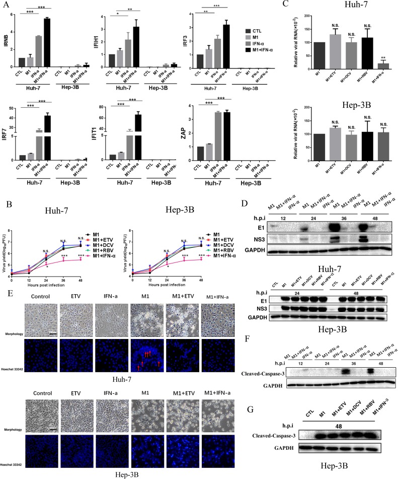
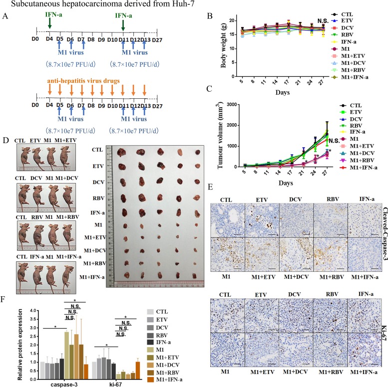
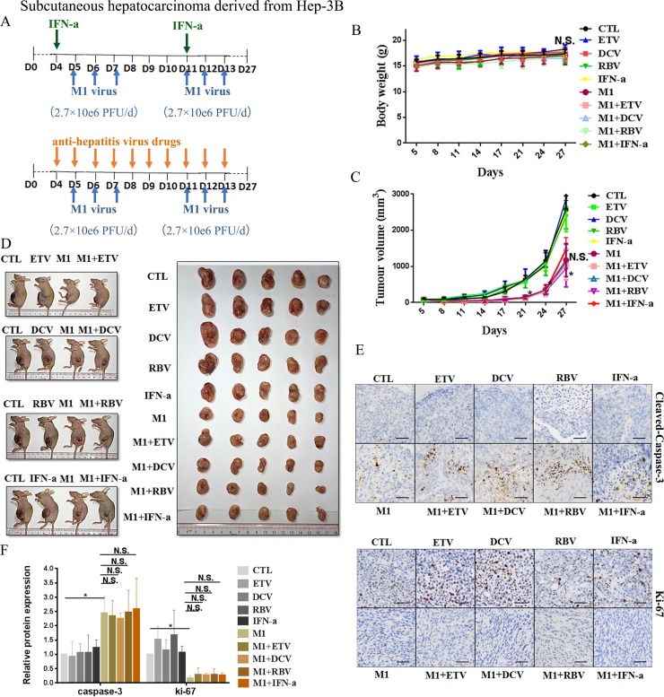
Alpha virus M1 is an oncolytic virus that targets zinc-finger antiviral protein (ZAP)-defective cancer cells, and may be useful for treatment of hepatocellular carcinoma (HCC). Most of HCC patients have hepatitis and need long-term antiviral medication. Thus, it is necessary to clarify whether anti-virus medicines influence oncolytic effect of M1. We examined the effect of drugs used to treat hepatitis B/C on M1-mediated oncolysis in vitro and in vivo. Interferon (IFN)-α induces expression of antiviral IFN-stimulated genes (ISGs) in HCC cells with moderate sensitivity to M1 virus. This leads to reduced replication of M1, and blocking of M1-mediated apoptosis. The antagonistic effect of IFN-α is positively related with the expressive level of ISGs. We also examined a population of 147 HCC patients. A total of 107 patients (73%) had low ZAP expression in liver tissues relative to adjacent tissues. Among these 107 patients, 77% were positive for hepatitis B and 2% were positive for hepatitis C. A combination of M1 virus and IFN should be avoided in those patients with HBV or HCV infection, of who ZAP expression is low but ISGs expression is moderate. In conclusion, this study provides a basis for anti-viral regimens for HCC patients with hepatitis B or C who are given oncolytic virus M1.
Keywords: ZAP; hepatocellular carcinoma; interferon; interferon-stimulated genes; oncolytic virus M1 virus.
Conflict of interest statement
The authors have declared that no conflicts of interestexists.
Figures

Similar articles
-
Sustained HCV clearance by interferon-based therapy reduces hepatocellular carcinoma in hepatitis B and C dually-infected patients.Antivir Ther. 2011;16(7):959-68. doi: 10.3851/IMP1842. Antivir Ther. 2011. PMID: 22024511
-
Interferon-α therapy after curative resection prevents early recurrence and improves survival in patients with hepatitis B virus-related hepatocellular carcinoma.J Surg Oncol. 2010 Dec 1;102(7):796-801. doi: 10.1002/jso.21741. J Surg Oncol. 2010. PMID: 20886584
-
Effect of the dose and duration of interferon-alpha therapy on the incidence of hepatocellular carcinoma in noncirrhotic patients with a nonsustained response to interferon for chronic hepatitis C.Oncology. 2001;61(2):134-42. doi: 10.1159/000055364. Oncology. 2001. PMID: 11528252 Clinical Trial.
-
Management of hepatitis B virus infection during treatment for hepatitis B virus-related hepatocellular carcinoma.World J Gastroenterol. 2015 Jul 21;21(27):8249-55. doi: 10.3748/wjg.v21.i27.8249. World J Gastroenterol. 2015. PMID: 26217076 Free PMC article. Review.
-
Impact of HBV therapy on the incidence of hepatocellular carcinoma.Liver Int. 2014 Feb;34 Suppl 1:139-45. doi: 10.1111/liv.12394. Liver Int. 2014. PMID: 24373091 Review.
Cited by
-
Advances in the clinical development of oncolytic viruses.Am J Transl Res. 2022 Jun 15;14(6):4192-4206. eCollection 2022. Am J Transl Res. 2022. PMID: 35836877 Free PMC article. Review.
-
Mathematical Modeling of Oncolytic Virus Therapy Reveals Role of the Immune Response.Viruses. 2023 Aug 25;15(9):1812. doi: 10.3390/v15091812. Viruses. 2023. PMID: 37766219 Free PMC article.
-
Intravenous injections of the oncolytic virus M1 as a novel therapy for muscle-invasive bladder cancer.Cell Death Dis. 2018 Feb 15;9(3):274. doi: 10.1038/s41419-018-0325-3. Cell Death Dis. 2018. PMID: 29449555 Free PMC article.
-
Current strategies to circumvent the antiviral immunity to optimize cancer virotherapy.J Immunother Cancer. 2021 Apr;9(4):e002086. doi: 10.1136/jitc-2020-002086. J Immunother Cancer. 2021. PMID: 33795384 Free PMC article. Review.
-
Secretome Screening Reveals Fibroblast Growth Factors as Novel Inhibitors of Viral Replication.J Virol. 2018 Jul 31;92(16):e00260-18. doi: 10.1128/JVI.00260-18. Print 2018 Aug 15. J Virol. 2018. PMID: 29899088 Free PMC article.
References
-
- Naik S, Russell SJ. Engineering oncolytic viruses to exploit tumor specific defects in innate immune signaling pathways. Expert Opin Biol Ther. 2009;9:1163–76. - PubMed
-
- Stojdl DF, Lichty BD, tenOever BR, Paterson JM, Power AT, Knowles S, Marius R, Reynard J, Poliquin L, Atkins H, Brown EG, Durbin RK, Durbin JE, et al. VSV strains with defects in their ability to shutdown innate immunity are potent systemic anti-cancer agents. Cancer Cell. 2003;4:263–75. - PubMed
-
- Siegel RL, Miller KD, Jemal A. Cancer statistics, 2015. CA Cancer J Clin. 2015;65:5–29. - PubMed
-
- European Association For The Study Of The Liver, European Organisation For Research And Treatment Of Cancer. EASL-EORTC clinical practice guidelines: management of hepatocellular carcinoma. J Hepatol. 2012;56:908–43. - PubMed
MeSH terms
Substances
LinkOut - more resources
Full Text Sources
Other Literature Sources
Medical
Molecular Biology Databases
