. 2017 Apr 11;8(15):25345-25361.
doi: 10.18632/oncotarget.15867.

MiR-148a increases glioma cell migration and invasion by downregulating GADD45A in human gliomas with IDH1 R132H mutations

Affiliations

MiR-148a increases glioma cell migration and invasion by downregulating GADD45A in human gliomas with IDH1 R132H mutations

Daming Cui et al. Oncotarget. .

Abstract

High-grade gliomas are severe tumors with poor prognosis. An R132H mutation in the isocitrate dehydrogenase (IDH1) gene prolongs the life of glioma patients. In this study, we investigated which genes are differentially regulated in IDH1 wild type (IDH1WT) or IDH1 R132H mutation (IDH1R132H) glioblastoma cells. Growth arrest and DNA-damage-inducible protein (GADD45A) was downregulated and microRNA 148a (miR-148a) was upregulated in in IDH1R132H human glioblastomas tissues. The relationship between GADD45A and miR-148a is unknown. In vitro experiments showed that GADD45A negatively regulates IDH1R132H glioma cell proliferation, migration, and invasion, and neurosphere formation in IDH1R132H glioblastoma stem cells (GSC). In addition, a human orthotopic xenograft mouse model showed that GADD45A reduced tumorigenesis in vivo. Our findings demonstrated that miR-148a promotes glioma cell invasion and tumorigenesis by downregulating GADD45A. Our findings provide novel insights into how GADD45A is downregulated by miR-148a in IDH1R132H glioma and may help to identify therapeutic targets for the effective treatment of high-grade glioma.

Keywords: GADD45A; invasion; miR-148a; migration; β-catenin.


Conflict of interest statement

CONFLICTS OF INTEREST

The authors declare no conflicts of interest.

Figures

Figure 1
Figure 1. GADD45A and miR-148a expression in normal tissues and IDH1WT or IDH1R132H glioma tissues
(A–B) qRT-PCR analysis of GADD45A and miR-148a mRNA expression in the three tissue types. (C) Kaplan-Meier analysis of the relationship between IDH1R132H (n=53) and IDH1WT (n=268) with patient survival in glioma patients (P<0.01, Log-rank test). (D) GADD45A immunostaining revealed lower protein expression in IDH1R132H glioma tissues compared with normal tissues and IDH1WT gliomas. Magnification: ×200. **P<0.01, ***P<0.001.
Figure 2
Figure 2. GADD45A inhibits cell proliferation in vitro
(A) Western blot analysis of IDH1WT and IDH1R132H protein expression in U87 and U251 glioblastoma cell lines and GSC 0308 cells after stable transfection with empty vector (EV), IDH1WT, and IDH1R132H. (B–C) qRT-PCR analysis of GADD45A and miR-148a expression in U87 and U251 glioblastoma cell lines and GSC 0308 cells stably transfected with EV, IDH1WT, and IDH1R132H. (D) Western blot analysis of GADD45A expression in U87, U251 glioblastoma cell lines and GSC 0308 stably transfected with EV, IDH1WT, or IDH1R132H. (E) GADD45A was silenced in U87 cells by three different siRNAs (siRNA#1–3) as shown by qRT-PCR. (F–K) The effect of GADD45A knockdown and overexpression on cell viability in IHD1WT or IDH1R132H U87, U251, and GSC 0308 cells. Non-transfected IHD1WT or IDH1R132H U87, U251 and 0308 cells were used as controls. *P<0.05, **P<0.01, ***P<0.001.
Figure 3
Figure 3. GADD45A inhibits tumor growth in vitro
(A) Implanted U87 MG-luc2 cells stably expressing pcDNA3.1-GADD45A or GADD45A-siRNA1# are visible in mouse brains as red–blue signals. Red indicates the highest Bioluminescence imaging (BLI) signal intensity. The BLI signal intensity increased from day 42, indicating progressive growth of the glioblastoma xenograft. (B) Average IVIS values of mice, data are expressed as mean ± SD. *P<0.05.
Figure 4
Figure 4. miR-148a binds GADD45A
(A-B) U87 cells were transfected with miR-148a mimics, mimics-NC, miR-148a inhibitor, or inhibitor-NC for 48 h. The miR-148a levels were determined by qRT-PCR. (C) The putative miR-148a binding sites in the GADD45A sequence. (D) The luciferase reporter plasmid containing wild type or mutant GADD45A 3′-UTR was co-transfected into HEK-293 T cells with miR-148a mimics or miR-148a mimics-NC. Luciferase activity was determined 48 h after transfection and was normalized to Renilla activity. (E–G) GADD45A expression in IDH1WT U87, U251, and GSC 0308 cells transfected with miR-148a inhibitor/ miR-148a inhibitor-NC, or miR-148a mimics/miR-148a mimics-NC for 48 h was measured by qRT-PCR and western blotting. (H–J) qRT-PCR analysis of GADD45A mrna and western blot analysis of GADD45A expression in IDH1R132H U87, U251, and 0308 cells transfected with miR-148a inhibitor/miR-148a inhibitor-NC or miR-148a mimics/miR-148a mimics-NC for 48 h. β-actin was used as a control. *P<0.05, **P<0.01, ***P<0.001.
Figure 5
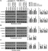
Figure 5. miR-148a partly stimulates β-catenin, MMP-9, and the epithelial-mesenchymal transition by downregulating GADD45A in IDH1WT glioblastoma cells
(A) Western blot analysis of β-catenin, N-cadherin, E-cadherin, and fibronectin expression in membrane extracts. Flotillin-2 was used as a membrane marker. (B) Western blot analysis of GADD45A, β-catenin, and MMP-9 expression in cytoplasmic extracts. β-actin was used as a control. (C) Western blot analysis of β-catenin and GADD45A expression in nuclear extracts. Lamin A was used as a control. All extracts were prepared from IDH1WT U87 and U251 glioblastoma cells. *P<0.05, **P<0.01.
Figure 6
Figure 6. miR-148a stimulates β-catenin, MMP-9, and the epithelial-mesenchymal transition by inhibiting GADD45A in IDH1R132H glioblastoma cells
(A) Western blot analysis of β-catenin, N-cadherin, E-cadherin, and fibronectin expression in membrane extracts. Flotillin-2 was used as a membrane marker. (B) Western blot analysis of GADD45A, β-catenin, and MMP-9 expression in cytoplasmic extracts. β-actin was used as a control. (C) Western blot analysis of β-catenin and GADD45A expression in nuclear extracts. Lamin A was used as a control. All extracts were prepared from IDH1R132H U87 and U251 glioblastoma cells. *P<0.05, **P<0.01, ***P<0.001.
Figure 7
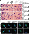
Figure 7. miR-148a partly increases cell migration and invasion and β-catenin distribution by downregulating GADD45A in IDH1WT glioblastoma cells
(A) Migration and invasion of IDH1WT U87 and U251 glioblastoma cells were measured using transwell assays. (B) Immunofluorescence staining of β-catenin in IDH1WT U87 and U251 cells. **P<0.01.
Figure 8
Figure 8. miR-148a increases cell migration and invasion and β-catenin distribution by inhibiting GADD45A in IDH1R132H U87 and U251 cells
(A) Migration and invasion of IDH1R132H U87 and U251 glioblastoma cells were measured using transwell assays. (B) Immunofluorescence staining of β-catenin in IDH1R132H U87 and U251 cells. GADD45A overexpression removed β-catenin from the nucleus and this effect was antagonized by miR-148a. *P<0.05, **P<0.01.
Figure 9
Figure 9. MiR-148a increases GSC neurosphere formation by downregulating GADD45A in IDH1WT and IDH1R132H cells
GSCs were transfected with pcDNA3.1-GADD45A without or with miR-148a inhibitor, miR-148a mimics, or controls and neurosphere formation was measured *P<0.05, **P<0.01.
Figure 10
Figure 10. GADD45A inhibits the effects of miR-148a on β-catenin, MMP-9, and EMT marker expression in IDH1R132H glioblastoma cells
(A) Western blot analysis of β-catenin, N-cadherin, E-cadherin, and fibronectin expression in membrane extracts. Flotillin-2 was used as a membrane marker. (B) Western blot analysis of GADD45A, β-catenin, and MMP-9 expression in cytoplasmic extracts. β-actin was used as a control. (C) Western blots analysis of β-catenin and GADD45A expression in nuclear extracts. Lamin A was used as a control. IDH1R132H U87 and U251 cells were transfected with miR-148a inhibitor before transfection with GADD45A siRNA1# or scrambled controls. GADD45A siRNA1# rescued the effects of miR-148a inhibition on protein expression. *P<0.05, **P<0.01.
Figure 11
Figure 11. GADD45A inhibits the effects of miR-148a on IDH1R132H GSC neurosphere formation
GSCs were transfected with miR-148a inhibitor before transfection with GADD45A siRNA1# or scrambled controls. GADD45A siRNA1# rescued the effects of miR-148a inhibition on neurosphere formation. *P<0.05, **P<0.01.

Similar articles

Cited by

References

    1. Ohgaki H, Kleihues P. Genetic Pathways to Primary and Secondary Glioblastoma. The American Journal of Pathology. 2007;170:1445–53. - PMC - PubMed
    1. Louis DN, Ohgaki H, Wiestler OD, Cavenee WK, Burger PC, Jouvet A, Scheithauer BW, Kleihues P. The 2007 WHO Classification of Tumours of the Central Nervous System. Acta Neuropathologica. 2007;114:97–109. - PMC - PubMed
    1. Purow B, Schiff D. Advances in the genetics of glioblastoma: are we reaching critical mass? Nat Rev Neurol. 2009;5:419–26. - PMC - PubMed
    1. Hadjipanayis CG, Van Meir EG. Tumor initiating cells in malignant gliomas: biology and implications for therapy. Journal of Molecular Medicine. 2009;87:363–74. - PMC - PubMed
    1. Chen R, Cohen AL, Colman H. Targeted Therapeutics in Patients With High-Grade Gliomas: Past, Present, and Future. Curr Treat Options Oncol. 2016;17:42. - PubMed

MeSH terms