. 2017 Apr 26;18(1):39.
doi: 10.1186/s12868-017-0357-0.

Effects of escitalopram and paroxetine on mTORC1 signaling in the rat hippocampus under chronic restraint stress

Affiliations

Effects of escitalopram and paroxetine on mTORC1 signaling in the rat hippocampus under chronic restraint stress

Mi Kyoung Seo et al. BMC Neurosci. .

Abstract

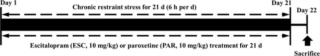
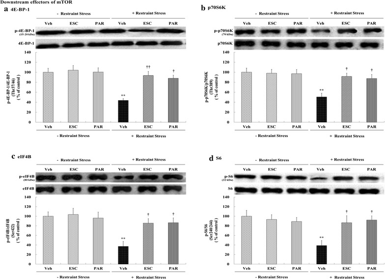
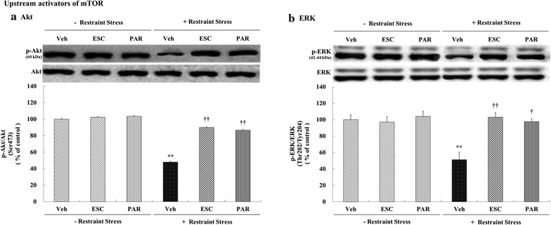
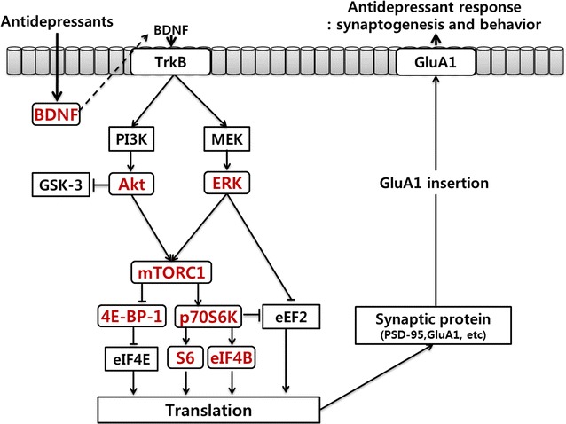
Background: Recent studies have suggested that the activation of mammalian target of rapamycin (mTOR) signaling may be related to antidepressant action. Therefore, the present study evaluated whether antidepressant drugs would exert differential effects on mTOR signaling in the rat hippocampus under conditions of chronic restraint stress. Male Sprague-Dawley rats were subjected to restraint stress for 6 h/days for 21 days with either escitalopram (10 mg/kg) or paroxetine (10 mg/kg) administered after the chronic stress procedure. Western blot analyses were used to assess changes in the levels of phospho-Ser2448-mTOR, phospho-Thr37/46-4E-BP-1, phospho-Thr389-p70S6 K, phospho-Ser422-eIF4B, phospho-Ser240/244-S6, phospho-Ser473-Akt, and phospho-Thr202/Tyr204-ERK in the hippocampus.

Results: Chronic restraint stress significantly decreased the levels of phospho-mTOR complex 1 (mTORC1), phospho-4E-BP-1, phospho-p70S6 K, phospho-eIF4B, phospho-S6, phospho-Akt, and phospho-ERK (p < 0.05); the administration of escitalopram and paroxetine increased the levels of all these proteins (p < 0.05 or 0.01). Additionally, chronic restraint stress reduced phospho-mTORC1 signaling activities in general, while escitalopram and paroxetine prevented these changes in phospho-mTORC1 signaling activities.

Conclusion: These findings provide further data that contribute to understanding the possible relationships among mTOR activity, stress, and antidepressant drugs.

Keywords: Antidepressants; Chronic restraint stress; Hippocampus; Neuroplasticity; mTOR signaling.


Figures

Fig. 1
Fig. 1
Schematic diagram of the experimental schedule. Escitalopram (ESC, 10 mg/kg) or paroxetine (PAR, 10 mg/kg) was administered 1 h prior to restraint stress for a total of 21 days (6 h/days). The rats were sacrificed on the 22nd day
Fig. 2
Fig. 2
Effects of antidepressants on levels of phospho-mTORC1 in the rat hippocampus. Rats (n = 6 animals/group) were given a daily injection of vehicle (Veh; 1 mL/kg), ESC (10 mg/kg), or PAR (10 mg/kg) for 21 days with or without restraint stress (6 h daily for 21 days). Levels of phosphorylated mTORC1 in brain homogenates from the hippocampus were detected by SDS-PAGE and Western blot analyses using anti-phospho-Ser2448-mTORC1 antibodies. A representative image and quantitative analysis normalized to the levels of total mTORC1 are shown. Results are expressed as a percentage of vehicle control and represent the mean ± standard error of the mean (SEM) of 6 animals per group. *p < 0.05 versus vehicle control; p < 0.05 versus stress + vehicle
Fig. 3
Fig. 3
Effects of antidepressants on the levels of mTORC1 downstream effectors (phospho-4E-BP-1, phospho-p70S6 K, phosphor-eIF4B, and phospho-S6) in the rat hippocampus. Rats (n = 6 animals/group) were given a daily injection of Veh (1 mL/kg), ESC (10 mg/kg), or PAR (10 mg/kg) for 21 days with or without restraint stress (6 h daily for 21 days). Levels of phosphorylated 4E-BP-1, p70S6 K, eIF4B, and S6 in brain homogenates from the hippocampus were detected by SDS-PAGE and Western blot analyses using anti-phospho-Thr37/46-4E-BP-1 (a), anti-phospho-Thr389-p70S6 K (b), anti-phospho-Ser422-eIF4B (c), and anti-phospho-Ser240/244-S6 (d) antibodies. A representative image and quantitative analysis normalized to the levels of total 4E-BP-1 (a), p70S6 K (b), eIF4B (c), and S6 (d) are shown. The results are expressed as a percentage of vehicle control and represent the mean ± SEM of 6 animals per group. **p < 0.01 versus vehicle control; p < 0.05 or †† p < 0.01 versus stress + vehicle
Fig. 4
Fig. 4
Effects of antidepressants on the levels of mTORC1 upstream activators (phospho-Akt and phospho-ERK) in the rat hippocampus. Rats (n = 6 animals/group) were given a daily injection of Veh (1 mL/kg), ESC (10 mg/kg), or PAR (10 mg/kg) for 21 days with or without restraint stress (6 h daily for 21 days). Levels of phosphorylated Akt and ERK in brain homogenates from the hippocampus were detected by SDS-PAGE and Western blot analyses using anti-phospho-Ser473-Akt (a) and anti-phospho-Thr202/Tyr204-ERK (b) antibodies. A representative image and quantitative analysis normalized to the levels of total Akt (a) and ERK (b) are shown. The results are expressed as the percentage of vehicle control and represent the mean ± SEM of 6 animals per group. **p < 0.01 versus vehicle controls; p < 0.05 or †† p < 0.01 versus stress + vehicle
Fig. 5
Fig. 5
Possible mechanisms underlying antidepressant-induced molecular changes related to antidepressant effects. Antidepressants increase BDNF [please spell out] expression. The release of BDNF and the stimulation of associated signaling cascades (PI3 K/Akt and MEK/ERK) activate mTORC1 signaling and translation which, in turn, increases synaptic protein levels and synaptogenesis. These effects contribute to the sustained antidepressant actions of antidepressants. TrkB tyrosine-related kinase B, PI3 K phosphoinositide 3-kinase, MEK MAP/ERK kinase, ERK extracellular signal-regulated kinases, GSK-3, glycogen synthase kinase-3, mTORC1 mammalian target of rapamycin complex 1, 4E-BP-1 4E-binding protein 1, p70S6 K p70ribosomal protein S6 kinase, eEF2 eukaryotic elongation factor 2, eIF4E eukaryotic translation initiation factor 4E, S6 small ribosomal protein 6, eIF4B eukaryotic translation initiation factor 4B, PSD-95 post-synaptic density 95, GluA1 glutamate ionotropic receptor AMPA type subunit 1, BDNF brain-derived neurotrophic factor. The molecular pathways shown in red illustrate novel observations from the present study while those in black are generally accepted signaling pathways involved in antidepressant action

References

    1. Lasserre AM, Marti-Soler H, Strippoli MP, Vaucher J, Glaus J, Vandeleur CL, et al. Clinical and course characteristics of depression and all-cause mortality: a prospective population-based study. J Affect Disord. 2016;189:17–24. doi: 10.1016/j.jad.2015.09.010. - DOI - PubMed
    1. Kessler RC, Berglund P, Demler O, Jin R, Koretz D, Merikangas KR, et al. The epidemiology of major depressive disorder: results from the National Comorbidity Survey Replication (NCS-R) JAMA. 2003;289:3095–3105. doi: 10.1001/jama.289.23.3095. - DOI - PubMed
    1. Hawton K, Casanas I, Comabella C, Haw C, Saunders K. Risk factors for suicide in individuals with depression: a systematic review. J Affect Disord. 2013;147:17–28. doi: 10.1016/j.jad.2013.01.004. - DOI - PubMed
    1. Richards D. Prevalence and clinical course of depression: a review. Clin Psychol Rev. 2011;31:1117–1125. doi: 10.1016/j.cpr.2011.07.004. - DOI - PubMed
    1. Duman RS. Pathophysiology of depression and innovative treatments: remodeling glutamatergic synaptic connections. Dialogues Clin Neurosci. 2014;16:11–27. - PMC - PubMed

Publication types

MeSH terms

Substances