. 2017 Apr 26;19(1):51.
doi: 10.1186/s13058-017-0844-3.

Cooperation of neurotrophin receptor TrkB and Her2 in breast cancer cells facilitates brain metastases

Affiliations

Cooperation of neurotrophin receptor TrkB and Her2 in breast cancer cells facilitates brain metastases

Cecilia Choy et al. Breast Cancer Res. .

Abstract

Background: Patients with primary breast cancer that is positive for human epidermal growth factor receptor 2 (Her2+) have a high risk of developing metastases in the brain. Despite gains with systemic control of Her2+ disease using molecular therapies, brain metastases remain recalcitrant to therapeutic discovery. The clinical predilection of Her2+ breast cancer cells to colonize the brain likely relies on paracrine mechanisms. The neural niche poses unique selection pressures, and neoplastic cells that utilize the brain microenvironment may have a survival advantage.

Methods: Tropomyosin-related kinase B (TrkB), Her2, and downstream targets were analyzed in primary breast cancer, breast-to-brain metastasis (BBM) tissues, and tumor-derived cell lines using quantitative real-time PCR, western blot, and immunohistochemical assessment. TrkB function on BBM was confirmed with intracranial, intracardiac, or mammary fat pad xenografts in non-obese diabetic/severe combined immunodeficiency mice. The function of brain-derived neurotrophic factor (BDNF) on cell proliferation and TrkB/Her2 signaling and interactions were confirmed using selective shRNA knockdown and selective inhibitors. The physical interaction of Her2-TrkB was analyzed using electron microscopy, co-immunoprecipitation, and in silico analysis. Dual targeting of Her2 and TrkB was analyzed using clinically utilized treatments.

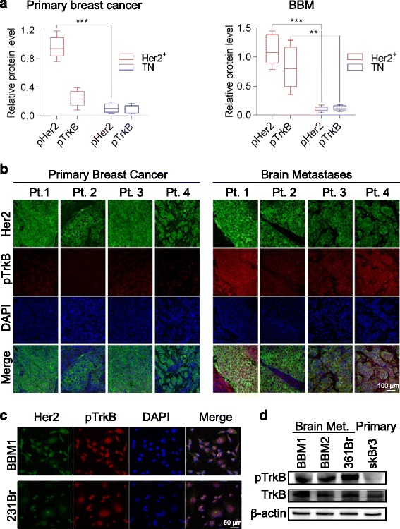
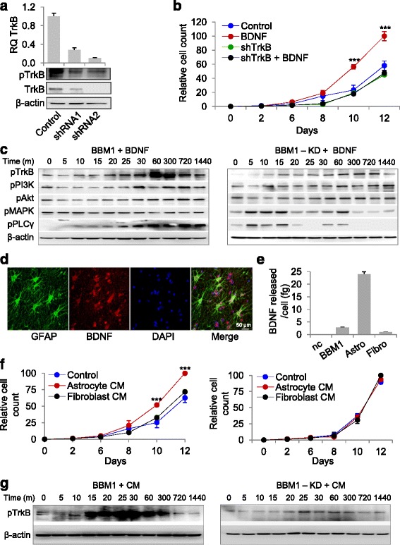
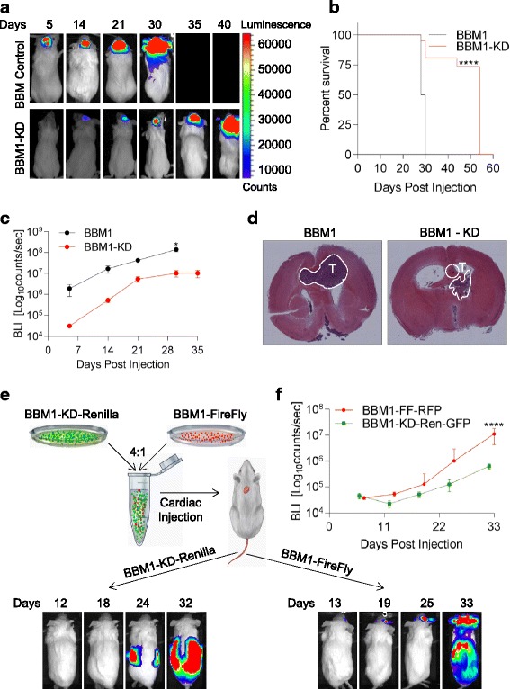
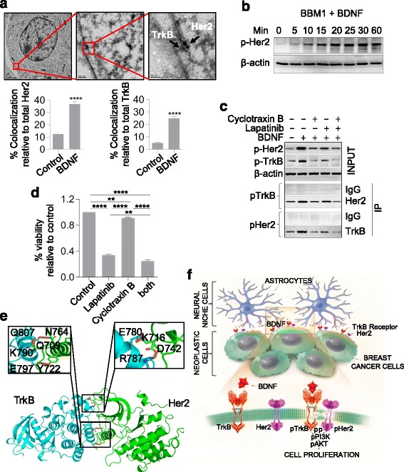
Results: We observed that patient tissues and cell lines derived from Her2+ human BBM displayed increased activation of TrkB, a neurotrophin receptor. BDNF, an extracellular neurotrophin, with roles in neuronal maturation and homeostasis, specifically binds to TrkB. TrkB knockdown in breast cancer cells led to decreased frequency and growth of brain metastasis in animal models, suggesting that circulating breast cancer cells entering the brain may take advantage of paracrine BDNF-TrkB signaling for colonization. In addition, we investigated a possible interaction between TrkB and Her2 receptors on brain metastatic breast cancer cells, and found that BDNF phosphorylated both its cognate TrkB receptor and the Her2 receptor in brain metastatic breast cancer cells.

Conclusion: Collectively, our findings suggest that heterodimerization of Her2 and TrkB receptors gives breast cancer cells a survival advantage in the brain and that dual inhibition of these receptors may hold therapeutic potential.

Keywords: Astrocyte; Brain-derived neurotrophic factor (BDNF); Breast cancer brain metastasis; Cyclotraxin B; Epidermal growth factor receptor 2 (Her2+); Glial cells; Lapatinib; Neural microenvironment; Neurotrophic factor; Neurotrophins; Tropomyosin-related kinase B (TrkB).


Figures

Fig. 1
Fig. 1
Human epidermal growth factor receptor 2 (Her2)-positive (Her2+) breast cancer brain metastasis (BBM) cells express high levels of phosphorylated neurotrophin receptor tropomyosin-related kinase B (p-TrkB). a Quantification of Her2 and TrkB in resected Her2+ (n = 8) and triple negative (TN) (n = 4) primary breast cancer tissue (left) and Her2+ (n = 3) and TN (n = 3) breast-to-brain metastases (*p < 0.05, **p < 0.01, ***p < 0.001; bars indicate SEM). b Representative immunofluorescence staining of primary breast cancer (left, n = 4) and breast-to-brain metastasis tissue (right, n = 4) from four patients (Pt). Tissue was stained for Her2 (green) and p-TrkB (red). Nuclear counter staining was done with 4',6-diamidino-2-phenylindole (DAPI, blue). c Immunofluorescence staining of p-TrkB in BBM1 and 231Br brain metastasis breast cancer cell lines. d Western blot analysis of p-TrkB and total TrkB in BBM1, BBM2, 361, and SkBr3 cells
Fig. 2
Fig. 2
Exogenous and astrocyte-released brain-derived neurotrophic factor (BDNF) binds Tropomyosin-related kinase B (TrkB) leading to increased tumor cell proliferation. a Real-time PCR and western blot analysis of TrkB knockdown with two different short hairpin (sh)RNAs in BBM1 cells. Control was non-transduced BBM1 cells. b Effect of exogenous BDNF (25 ng/mL) on BBM1 (BDNF) and TrkB knockdown BBM1 (shTrkB) cell proliferation over 12 days in vitro. Control was non-treated cells (n = 3; ****p < 0.0001; bars indicate SEM). c Western blot analysis of PI3K pathway activation by exogenous BDNF (25 ng/mL) over 24 h in BBM1 and TrkB knockdown BBM1 (BBM1-KD) cells. d Immunohistochemical staining of glial fibrillary acidic proteins (GFAP) and BDNF in the peritumoral region in a human breast cancer brain metastasis (BBM) specimen. GFAP (green), BDNF (red), DAPI (blue). e ELISA quantification of BDNF released from BBM1 cells, astrocytes, and fibroblasts (nc medium only; ****p < 0.0001). f Proliferation of BBM1 (left) and TrkB knockdown BBM1 (BBM1-KD) (right) cultured with control DMEM (Control), astrocyte-conditioned medium (Astrocyte CM), or fibroblast-conditioned medium (Fibroblast CM) for 12 days in vitro (n = 3; **p < 0.01, ****p < 0.0001; bars indicate SEM). g Phosphorylation of TrkB in BBM1 and TrkB knockdown BBM1 (BBM1-KD) cells grown in astrocyte-conditioned medium (CM)
Fig. 3
Fig. 3
Tropomyosin-related kinase B (TrkB) knockdown disrupts colonization and metastatic efficiency of breast cancer brain metastasis (BBM) xenografts. a Representative bioluminescence imaging (BLI) of intracranially injected BBM1 or BBM1-knockdown (BBM1-KD) cells in NOD-SCID mice. b Survival curve of mice injected intracranially with BBM1 or BBM1-KD cells (****p < 0.0001). c Tumor growth over 35 days (n = 6; *p < 0.05; bars indicate SEM). d Hematoxylin and eosin stained brain sections from mice euthanized 28 days after intracranial injection of BBM1 or BBM1-KD cells: tiled images (×5 magnification). White perforated lines indicate locations of the tumor. e The schema for BBM1 and BBM1-KD co-injection study (top). Representative BLI images of mice that received cardiac injections (bottom). f BLI of brain metastases arising from BBM1 cells in mice that received cardiac injections shown in e (n = 6; ****p < 0.0001; bars indicate SEM). Ren renilla, RFP red fluorescent protein, GFP green fluorescent protein
Fig. 4
Fig. 4
Tropomyosin-related kinase B (TrkB) and human epidermal growth factor receptor 2 (Her2) heterodimerize and activate upon brain-derived neurotrophic factor (BDNF) administration. a Representative post-embedding electron microscopy image (top) of TrkB and Her2 clustering at the cell membrane upon BDNF treatment; t = 30 minutes, TrkB = 10 nm, Her2 = 20 nm. Quantification (bottom) of co-localized Her2 or TrkB receptors relative to total Her2 or TrkB receptors, respectively (bars indicate SEM). b Western blot analysis of Her2 activated by exogenous BDNF over 1 h in BBM1 cells. c Co-immunoprecipitation of TrkB and Her2 from BBM1 cells grown in the presence of lapatinib (50 μM) and/or cyclotraxin B (20 μM). Her2 and TrkB immunoprecipitations were analyzed by western blotting with anti-TrkB and anti-Her2 antibodies, respectively. d Molecular model of TrkB (left) and Her2 (right). Insets indicate amino acids on TrkB and Her2 involved in possible hydrogen bonding. e Quantification of viable BBM1 cells grown in the presence of Her2 and TrkB inhibitors lapatinib (50 μM) and cyclotraxin B (20 μM), respectively. f Predicted model of paracrine signaling between breast cancer cells and the brain microenvironment. Her2 receptor (orange) and TrkB receptor (purple) on the cell surface membrane (green) are phosphorylated upon binding of BDNF (red) secreted by surrounding astrocytes (blue)

References

    1. Jemal A, Bray F, Center MM, Ferlay J, Ward E, et al. Global cancer statistics. CA Cancer J Clin. 2011;61:69–90. doi: 10.3322/caac.20107. - DOI - PubMed
    1. Lu X, Kang Y. Organotropism of breast cancer metastasis. J Mammary Gland Biol Neoplasia. 2007;12:153–62. doi: 10.1007/s10911-007-9047-3. - DOI - PubMed
    1. Palmieri D. An introduction to brain metastasis. In: Palmieri D, editor. Central nervous system metastasis, the biological basis, and clinical considerations. Dordrecht, The Netherlands: Springer; 2012. pp. 1–13.
    1. Lorger M, Felding-Habermann B. Capturing changes in the brain microenvironment during initial steps of breast cancer brain metastasis. Am J Pathol. 2010;176:2958–71. doi: 10.2353/ajpath.2010.090838. - DOI - PMC - PubMed
    1. Dougherty KD, Dreyfus CF, Black IB. Brain-derived neurotrophic factor in astrocytes, oligodendrocytes, and microglia/macrophages after spinal cord injury. Neurobiol Dis. 2000;7:574–85. doi: 10.1006/nbdi.2000.0318. - DOI - PubMed

Publication types

MeSH terms