. 2017 Apr 26;8(1):99.
doi: 10.1186/s13287-017-0554-x.

The crosstalk between vascular MSCs and inflammatory mediators determines the pro-calcific remodelling of human atherosclerotic aneurysm

Affiliations

The crosstalk between vascular MSCs and inflammatory mediators determines the pro-calcific remodelling of human atherosclerotic aneurysm

Carmen Ciavarella et al. Stem Cell Res Ther. .

Abstract

Background: Human mesenchymal stem cells (MSCs) possess well-known reparative abilities, but any defect of the immunomodulatory activity and/or the differentiation process may determine the development of human diseases, including those affecting the vascular wall. MSCs residing within the human aortic wall represent a potential cell mediator of atherosclerotic aneurysm development.

Methods: MSCs isolated from healthy and aneurysm aortas were characterized by flow cytometer and tested for differentiation properties. Healthy aorta (ha)-MSCs were then subjected to inflammatory stimuli to evaluate the microenvironmental impact on their function and involvement in vascular remodelling.

Results: Abdominal aortic aneurysm (AAA)-MSCs were isolated from calcified and inflamed aortas of 12 patients with high serum levels of MMP-9 protein. AAA-MSCs expressed typical mesenchymal markers and, in line with the histological analysis, elevated levels of OPN, an osteogenic marker also involved in vascular remodelling. AAA-MSCs were highly osteogenic and underwent intense calcium deposition under proper stimulation; moreover, AAA-MSCs were able to differentiate into tubule-like structures in Matrigel, even if the lack of CD146 and the reduced structural stability suggested an inefficient maturation process. We further demonstrated an association between osteogenesis and inflammation; indeed, ha-MSCs cultured with either cytokines (TNF-α, IL-1β) or AAA-PBMCs showed increased expression of MMP-9 and osteogenic markers, to the detriment of the adipogenic regulator PPAR-γ. Interestingly, the culture with inflammatory cells highly stimulated ha-MSCs towards the osteogenic commitment.

Conclusions: AAA-MSCs displayed high osteogenic potential and pathological angiogenesis that represent crucial steps for AAA progression; we showed that the inflammatory process critically addresses human vascular MSCs towards a pathological behaviour, inducing vascular bone matrix deposition and remodelling. Inhibition of this pathway may represent a pharmacological approach against arterial calcification.

Keywords: Angiogenesis; Atherosclerotic aneurysm; Calcification; Human aortic mesenchymal stem cells; Inflammation; Vascular remodelling.


Figures

Fig. 1
Fig. 1
Clinical background of study patients. a Clinical data of AAA patients; data are reported as mean ± standard deviation (n = 12). b Evaluation of calcium content within the aortic wall: representative non-contrast angio-CT acquisition of AAA wall corresponding to scores 1 (upper panel) and 3 (lower panel). c Alizarin Red staining of healthy vs AAA tissue sections showed  the presence of calcium depositions only in the affected aorta (black stars), reflecting a diffuse localization within the whole arterial wall with more intensive staining at the media level. d Serum levels of MMP-9 protein in AAA patients compared with pooled sera from healthy donors. The assay was executed in duplicate and data are reported as mean ± standard deviation of two independent assays. *p < 0.05. AAA abdominal aortic aneurysm, CV cardiovascular, CT computed tomography, LDL low density lipoprotein, HDL high density lipoprotein, MMP-9 matrix metalloproteinase-9
Fig. 2
Fig. 2
Mesenchymal immunophenotype of AAA-MSCs. Representative images showing flow cytometry for CD44, CD90, CD73 and CD105 confirmed the mesenchymal phenotype of MSCs isolated from the AAA wall (black histograms)
Fig. 3
Fig. 3
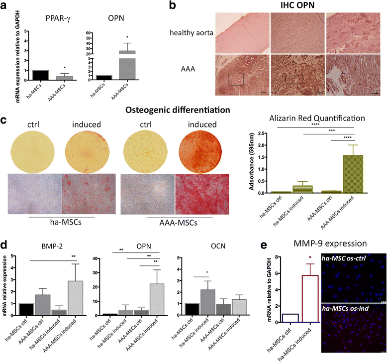
AAA-MSCs possess elevated osteogenic potential. AAA-MSCs were compared with ha-MSCs for expression of lineage-specific markers at time 0 and after stimulation with osteogenic medium. a Significant down-regulation of PPAR-γ and pronounced expression of OPN in AAA-MSCs. Results are expressed as fold changes relative to ha-MSCs. b In-situ detection of OPN protein was carried out on healthy and aneurysm aortic tissues. Diffuse positivity to OPN was detected on the whole AAA tissue (boxes), and predominantly on SMCs and inflammatory cells (arrows and stars). Scale bar: 100 μm. c Representative Alizarin Red staining of the mineralization process on ha-MSCs and AAA-MSCs, after exposure to osteogenic induction medium for 21 days. Quantitative data are expressed as mean ± standard deviation and compared with undifferentiated ha-MSCs. d Real-time PCR analysis of osteogenic markers in ha-MSCs and AAA-MSCs after osteogenic stimulation. Results are expressed as fold changes relative to undifferentiated ha-MSCs. e MMP-9 mRNA and protein detection on ha-MSCs after osteogenic differentiation; increased MMP-9 transcription (6-fold) and staining were observed on differentiated vs undifferentiated ha-MSCs. Scale bar 50 μm. *p < 0.05, **p < 0.01, ***p < 0.001, ****p < 0.0001. BMP-2 bone morphogenetic protein-2, IHC immunohistochemistry, OPN osteopontin, OCN osteocalcin, PPAR-γ peroxisome proliferation activated receptor gamma, ha-MSC healthy aortic MSC, AAA-MSC abdominal aortic aneurysm MSC, MSC mesenchymal stem cell, os-ind osteogenic-induced, MMP-9 matrix metalloproteinase-9, ctrl control
Fig. 4
Fig. 4
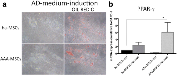
Adipogenic differentiation of AAA-MSCs. a Representative Oil Red O staining of ha-MSCs and AAA-MSCs showed the formation of lipid droplets after induction with adipogenic medium for 14 days. b Real-time PCR performed on differentiated MSCs revealed significant up-regulation of PPAR-γ in AAA-MSCs following induction. Results are expressed as fold changes relative to undifferentiated ha-MSCs and are representative of at least three independent experiments. *p < 0.05. AD adipogenic, PPAR-γ peroxisome proliferation activated receptor gamma, ha-MSC healthy aortic MSC, AAA-MSC abdominal aortic aneurysm MSC, MSC mesenchymal stem cell, ctrl control
Fig. 5
Fig. 5
Impaired endothelial differentiation in AAA-MSCs. a Representative pictures of ha-MSCs and AAA-MSCs after culture in Matrigel for 6 hours. Scale bar: 50 μm. b Morphometric analysis of neo-vessels performed on Matrigel-differentiated MSCs after 6 hours, according to total tubule length and number of branching points and total tubes (Wimasis Image Analysis software). Results are expressed as fold changes relative to ha-MSC control; statistical analysis was performed by two-way ANOVA with multiple comparisons among all experimental conditions. *p < 0.05. ha-MSC healthy aortic MSC, AAA-MSC abdominal aortic aneurysm MSC, MSC mesenchymal stem cell, ctrl control, VEGF vascular endothelial growth factor
Fig. 6
Fig. 6
Low expression of CD146 in AAA-MSC newly formed vessels. a Flow cytometry analysis of CD31 and CD146 proteins in ha-MSC and AAA-MSC 2D culture before seeding in Matrigel. b Gene expression profiles of angiogenic markers CD31, α-SMA and CD146 in healthy and AAA-MSCs after Matrigel culture for 6 hours. Results are expressed as fold changes relative to ha-MSC control; statistical analysis performed by two-way ANOVA with multiple comparisons among all experimental conditions. *p < 0.05, ***p < 0.001. c CD146 protein expression in ha-MSC and AAA-MSC newly formed vessels, as detected by flow cytometry after Matrigel dissociation. a, c Black histograms, untreated ctrl; white histograms, VEGF-induced condition. ha-MSC healthy aortic MSC, AAA-MSC abdominal aortic aneurysm MSC, MSC mesenchymal stem cell, ctrl control, VEGF vascular endothelial growth factor
Fig. 7
Fig. 7
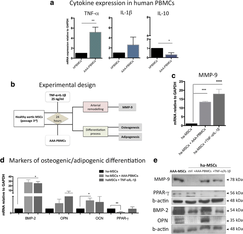
ha-MSCs exposed to inflammatory conditions assume a pathological phenotype. a PBMCs isolated from AAA patients showed a molecular signature reporting high levels of inflammatory cytokines (TNF-α and IL-1β) and reduced anti-inflammatory IL-10. Results are expressed as fold changes relative to healthy PBMCs. b According to the experimental design, ha-MSCs were exposed to inflammatory mediators (cytokines and PBMCs) for 24 hours, then investigated in terms of vascular remodelling and differentiation properties. ha-MSCs exposed to inflammation underwent increased transcription of (c) MMP-9 and (d) osteogenic lineage-specific markers (BMP-2, OPN, OCN), to the detriment of the adipogenic transcriptional factor PPAR-γ. Results are expressed as fold changes relative to unexposed ha-MSCs. *p < 0.05, **p < 0.01, ***p < 0.001, ****p < 0.0001. e Western blot analysis of MMP-9 was performed on serum-free conditioned media, whereas PPAR-γ, BMP-2 and OPN were detected in cell lysates. PBMC peripheral blood mononuclear cell, TNF-α tumour necrosis factor alpha, IL interleukin, ha-PMSC healthy aortic PMSC, AAA-PMSC abdominal aortic aneurysm PMSC, MSC mesenchymal stem cell, MMP-9 matrix metalloproteinase-9, BMP-2 bone morphogenetic protein-2, OPN osteopontin, OCN osteocalcin, PPAR-γ peroxisome proliferation activated receptor gamma, ctrl control
Fig. 8
Fig. 8
Inflammation enhances the mineralization process in ha-MSCs. a After inflammatory stimulation, ha-MSCs were cultured with specific osteogenic and adipogenic induction media for 21 and 14 days, respectively. b Calcium mineralization process was significantly marked under inflammatory conditions, mainly after AAA-PBMC influence as showed by Alizarin Red staining. Quantification values are represented as mean ± standard deviation and compared with induced ha-MSCs. c MMP-9 detection on osteogenic differentiated ha-MSCs was performed by immunofluorescence, revealing an appreciable staining only after osteogenic induction; 20× magnification. d Oil Red O staining of lipid droplets in ha-MSCs was reduced after priming cells with inflammatory cytokines, as shown by e PPAR-γ mRNA. Results expressed as fold changes relative to induced ha-MSCs. *p < 0.05. f Representative western blot analysis of PPAR-γ protein and relative densitometry after β-actin normalization (ImageJ software). PBMC peripheral blood mononuclear cell, TNF-α tumour necrosis factor alpha, IL interleukin, ha-MSC healthy aortic MSC, AAA-MSC abdominal aortic aneurysm MSC, MSC mesenchymal stem cell, MMP-9 matrix metalloproteinase-9, PPAR-γ peroxisome proliferation activated receptor gamma, ctrl control

Similar articles

Cited by

References

    1. Barry FP, Murphy JM. Mesenchymal stem cells: clinical applications and biological characterization. Int J Biochem Cell Biol. 2004;36:568–84. doi: 10.1016/j.biocel.2003.11.001. - DOI - PubMed
    1. Zuk PA, Zhu M, Ashjian P, De Ugarte DA, Huang JI, Mizuno H, et al. Human adipose tissue is a source of multipotent stem cells. Mol Biol Cell. 2002;13(12):4279–95. doi: 10.1091/mbc.E02-02-0105. - DOI - PMC - PubMed
    1. Ergün S, Tilki D, Klein D. Vascular wall as a reservoir for different types of stem and progenitor cells. Antioxid Redox Signal. 2011;15:981–95. doi: 10.1089/ars.2010.3507. - DOI - PubMed
    1. Kolf CM, Cho E, Tuan RS. Mesenchymal stromal cells. Biology of adult mesenchymal stem cells: regulation of niche, self-renewal and differentiation. Arthritis Res Ther. 2007;9:204. doi: 10.1186/ar2116. - DOI - PMC - PubMed
    1. Gebler A, Zabel O, Seliger B. The immunomodulatory capacity of mesenchymal stem cells. Trends Mol Med. 2012;18:128–34. doi: 10.1016/j.molmed.2011.10.004. - DOI - PubMed

MeSH terms