. 2017 Jul 7;121(2):113-124.
doi: 10.1161/CIRCRESAHA.116.310494. Epub 2017 Apr 26.

Concurrent Isolation of 3 Distinct Cardiac Stem Cell Populations From a Single Human Heart Biopsy

Affiliations

Concurrent Isolation of 3 Distinct Cardiac Stem Cell Populations From a Single Human Heart Biopsy

Megan M Monsanto et al. Circ Res. .

Abstract

Rationale: The relative actions and synergism between distinct myocardial-derived stem cell populations remain obscure. Ongoing debates on optimal cell population(s) for treatment of heart failure prompted implementation of a protocol for isolation of multiple stem cell populations from a single myocardial tissue sample to develop new insights for achieving myocardial regeneration.

Objective: Establish a robust cardiac stem cell isolation and culture protocol to consistently generate 3 distinct stem cell populations from a single human heart biopsy.

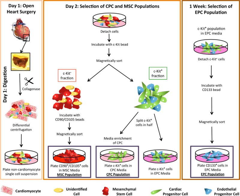
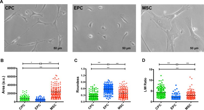
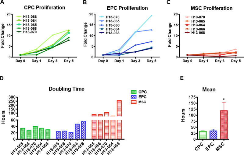
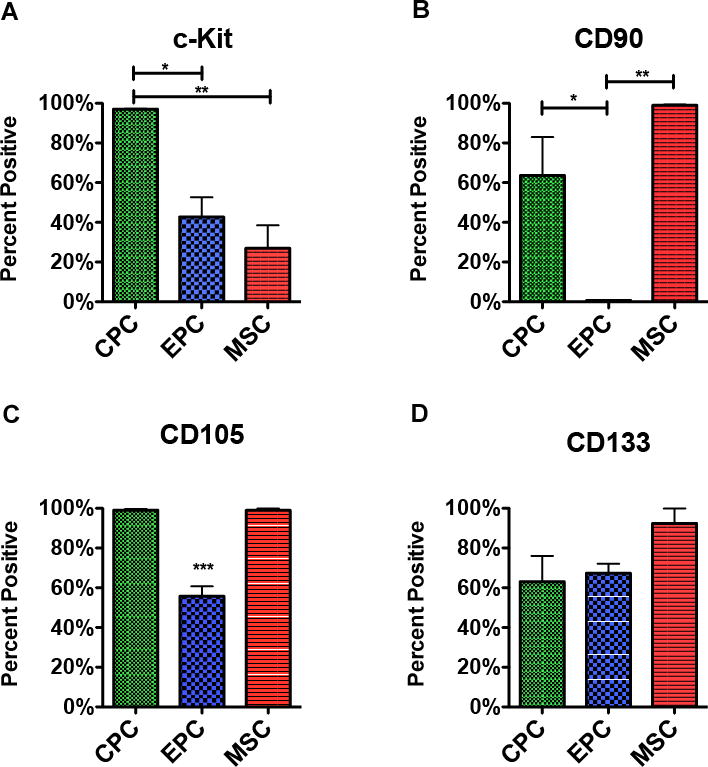
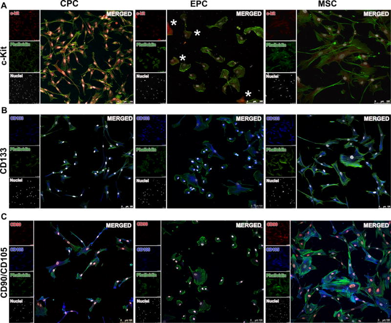
Methods and results: Isolation of 3 endogenous cardiac stem cell populations was performed from human heart samples routinely discarded during implantation of a left ventricular assist device. Tissue explants were mechanically minced into 1 mm3 pieces to minimize time exposure to collagenase digestion and preserve cell viability. Centrifugation removes large cardiomyocytes and tissue debris producing a single cell suspension that is sorted using magnetic-activated cell sorting technology. Initial sorting is based on tyrosine-protein kinase Kit (c-Kit) expression that enriches for 2 c-Kit+ cell populations yielding a mixture of cardiac progenitor cells and endothelial progenitor cells. Flowthrough c-Kit- mesenchymal stem cells are positively selected by surface expression of markers CD90 and CD105. After 1 week of culture, the c-Kit+ population is further enriched by selection for a CD133+ endothelial progenitor cell population. Persistence of respective cell surface markers in vitro is confirmed both by flow cytometry and immunocytochemistry.

Conclusions: Three distinct cardiac cell populations with individualized phenotypic properties consistent with cardiac progenitor cells, endothelial progenitor cells, and mesenchymal stem cells can be successfully concurrently isolated and expanded from a single tissue sample derived from human heart failure patients.

Keywords: adult stem cells; biopsy; centrifugation; endothelial progenitor cells; heart failure; mesenchymal stem cells.


Conflict of interest statement

AUTHOR CONTRIBUTIONS

M. Monsanto and M. Sussman designed experiments. M. Monsanto, K. White, T. Kim, B. Wang, K. Fisher, F. Khalafalla, and S. Mohsin performed experiments and analyzed data. A. Casillas, K. Broughton, T. Kim and M. Monsanto helped in the schematic design. M. Monsanto, K. Ilves, and M. Sussman wrote the article. All authors read and approved the final article.

Figures

Figure 1
Figure 1. Cardiac stem cell isolation protocol
Schematic representation of the process used to isolate cardiac cells from LVAD tissue. Following open-heart surgery the tissue plug is digested to the single cell level and, following centrifugation to remove the cardiomyocytes, plated overnight at 37°C in CO2 incubator. Day 2, the single cell suspension is incubated with microbeads conjugated to c-Kit and magnetically sorted. C-Kit+ cells are split in half with a portion being plated in CPC media and the other half being plated in EPC media. The c-Kit fraction is further sorted for CD90 and CD105 with positive cells being plated in MSC media. 1 week later, the c-Kit+ cells plated in EPC media are further enriched for CD133. Red cells indicate mesenchymal stem cells (MSC); green cells indicate cardiac progenitor cells (CPC); blue cells indicate endothelial progenitor cells (EPC); yellow cells indicate non-specific cell population.
Figure 2
Figure 2. Isolation of three distinct cardiac stem cell populations from LVAD patients
A, Representative phase contrast images for the three isolated cardiac cell populations. B-D, Cell morphometric parameters measuring area (B), roundness (C), and length-to-width (L/W) ratio (D) (n=4–5 patients, minimum of 30 cells traced per cell type per patient). Data are presented as 1 way ANOVA, *p<0.01, **p<0.001, ***p<0.0001. Scale bar, 50 uM.
Figure 3
Figure 3. Cardiac stem cell populations exhibit distinct growth kinetics
A-C, Cardiac cell proliferation measured using CyQuant assays at day 0, day 1, day 3, and day 5 for CPC (A), EPC (B), and MSC (C). D, Bar graph of doubling times calculated using periods of exponential cell growth for each cell line. E, Bar graph of mean doubling time for each cell type (n=5 patients). Data are presented as 1 Way ANOVA, *p<0.05.
Figure 4
Figure 4. Flow cytometry analysis of markers expressed upon in vitro culture
A-D, Single cell suspensions stained with antibodies for the markers used in cell isolation. Flow cytometry analysis of cardiac cell populations sorted for the presence of c-Kit (A), CD90 (B), CD105 (C), and CD133 (D) (n=3 patients per marker). Data are presented as 1 way ANOVA, *p<0.05, **p<0.01, ***p<0.001.
Figure 5
Figure 5. Immunofluorescence imaging of stem cell markers
A, Representative florescence microscopy showing c-Kit localization in CPC, EPC, and MSC (red, c-Kit; green, phalloidin; white, nuclei (DAPI)). B, Immunofluorescence labeling of the mesenchymal markers CD90 and CD105 showing varying degree of co-localization for these two markers in the three cell populations (red, CD90; blue, CD105; green, phalloidin; white, nuclei (DAPI)). C, CD133 expression revealed by immunofluorescence in the cardiac cells (blue, CD133; green, phalloidin; white, nuclei (DAPI)). Scale bar, 100 um. Asterisks (*) indicate cells positive for c-Kit.
Figure 6
Figure 6. Supervised cluster analysis of gene expression levels in cardiac stem cells
A-B, Heatmaps representing differentially expressed genes in three different cell types. A, Cluster of genes showing specific up-regulation in EPC, but not CPC and MSC. B, Second cluster of genes showing specific down-regulation in EPC compared to CPC and MSC (n=5 patients).
Figure 7
Figure 7. In vitro lineage assessment and comparison to established cell lines
A-C, Representative images of tubular network formation when plated on growth factor reduced matrigel for CPC (A), EPC (B), and MSC (C). D–F, Bar graphs using established cell lines, HUVECs, bMSCs and adult CM, to assess the potential of cardiac stem cells to commit to an angiogenic (D), smooth muscle (E), and cardiogenic fate (F) (n=3 patients). Data are presented as 1 Way ANOVA, **P<0.001, ***p<0.0001, versus cell type used for normalization. Scale bar, 200 um. CM indicates Cardiomyocytes; GATA4, GATA binding protein 4; PECAM-1, Platelet endothelial cell adhesion molecule; and SMA, α-smooth muscle actin (SMA).

Comment in

Similar articles

Cited by

References

    1. Sanganalmath SK, Bolli R. Cell therapy for heart failure: a comprehensive overview of experimental and clinical studies, current challenges, and future directions. Circulation research. 2013;113:810–34. - PMC - PubMed
    1. Dixit P, Katare R. Challenges in identifying the best source of stem cells for cardiac regeneration therapy. Stem Cell Res Ther. 2015;6:26. - PMC - PubMed
    1. Assmus B, Schachinger V, Teupe C, Britten M, Lehmann R, Dobert N, Grunwald F, Aicher A, Urbich C, Martin H, Hoelzer D, Dimmeler S, Zeiher AM. Transplantation of Progenitor Cells and Regeneration Enhancement in Acute Myocardial Infarction (TOPCARE-AMI) Circulation. 2002;106:3009–17. - PubMed
    1. Clifford DM, Fisher SA, Brunskill SJ, Doree C, Mathur A, Watt S, Martin-Rendon E. Stem cell treatment for acute myocardial infarction. Cochrane Database Syst Rev. 2012:CD006536. - PubMed
    1. Hare JM, et al. Comparison of allogeneic vs autologous bone marrow-derived mesenchymal stem cells delivered by transendocardial injection in patients with ischemic cardiomyopathy: the POSEIDON randomized trial. JAMA. 2012;308:2369–79. - PMC - PubMed