. 2017 Jul 25;136(4):367-383.
doi: 10.1161/CIRCULATIONAHA.117.028388. Epub 2017 Apr 26.

A Novel α-Calcitonin Gene-Related Peptide Analogue Protects Against End-Organ Damage in Experimental Hypertension, Cardiac Hypertrophy, and Heart Failure

Affiliations

A Novel α-Calcitonin Gene-Related Peptide Analogue Protects Against End-Organ Damage in Experimental Hypertension, Cardiac Hypertrophy, and Heart Failure

Aisah A Aubdool et al. Circulation. .

Abstract

Background: Research into the therapeutic potential of α-calcitonin gene-related peptide (α-CGRP) has been limited because of its peptide nature and short half-life. Here, we evaluate whether a novel potent and long-lasting (t½ ≥7 hours) acylated α-CGRP analogue (αAnalogue) could alleviate and reverse cardiovascular disease in 2 distinct murine models of hypertension and heart failure in vivo.

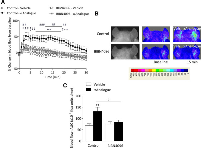
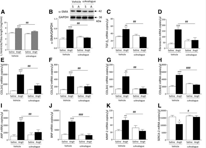
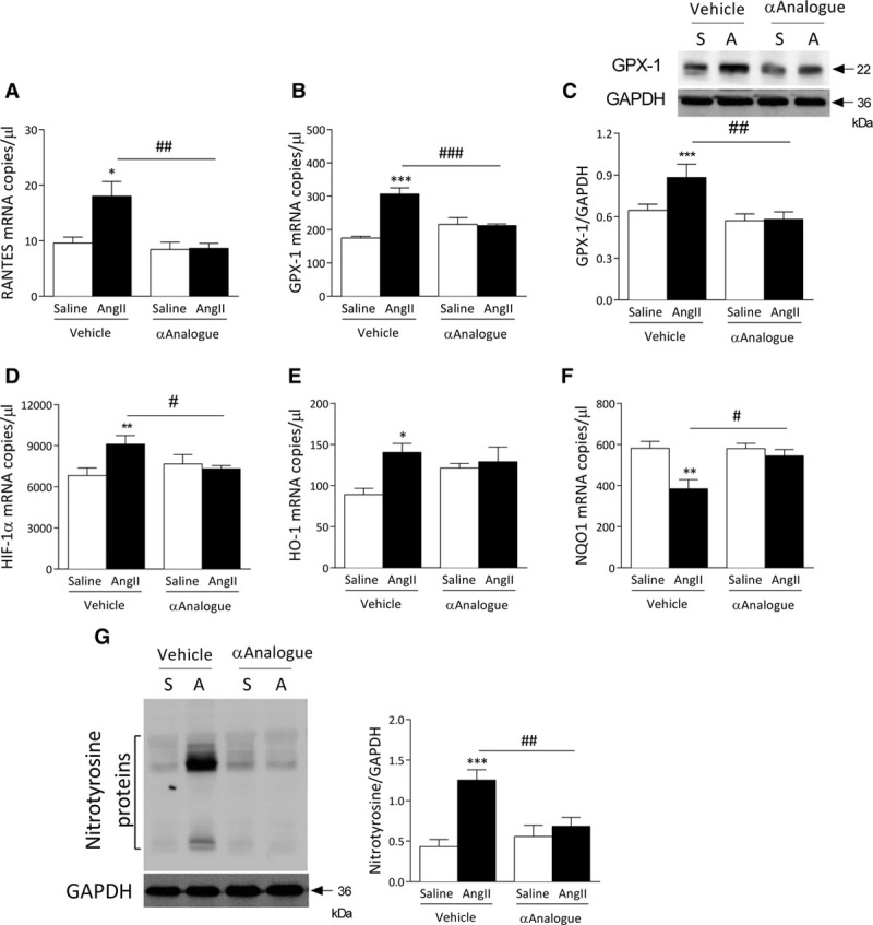
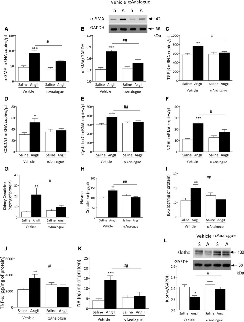
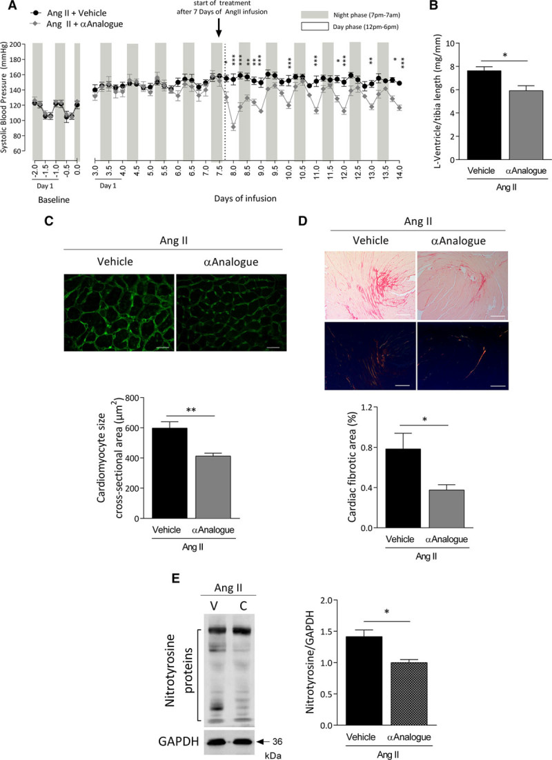
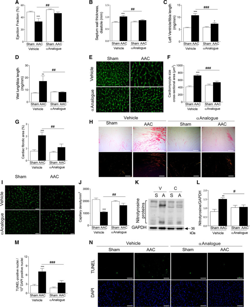
Methods: The ability of the αAnalogue to act selectively via the CGRP pathway was shown in skin by using a CGRP receptor antagonist. The effect of the αAnalogue on angiotensin II-induced hypertension was investigated over 14 days. Blood pressure was measured by radiotelemetry. The ability of the αAnalogue to modulate heart failure was studied in an abdominal aortic constriction model of murine cardiac hypertrophy and heart failure over 5 weeks. Extensive ex vivo analysis was performed via RNA analysis, Western blot, and histology.

Results: The angiotensin II-induced hypertension was attenuated by cotreatment with the αAnalogue (50 nmol·kg-1·d-1, SC, at a dose selected for lack of long-term hypotensive effects at baseline). The αAnalogue protected against vascular, renal, and cardiac dysfunction, characterized by reduced hypertrophy and biomarkers of fibrosis, remodeling, inflammation, and oxidative stress. In a separate study, the αAnalogue reversed angiotensin II-induced hypertension and associated vascular and cardiac damage. The αAnalogue was effective over 5 weeks in a murine model of cardiac hypertrophy and heart failure. It preserved heart function, assessed by echocardiography, while protecting against adverse cardiac remodeling and apoptosis. Moreover, treatment with the αAnalogue was well tolerated with neither signs of desensitization nor behavioral changes.

Conclusions: These findings, in 2 distinct models, provide the first evidence for the therapeutic potential of a stabilized αAnalogue, by mediating (1) antihypertensive effects, (2) attenuating cardiac remodeling, and (3) increasing angiogenesis and cell survival to protect against and limit damage associated with the progression of cardiovascular diseases. This indicates the therapeutic potential of the CGRP pathway and the possibility that this injectable CGRP analogue may be effective in cardiac disease.

Keywords: heart failure; hypertension; inflammation; oxidative stress; receptors, calcitonin gene-related peptide.


Figures

Figure 1.
Figure 1.
α-CGRP analogue (αAnalogue) increases vascular blood flow via CGRP receptors. Blood flow monitored using Full-field Laser Perfusion Imager in the ear vasculature of mice pretreated with control (saline) or CGRP receptor antagonist BIBN4096 (0.3 mg/kg, IV) at baseline and following intradermal injection of αAnalogue (100 pmol, daily) or vehicle (Veh) (15 µL, n=6). A, Blood flow responses expressed as % change from baseline. B, Representative Full-field Laser Perfusion Imager pictures alongside gray/black photo showing blood flow at baseline and 15 minutes after treatment. C, Blood flow assessed by area under the curve (AUC) for 30 minutes following vehicle or αAnalogue administration (n=6). Data showed as mean±SEM. *P<0.05, **P<0.01, ***P<0.001 versus vehicle-treated; #P<0.05, ##P<0.01, ###P<0.001 for αAnalogue treated (A, repeated-measures 2-way ANOVA + Bonferroni post hoc test; C, 2-way ANOVA + Bonferroni post hoc test). α-CGRP indicates α-calcitonin gene–related peptide.
Figure 2.
Figure 2.
Daily systemic treatment with α-CGRP analogue (αAnalogue) protects against angiotensin II (AngII)–induced hypertension and vascular damage. Mice were infused with AngII (A, 1.1 mg·kg–1·d-–1) or control (S, saline) for 14 days and treated daily with vehicle (V) or αAnalogue (50 nmol/kg, SC). A, Systolic blood pressure was measured by radiotelemetry. Results expressed as 6-hour average. Mice experience a 12/12 hour light/dark cycle, with the dark cycle shown in the gray striped area. Arrow represents the start of daily treatment. B, Protein expression of total eNOS in aorta (n=4–5). C, Representative images of Masson trichrome-stained aortic sections. D, Quantification of smooth muscle wall width (n=4–5; scale bars, 100 µm). Protein expression of NADPH oxidase-2 (NOX-2) (E), heme oxygenase-1 (HO-1) (F), nitrotyrosine in aorta (n=4–6) (G). H, Protein expression of nitrotyrosine in mesenteric vessels (n=6–7). Results shown as mean±SEM. *P<0.05, **P<0.01, ***P<0.001 versus vehicle-treated saline-infused; #P<0.05, ##P<0.01, ###P<0.001 for αAnalogue-treated AngII-infused versus vehicle-treated AngII-infused (A, repeated-measures 2-way ANOVA + Bonferroni post hoc test; B through H, 2-way ANOVA + Bonferroni post hoc test). α-CGRP indicates α-calcitonin gene–related peptide; and eNOS, endothelial nitric oxide synthase.
Figure 3.
Figure 3.
α-CGRP analogue (αAnalogue) protects against angiotensin II (AngII)–induced cardiac hypertrophy and fibrosis. Mice were treated as in Figure 2 (AngII, A; Saline, S). A, Left ventricle weight normalized to tibia length ratio (mg/mm). B, Protein expression of α-smooth muscle actin (α-SMA) in heart (n=5). mRNA expression measured by real-time quantitative polymerase chain reaction for transforming growth factor-β1 (TGF-β1) (C), fibronectin (D), collagen type 1 α1 (COL1A1) (E), collagen type 1 α2 (COL1A2) (F), collagen type 3 α1 (COL3A1) (G), collagen type 4 α1 (COL4A1) (H), atrial natriuretic peptide (ANP) (I), brain natriuretic peptide (BNP) (J), matrix metalloproteinase-2 (MMP-2) (K), and sarcoplasmic reticulum Ca2+ ATPase-2 (SERCA-2) (L) in heart (n=5–11). Results expressed as copy numbers per microliter normalized to hypoxanthine-guanine phosphoribosyltransferase, B2M and β-actin, and showed as mean±SEM. *P<0.05, **P<0.01, ***P<0.001 versus vehicle-treated saline-infused; #P<0.05, ##P<0.01, ###P<0.001 versus vehicle-treated AngII-infused (2-way ANOVA + Bonferroni post hoc test). α-CGRP indicates α-calcitonin gene–related peptide.
Figure 4.
Figure 4.
Daily systemic treatment with α-CGRP analogue (αAnalogue) protects against angiotensin II (AngII)–induced cardiac inflammation and oxidative stress. Mice were treated as in Figure 2 (AngII, A; Saline, S). mRNA expression measured by real-time quantitative polymerase chain reaction (n=5–11) for RANTES (A) and glutathione peroxidase-1 (GPX-1) (n=6–11) (B). C, Protein expression of GPX-1 in heart (n=5). mRNA expression for hypoxia-inducible factor-1 (HIF-1α) (D), heme oxygenase-1 (HO-1) (E), and NADPH dehydrogenase quinone-1 (NQO1) (n=5–11) (F). Results expressed as copy numbers per microliter normalized to hypoxanthine-guanine phosphoribosyltransferase, B2M and β-actin, and showed as mean±SEM. G, Protein expression of nitrotyrosine (n=5) in heart (n=6–7). *P<0.05, **P<0.01, ***P<0.001 versus vehicle-treated saline-infused; #P<0.05, ##P<0.01, ###P<0.001 versus vehicle-treated AngII-infused (2-way ANOVA + Bonferroni post hoc test). α-CGRP indicates α-calcitonin gene–related peptide.
Figure 5.
Figure 5.
Daily systemic treatment with α-CGRP analogue (αAnalogue) protects against angiotensin II (AngII)–induced renal fibrosis, dysfunction, and inflammation. Mice were treated as in Figure 2 (AngII, A; Saline, S). mRNA (A) and protein expression of α-SMA (B) in kidney. mRNA expression of TGF-β1 (C), collagen type 1a1 (COL1A1) (D), cystatin C (E), and neutrophil gelatinase-associated lipocalin (NGAL) (F) in kidney. Creatinine levels in kidney (G) and plasma (H) (n=6–8). IL-6 (I), TNF-α (J), and noradrenaline (NA) (K) levels in kidney (n=6–9). L, Protein expression of klotho in kidney. mRNA expression measured by real-time quantitative polymerase chain reaction (n=6–8), expressed as copy numbers per microliter normalized to hypoxanthine-guanine phosphoribosyltransferase, B2M, and β-actin. Results shown as mean±SEM. *P<0.05, **P<0.01, ***P<0.001 versus vehicle-treated saline-infused; #P<0.05, ##P<0.01 versus vehicle-treated AngII-infused (2-way ANOVA + Bonferroni post hoc test). α-CGRP indicates α-calcitonin gene–related peptide; IL-6, interleukin 6; α-SMA; α-smooth muscle actin; TGF-β1; transforming growth factor-β1; and TNF-α, tumor necrosis factor-α.
Figure 6.
Figure 6.
Effects of daily systemic treatment with α-CGRP analogue (αAnalogue) on CGRP receptor expression in angiotensin II (AngII)–induced hypertension. Mice were treated as in Figure 2 (AngII, A; saline, S, V, vehicle; α-CGRP analogue; C). Protein expression of calcitonin receptor-like receptor (CLR) (A) and receptor-associated membrane protein-1 (RAMP1) (B) in aorta (n=6). Protein expression of CLR (C) and RAMP1 (D) in mesenteric vessels (n=6). Protein expression of CLR (E) and RAMP1 (F) in heart (n=6–7). Protein expression of CLR (G) and RAMP1 (H) in kidney (n=5–6). Results showed as mean±SEM.*P<0.05, ***P<0.001 versus vehicle-treated saline-infused; #P<0.05, ###P<0.001 versus vehicle-treated AngII-infused (2-way ANOVA + Bonferroni post hoc test). α-CGRP indicates α-calcitonin gene–related peptide.
Figure 7.
Figure 7.
α-CGRP analogue (αAnalogue) limits angiotensin II (AngII)–induced hypertension. Mice were infused with AngII (1.1 mg·kg–1·d–1 for 14 days) and treated daily with vehicle or αAnalogue (50 nmol/kg, SC) at day 7 to 14 of infusion (n=4). A, Systolic blood pressure was measured by radiotelemetry. Results expressed as 6-hour average. Mice experience a 12/12 hour light/dark cycle, with the dark cycle shown in the gray striped area. Arrow represents the start of daily treatment. B, Left ventricle weight normalized to tibia length ratio (mg/mm). Representative heart sections (Top) and analysis (Bottom) showing cardiac hypertrophy by cardiomyocyte borders outlined using wheat germ agglutinin (scale bars, 20 µm) (C) and cardiac fibrosis by Picrosirius Red staining (scale bars, 200 µm) (D). E, Protein expression of nitrotyrosine in heart. Results showed as mean±SEM.*P<0.05, **P<0.01, ***P<0.001 versus vehicle-treated (A, repeated-measures 2-way ANOVA + Bonferroni post hoc test; B through E, 2-way ANOVA + Bonferroni post hoc test). α-CGRP indicates α-calcitonin gene–related peptide.
Figure 8.
Figure 8.
α-CGRP analogue (αAnalogue) preserves heart function post–AAC-induced cardiac hypertrophy and heart failure. Mice were treated daily with vehicle and αAnalogue (50 nmol/kg, SC) postsurgery for 5 weeks (n=6–8). Ejection fraction (%) (A), septum wall thickness at diastole (mm) (B), left ventricle mass normalized to tibia length (mg/mm) (C), and wet lung mass (mg) normalized to tibia length (D). Representative images (E) and quantification of cardiomyocyte cross-sectional area (F) in heart using wheat germ agglutinin staining (scale bars, 20 µm). Quantification (G) and representative images of fibrosis in heart (H) using picrosirius red staining (scale bars, 200 µm). Representative images (I) and quantification of capillary density in heart (J) using isolectin-B4 staining (scale bars, 20 µm). K and L, Protein expression of nitrotyrosine in heart. Quantification (M) and representative images of apoptosis (N) using TUNEL staining (scale bars, 50 µm). Results showed as mean±SEM. *P<0.05, **P<0.01, ***P<0.001 versus vehicle-treated sham mice; #P<0.05, ##P<0.01, ###P<0.001 versus vehicle-treated AAC mice (2-way ANOVA + Bonferroni post hoc test). AAC indicates abdominal aorta constriction; α-CGRP, α-calcitonin gene–related peptide; and TUNEL, terminal deoxynucleotidyl transferase dUTP nick-end labeling.

Comment in

Similar articles

Cited by

References

    1. Russell FA, King R, Smillie SJ, Kodji X, Brain SD. Calcitonin gene-related peptide: physiology and pathophysiology. Physiol Rev. 2014;94:1099–1142. doi: 10.1152/physrev.00034.2013. - PMC - PubMed
    1. Diener HC, Charles A, Goadsby PJ, Holle D. New therapeutic approaches for the prevention and treatment of migraine. Lancet Neurol. 2015;14:1010–1022. doi: 10.1016/S1474-4422(15)00198-2. - PubMed
    1. Fujioka S, Sasakawa O, Kishimoto H, Tsumura K, Morii H. The antihypertensive effect of calcitonin gene-related peptide in rats with norepinephrine- and angiotensin II-induced hypertension. J Hypertens. 1991;9:175–179. - PubMed
    1. Supowit SC, Katki KA, Hein TW, Gupta P, Kuo L, Dickerson IM, Dipette DJ. Vascular reactivity to calcitonin gene-related peptide is enhanced in subtotal nephrectomy-salt induced hypertension. Am J Physiol Heart Circ Physiol. 2011;301:H683–H688. doi: 10.1152/ajpheart.00598.2009. - PMC - PubMed
    1. Hobara N, Gessei-Tsutsumi N, Goda M, Takayama F, Akiyama S, Kurosaki Y, Kawasaki H. Long-term inhibition of angiotensin prevents reduction of periarterial innervation of calcitonin gene-related peptide (CGRP)-containing nerves in spontaneously hypertensive rats. Hypertens Res. 2005;28:465–474. doi: 10.1291/hypres.28.465. - PubMed

MeSH terms

Substances