Review
. 2017 May-Jun;14(3):143-160.
doi: 10.21873/cgp.20027.

Long Non-coding RNAs and their Role in Metastasis

Affiliations
Review

Long Non-coding RNAs and their Role in Metastasis

Ulrich H Weidle et al. Cancer Genomics Proteomics. 2017 May-Jun.

Abstract

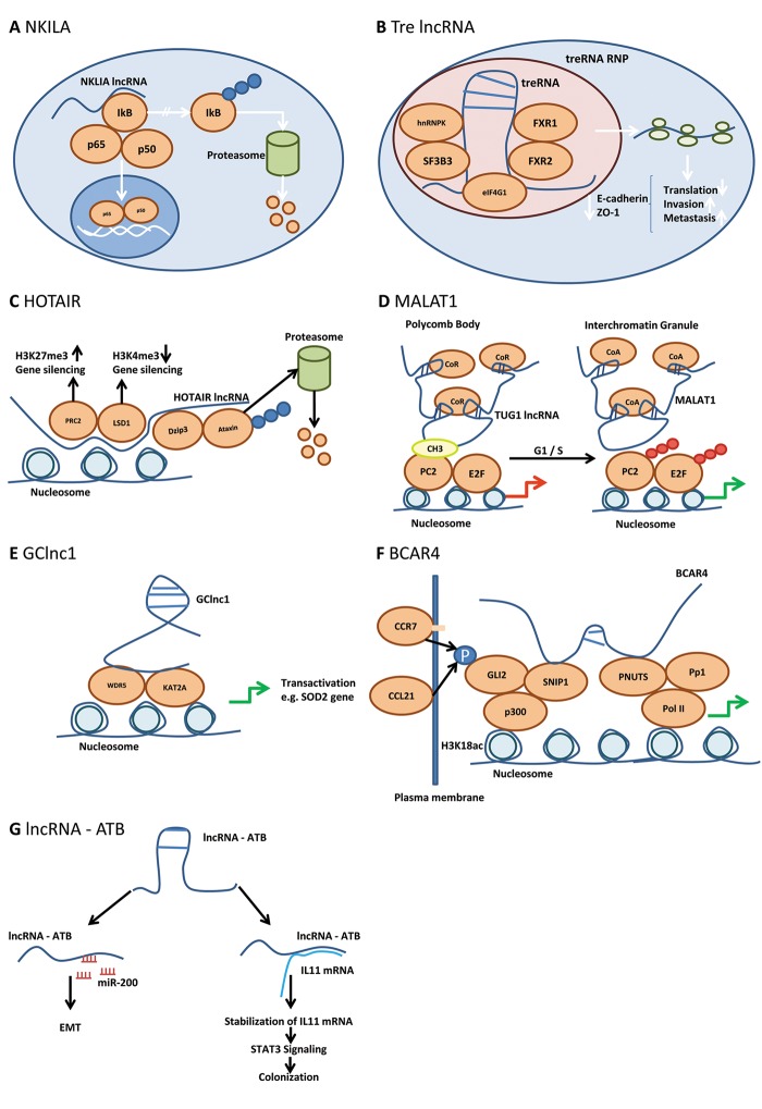
The perception of long non-coding RNAs as chunk RNA and transcriptional noise has been steadily replaced by their role as validated targets for a diverse set of physiological processes in the past few years. However, for the vast majority of lncRNAs their precise mode of action and physiological function remain to be uncovered. A large body of evidence has revealed their essential role in all stages of cancirogenesis and metastasis. In this review we focus on the role of lncRNAs in metastasis. We grouped selected lncRNAs into three categories based on in vitro and in vivo mode of action-related studies and clinical relevance for metastasis. Grouped according to their mode of action, in category I we discuss lncRNAs such as CCAT2, DREH, LET, NKILA, treRNA, HOTAIR, H19, FENDRR, lincROR, MALAT, GClnc1, BCAR4, SCHLAP1 and lncRNA ATP, all lncRNAs with in vitro and in vivo metastasis-related data and clinical significance. In category II we discuss lncRNAs CCAT1, PCAT1, PTENgp1, GPLINC, MEG3, ZEB2-AS, LCT13, ANRIL, NBAT1 and lncTCF7 all characterized by their mode of action in vitro and clinical significance, but pending or preliminary in vivo data. Finally, under category III, we discuss lncRNAs BANCR, FRLnc1, SPRY4-IT1 and LIMT with partially or poorly-resolved mode of action and varying degree of validation in clinical metastasis. Finally we discuss metastasis-related translational aspects of lncRNAs.

Keywords: Chromatin remodeling; DNA-RNA interaction; RNA-RNA interaction; decoy function; epigenetic modification; guide function; nucleic acid-based therapy; protein-RNA interaction; review; scaffold function; signaling networks.


Figures

Figure 1
Figure 1. Mode of action of lncNKILA, Tre lncRNA, HOTAIR, MALAT1, GClncRNA, BCAR4 and lncRNA-ATP. (A) NKILA acts as a negative regulator of NFĸB signaling. IĸB, inhibitor of ĸB; NFĸB, nuclear factor ĸ; NKILA, NFĸB-activating lncRNA; p50 and p65, subunits of NFĸB. Blue circles indicate polyubiquitinylation. (B) Tre lncRNA inhibits translation of pro-metastatic mRNAs via RNP formation by a poorly resolved mode of action. eIF-4G1, translation initiation factor 4G1; FXR1, FXR3, fragile X mental retardation syndrome-related proteins 1, 3; hnRNPK, heterogeneous ribonucleoprotein complex K; SF3B3, splicing factor 3B subunit 3; treRNA, translational regulatory RNA; treRNA RNP, tre RNA ribonucleoprotein complex. (C) HOTAIR represses gene expression by recruitment of PRC2 and LSD1 by increasing the repression code H3K27me3 and decreasing the activation code H3K4me3. DZIP3, DAZ interacting protein 3 (E3 ubiquitin-protein ligase); HOTAIR, HOX antisense intergenic RNA; H3K4me3, histone 3 trimethylated lysine 4; H3K27me3, histone 3 trimethylated lysine 27; LSD1, lysine-specific demethylase 1; PRC2, polycomb repressive complex 2. Blue circles indicate polyubiquitinylation. (D) MALAT1 relieves gene repression in polycomb bodies by replacement of TUG1 lncRNA and recruitment of co-activators in Interchromatin granules thus promoting G1/S transition. CoA, co-activator; CoR, co-repressor; CH3, methyl group; E2F, transcription factor 2F; MALAT1, metastasisassociated lung adenocarcinoma transcript; Pc2, polycomb 2; Pc2-CH3, methylated Pc2; SUMO, sumoylated; lnc RNA TUG1, lnc RNA taurine-upregulated gene 1. Red/green arrow: inhibition/activation of transcription; red circles indicate sumoylation. (E) GClncRNA recruits histone modifiers such as WDR5 and KAT2A for transactivation of transcription of selected genes. GC lncRNA, Gastric cancer lncRNA; KAT2A, lysine acetyltransferase 2A; WDR5, WD repeat containing protein 5; SOD2, superoxide dismutase 2. Green arrow, indicates activation of transcription. (F) BCAR4 mediates activation of phospho-GLI-2 dependent target genes. BCAR4, Breast cancer anti-estrogen resistance; CCL21, chemokine (C-C motif) ligand 21; CCR7, CCchemokine receptor 7; GLI2, glioma-associated oncogene family zinc finger 2; p, phosphorylated; PNUTS, putative protein phosphatase 1 nuclear; Pol II, polymerase II; Pp1, phosphatase 1; p300, E1A binding protein 300; SNIP1, smad nuclear interacting protein 1. Black arrows indicate phosphorylation of GLI2 by CCR7/CCL21 interaction; green arrows indicate activation of transcription. (G) lncRNA – ATP mimics the prometastatic role of TGFβ biy inducing miR200 family members and promotes IL11 signaling. EMT, Epithelial mesenchymal transition; IL11, interleukin 11; lnc RNA – ATP, lncRNA activated by TGFβ; STAT3, signal transducer and activator of transcription 3; TGFβ, transforming growth factor β; blue circled P, phosphorylation.
Figure 2
Figure 2. RNA steady-state levels of selected RNAs in different tumor entities. (A) H19; (B) MALAT1; (C) HOTAIR; (D) BCAR4; (E) MEG3. Expression in tumor/matching normal samples is shown for selected cohorts derived from The Cancer Genome Atlas (TCGA) database. Bladder cancer (408/19), breast cancer (1,100/112), colon cancer (475/41), liver cancer (373/50), lung adenocarcinoma (517/59), lung squamous carcinoma (501/51), prostate cancer (498/52) and stomach cancer (415/35). Expression is measured as normalized read counts, the red lines indicate low versus higher expression (~100 counts). Expression data are shown as box blots where the line in the middle of the box represents the data median, the rectangles show the upper and lower quartile (therefore, 50% of all data points are included in the rectangle). All other data points, except for outliers lie within the upper and lower whiskers. A value of -10 indicates no expression in the corresponding sample. For some tumor types not a single sample expressing the corresponding lncRNA was found.

References

    1. Fidler IJ. The pathogenesis of cancer metastases: the 'seed and soil' hypothesis revisited. Nat Rev Cancer. 2003;3:453–458. - PubMed
    1. Langley RR, Fidler IJ. The seed and soil hypothesis revisited – the role of tumor-stroma interactions in metastasis to different organs. Int J Cancer. 2011;128:2527–2535. - PMC - PubMed
    1. Peinado H, Lavotshkin S, Lyden D. The secreted factors responsible for pre-metastatic niche formation: old sayings and new thoughts. Sem Cancer Biol. 2011;21:139–146. - PubMed
    1. Psaila B, Lyden D. The metastatic niche: adapting the foreign soil. Nat Rev Cancer. 2009;9:285–293. - PMC - PubMed
    1. Massague J, Obenauf AC. Metastatic colonization by circulating tumor cells. Nature. 2016;529:298–306. - PMC - PubMed

MeSH terms

Substances