. 2017 Jun 1;130(11):1965-1978.
doi: 10.1242/jcs.199760. Epub 2017 Apr 26.

Transient mechanical strain promotes the maturation of invadopodia and enhances cancer cell invasion in vitro

Affiliations

Transient mechanical strain promotes the maturation of invadopodia and enhances cancer cell invasion in vitro

Alexander N Gasparski et al. J Cell Sci. .

Abstract

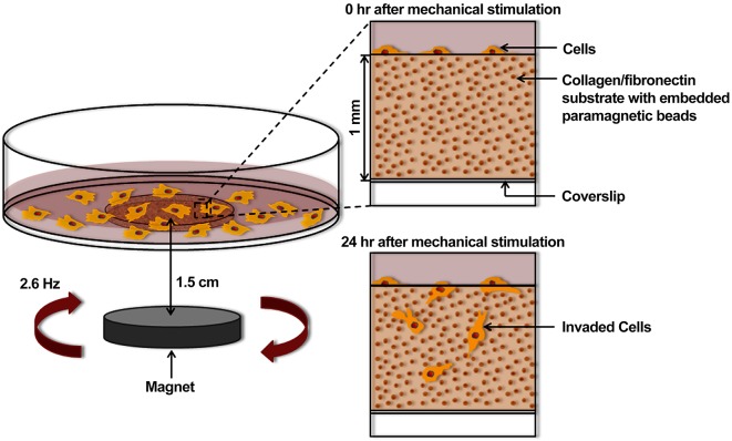
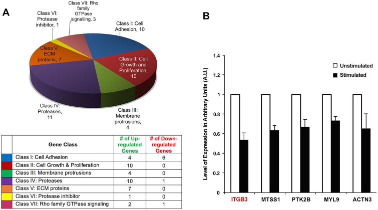
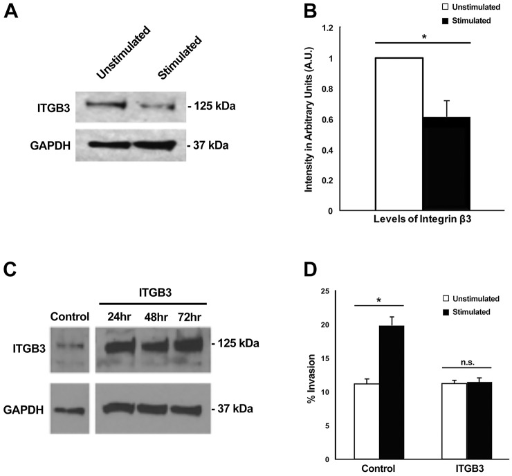
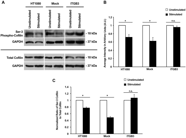
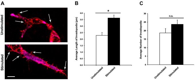
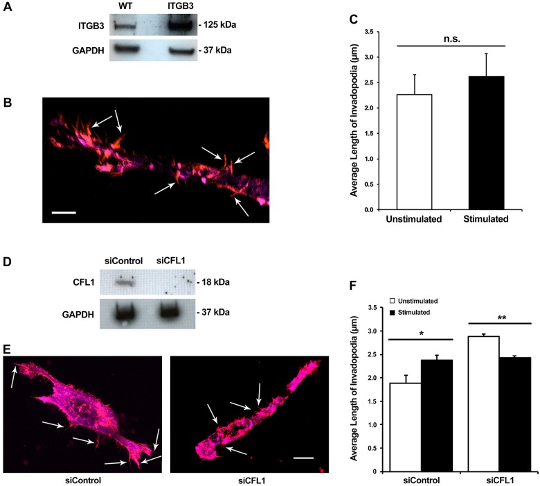
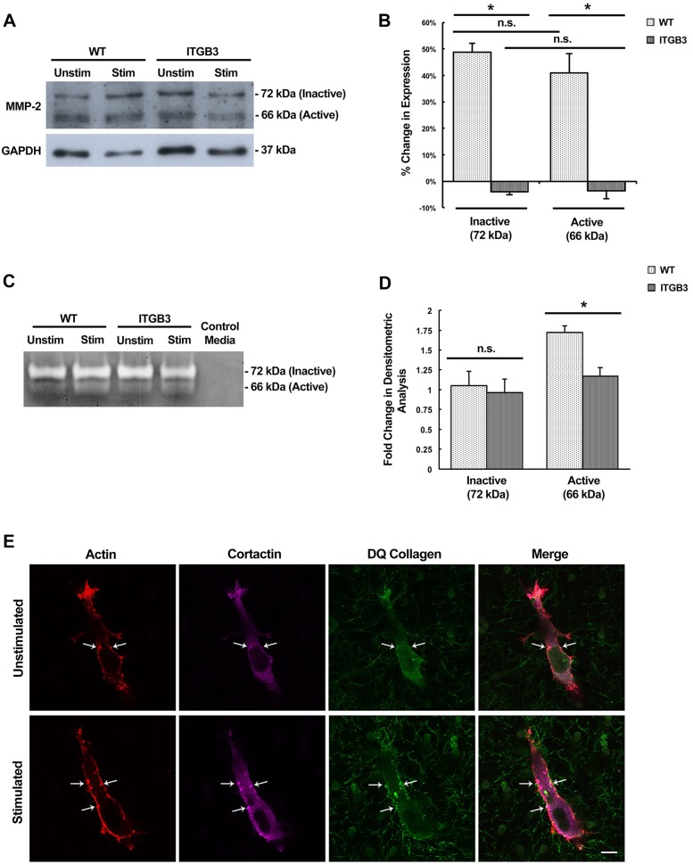
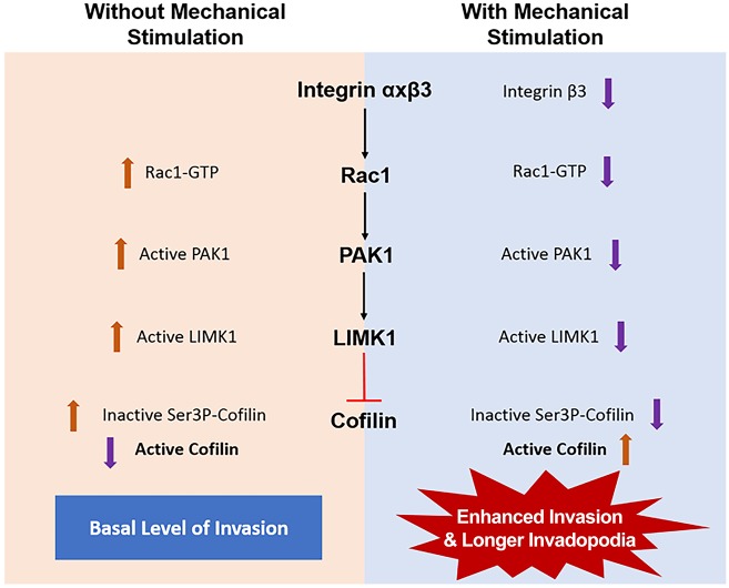
Cancer cell invasion is influenced by various biomechanical forces found within the microenvironment. We have previously found that invasion is enhanced in fibrosarcoma cells when transient mechanical stimulation is applied within an in vitro mechano-invasion assay. This enhancement of invasion is dependent on cofilin (CFL1), a known regulator of invadopodia maturation. Invadopodia are actin-rich structures present in invasive cancer cells that are enzymatically active and degrade the surrounding extracellular matrix to facilitate invasion. In this study, we examine changes in gene expression in response to tugging on matrix fibers. Interestingly, we find that integrin β3 expression is downregulated and leads to an increase in cofilin activity, as evidenced by a reduction in its Ser3 phosphorylation levels. As a result, invadopodia lengthen and have increased enzymatic activity, indicating that transient mechanical stimulation promotes the maturation of invadopodia leading to increased levels of cell invasion. Our results are unique in defining an invasive mechanism specific to the invasive process of cancer cells that is triggered by tugging forces in the microenvironment, as opposed to rigidity, compression or stretch forces.

Keywords: Cancer; Cell invasion; Cell mechanics; Cofilin; Invadopodia.


Conflict of interest statement

Competing interestsThe authors declare no competing or financial interests.

Figures

Fig. 1.
Fig. 1.
Design of an in vitro mechano-invasion assay. A 1 mm deep well is created in a 60 mm cell culture dish by drilling a hole in the bottom of the dish and attaching an activated glass coverslip with vacuum grease. The resulting well is filled with a collagen type I and fibronectin matrix containing 1 µm carboxylated paramagnetic beads, which covalently attach to the fibers upon polymerization. HT1080 fibrosarcoma cells are seeded onto the surface of the matrix and either cultured 1.5 cm above a rotating magnet or outside of the magnetic field (unstimulated). After 24 h, cells invade into the matrix and are counted to determine the percentage invasion.
Fig. 2.
Fig. 2.
Differentially expressed genes resulting from mechanical stimulation. (A) qPCR array analysis of genes found to have a ±1.35-fold differential expression upon mechanical stimulation. The genes were classified into seven groups. The pie chart illustrates the number of differentially expressed genes within each class and the table contains the number of up- and down-regulated genes within each class. (B) qPCR confirmation of select genes with downregulated expression upon stimulation. These differentially expressed genes were selected for further confirmation by qPCR. Three biological replicates were used for qPCR of ITGB3, MTSS1, MYL9 and ACTN3; four biological replicates were used for qPCR of PTK2B. For every biological replicate, two technical replicates were performed; values represent mean±s.e.m. All results were P<0.05 (two-tailed t-test).
Fig. 3.
Fig. 3.
Downregulation of integrin β3 expression upon mechanical stimulation enhances cell invasion. (A) Western blot of lysates, examining ITGB3 expression with and without stimulation. (B) Quantification of ITGB3 protein levels with and without stimulation. Results are mean±s.e.m. (n=4). *P=0.0059 (Student's t-test). (C) Integrin β3 protein levels when overexpressed (ITGB3) via nucleofection compared to mock nucleofected cells (control). (D) After 48 h with and without stimulation, the percentage of cell invasion was examined in mock nucleofected cells (control) and cells with integrin β3 overexpressed (ITGB3). Percentage invasion was determined by dividing the number of invaded cells (cells below the matrix surface) by the total number of cells. Results are mean±s.e.m. (n=4). *P<0.05; n.s., not significant (two-factor ANOVA).
Fig. 4.
Fig. 4.
The decrease in levels of Ser3 phospho-cofilin upon mechanical stimulation is dependent on the downregulation of integrin β3 expression. (A) Lysates from wild-type (HT1080), mock nucleofected (Mock) and overexpressing integrin β3 (ITGB3) cells cultured with or without stimulation were used for analysis of Ser3 phospho-cofilin and total cofilin levels. GAPDH served as a loading control. (B) Quantification of Ser3 phospho-cofilin levels with and without stimulation. Results are mean±s.e.m. from three biological replicates. *P<0.05; n.s., not significant (two-tailed t-test). (C) The normalized ratio of Ser3 phospho-cofilin to total cofilin based on data obtained in A. Results are mean±s.e.m. (n=3). *P<0.05; n.s., not significant (two-tailed t-test). In B and C, the data was normalized within each condition.
Fig. 5.
Fig. 5.
Mechanical stimulation produces an increase in the length of invadopodia without affecting the number of invadopodia. (A) Representative confocal fluorescent images of HT1080 cells fixed within the collagen–fibronectin matrix with and without mechanical stimulation. Red indicates actin and magenta indicates cortactin. Arrows indicate invadopodia, based on cortactin and actin colocalization. Scale bar: 10 µm. (B) The average length of invadopodia per cell after 48 h with and without stimulation. Results are mean±s.e.m. (n=16). *P<0.05 (two-tailed t-test). (C) The average number of invadopodia per cell with and without stimulation. Results are mean±s.e.m. (n=16). n.s., not significant (two-tailed t-test).
Fig. 6.
Fig. 6.
Overexpression of integrin β3 and downregulation of cofilin expression both negatively affect the lengthening of invadopodia upon stimulation. (A) Western blot confirming overexpression of integrin β3 in protein lysates from control vector (WT) and integrin β3 (ITGB3)-overexpressing cells after 48 h. (B) A representative confocal fluorescent image of a fixed cell with integrin β3 overexpression residing within the collagen–fibronectin matrix. Red indicates actin and magenta indicates cortactin. Invadopodia are indicated by the arrows. Scale bar: 5 µm. (C) Measurement of invadopodia length with and without stimulation in cells that overexpress integrin β3. Results are mean±s.e.m. from four biological replicates. n.s., not significant (two-tailed t-test). (D) Western blot of cofilin knockdown by cofilin-specific siRNA (siCFL1). siControl, control siRNA. (E) Representative confocal fluorescent images of control siRNA and siCFL1 nucleofected cells chemically fixed within the matrix. Red indicates actin and magenta indicates cortactin. Arrows indicate individual invadopodia. Scale bar: 10 µm. (F) Invadopodia length with and without stimulation in cells nucleofected with control siRNA and siCFL1. Results are mean±s.e.m. from four biological replicates. *P<0.05, **P<0.01 (two-tailed t-test).
Fig. 7.
Fig. 7.
MMP-2 protein expression and enzymatic activity is enhanced upon mechanical stimulation and inhibited by integrin β3 overexpression. (A) Western blot analysis of protein lysates from control vector (WT) and integrin β3 (ITGB3)-overexpressing cells after 48 h. Two isoforms of MMP-2 are detected: the inactive form (72 kDa) and the active form (66 kDa). GAPDH served as the loading control. (B) The change in expression of MMP-2 inactive and active isoforms upon stimulation in WT and ITGB3 cells. Results are mean±s.e.m. from three biological replicates. (C) Zymography of conditioned medium collected from WT and ITGB3 cells with and without mechanical stimulation. Control medium served as a negative control. Unstained bands (white) indicate enzymatic proteolysis. (D) Quantification of zymogram band intensities in WT and ITGB3 cells. Results are mean±s.e.m. from three biological replicates. (E) Representative confocal fluorescent images of DQ collagen-containing matrices from stimulated and unstimulated cells. Punctate structures with actin, cortactin and DQ collagen colocalization are invadopodia. Select invadopodia are indicated by the arrows. *P<0.01; n.s., not significant (two-tailed t-test). Scale bar: 10 µm.
Fig. 8.
Fig. 8.
Potential pathway for enhanced invasion upon mechanical stimulation. Signaling downstream of integrin β3, through sequential activation of Rac1, PAK1 and LIMK1, results in inhibition of cofilin activity by phosphorylation at the Ser3 position. Upon mechanical stimulation, integrin β3 is downregulated thereby decreasing the levels of downstream kinases. A decrease in the activation of LIMK1 also decreases phosphorylation of cofilin. This leads to an increase in levels of active cofilin producing longer and more-mature invadopodia that enhance cell invasion.

Similar articles

Cited by

References

    1. Albiges-Rizo C., Destaing O., Fourcade B., Planus E. and Block M. R. (2009). Actin machinery and mechanosensitivity in invadopodia, podosomes and focal adhesions. J. Cell Sci. 122, 3037-3049. 10.1242/jcs.052704 - DOI - PMC - PubMed
    1. Alexander N. R., Branch K. M., Parekh A., Clark E. S., Iwueke I. C., Guelcher S. A. and Weaver A. M. (2008). Extracellular matrix rigidity promotes invadopodia activity. Curr. Biol. 18, 1295-1299. 10.1016/j.cub.2008.07.090 - DOI - PMC - PubMed
    1. Arber S., Barbayannis F. A., Hanser H., Schneider C., Stanyon C. A., Bernard O. and Caroni P. (1998). Regulation of actin dynamics through phosphorylation of cofilin by LIM-kinase. Nature 393, 805-809. 10.1038/31729 - DOI - PubMed
    1. Antelmi E., Cardone R. A., Greco M. R., Rubino R., Di Sole F., Martino N. A., Casavola V., Carcangiu M. L., Moro L. and Reshkin S. J. (2013). β1 integrin binding phosphorylates ezrin at T567 to activate a lipid raft signalsome driving invadopodia activity and invasion. PLoS ONE 8, e75113 10.1371/journal.pone.0075113 - DOI - PMC - PubMed
    1. Artym V. V., Zhang Y., Seillier-Moiseiwitsch F., Yamada K. M. and Mueller S. C. (2006). Dynamic interactions of cortactin and membrane type 1 matrix metalloproteinase at invadopodia: defining the stages of invadopodia formation and function. Cancer Res. 66, 3034-3043. 10.1158/0008-5472.CAN-05-2177 - DOI - PubMed

MeSH terms