. 2017 Jun 1;198(11):4312-4326.
doi: 10.4049/jimmunol.1700149. Epub 2017 Apr 26.

Induction of Adaptive Immunity Leads to Nigrostriatal Disease Progression in MPTP Mouse Model of Parkinson's Disease

Affiliations

Induction of Adaptive Immunity Leads to Nigrostriatal Disease Progression in MPTP Mouse Model of Parkinson's Disease

Goutam Chandra et al. J Immunol. .

Abstract

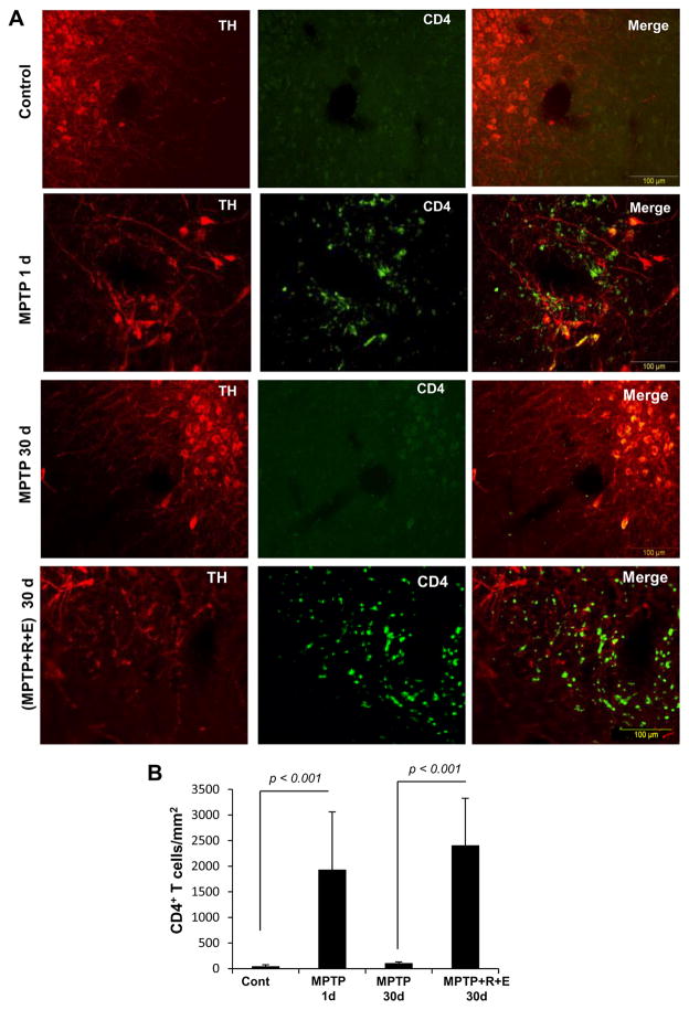
Although the 1-methyl-4-phenyl-1,2,3,6-tetrahydropyridine (MPTP) mouse model is the most widely used animal model for Parkinson's disease (PD), it is known that nigrostriatal pathologies do not persist in the acute MPTP mouse model. This study highlights the importance of adaptive immunity in driving persistent and progressive disease in acute MPTP-intoxicated mice. Although marked infiltration of T cells into the nigra was found on 1 d of MPTP insult, T cell infiltration decreased afterward, becoming normal on 30 d of insult. Interestingly, twice-weekly supplementation of RANTES and eotaxin, chemokines that are involved in T cell trafficking, drove continuous T cell infiltration to the nigra and incessant glial inflammation. Supplementation of RANTES and eotaxin was also associated with the induction of nigral α-synuclein pathology, persistent loss of dopaminergic neurons and striatal neurotransmitters, and continuous impairment of motor functions in MPTP-intoxicated mice. In contrast, supplementation of TNF-α and IL-1β, widely studied proinflammatory cytokines, did not induce persistent disease in MPTP-insulted mice. Our results suggest that induction of adaptive immunity by RANTES and eotaxin could hold the key for driving persistent nigrostriatal pathologies in the MPTP mouse model, and that targeting these factors may halt disease progression in PD patients.


Figures

Figure 1
Figure 1. Upregulation of RANTES and eotaxin in MPTP-intoxicated mice and monkeys
Male C57/BL6 mice (6–8 week old) were insulted with 20 mg/kg body wt MPTP (four injections at every 2 h interval). After 1, 3, 7, 30, and 60 d of MPTP intoxication, the mRNA expression of RANTES and eotaxin was monitored in nigra by semi-quantitative RT-PCR (A) and real-time PCR (B, RANTES; C, eotaxin). Levels of RANTES (D) and eotaxin (E) were also measured in serum by ELISA. Results are mean ± SEM of four mice (n=4) per group. ap < 0.001 vs control; bp < 0.05 vs MPTP-1d; cp < 0.001 vs MPTP-1d. F) Ventral midbrain sections (Coordinates: Anteroposterior −4.04 mm from Bregma, dorsoventral 3.75 mm, mediolateral 1.25 mm) were double-labeled for CD4 and TH. G) CD4-positive cells were counted in two nigral sections (two images per slide) of each of five mice (n=5) per group. ap<0.001 vs. control; bp<0.001 vs. MPTP-1d.
Figure 2
Figure 2
Schematic presentation of treatment schedule of MPTP-intoxicated mice with RANTES and eotaxin and associated experiments.
Figure 3
Figure 3. Supplementation of RANTES (R) and eotaxin (E) maintains T cell gradient in the nigra of MPTP-intoxicated mice
Male C57/BL6 mice (6–8 week old) were insulted with 20 mg/kg body wt MPTP (four injections at every 2 h interval). From 3d of the last injection of MPTP, mice received the combination of R (100 ng/mouse) and E (100 ng/mouse) via i.p. injection every 3.5 d interval. A) At different d of MPTP intoxication, ventral midbrain sections (Coordinates: Anteroposterior −4.04 mm from Bregma, dorsoventral 3.75 mm, mediolateral 1.25 mm) were double-labeled for CD4 and TH. B) CD4-positive cells were counted in two nigral sections (two images per slide) of each of five mice (n=5) per group.
Figure 4
Figure 4. Supplementation of RANTES (R) and eotaxin (E) induces chronic inflammation in the nigra of MPTP mouse model
Male C57/BL6 mice (6–8 week old) were insulted with 20 mg/kg body wt MPTP (four injections at every 2 h interval). From 3d of the last injection of MPTP, mice received the combination of R (100 ng/mouse) and E (100 ng/mouse) via i.p. injection every 3.5 d interval. Control groups with (R+E), but without MPTP, were also included. On 7, 30 and 60 d of MPTP intoxication, the mRNA expression of iNOS, IL-1β, GFAP, and CD11b was monitored by semi-quantitative RT-PCR (A) and real-time PCR (B, iNOS and IL-1β; C, GFAP and CD11b). Results are mean ± SEM of four mice (n=4) per group. ap < 0.001 vs control; bp < 0.001 vs MPTP-1d; cp < 0.001 vs MPTP-30d. Protein expression was also analyzed by Western blot (D). Actin was run as control. Bands were scanned and values (E, GFAP/Actin and Iba1/Actin; F, iNOS/Actin and IL-1β/Actin) are presented as relative to control. Results are mean ± SEM of four mice (n=4) per group. ap < 0.001 vs control; bp < 0.001 vs MPTP-1d; cp < 0.001 vs MPTP-30d.
Figure 5
Figure 5. Supplementation of RANTES (R) and eotaxin (E) induces chronic microglial activation in the nigra of MPTP mouse model
Male C57/BL6 mice (6–8 week old) were insulted with 20 mg/kg body wt MPTP (four injections at every 2 h interval). From 3d of the last injection of MPTP, mice received the combination of R (100 ng/mouse) and E (100 ng/mouse) via i.p. injection every 3.5 d interval. A) On 7, 30 and 60 d of MPTP intoxication, nigral sections were double-labeled for Iba-1 (microglia) and iNOS. Cells positive for Iba-1 (B) and iNOS (C) were counted in two nigral sections (two images per slide) of each of five mice (n=5) per group. ap < 0.001 vs control; bp < 0.01 vs MPTP-1d; cp < 0.001 vs MPTP-1d; dp < 0.001 vs MPTP-30d.
Figure 6
Figure 6. Microglial activation and infiltration of CD4-ir T lymphocytes in the nigra of MPTP-treated mice
Male C57/BL6 mice (6–8 week old) were insulted with 20 mg/kg body wt MPTP (four injections at every 2 h interval). From 3d of the last injection of MPTP, mice received the combination of R (100 ng/mouse) and E (100 ng/mouse) via i.p. injection every 3.5 d interval. A) On 30 d of MPTP intoxication, nigral sections were triple-immunostained with TH (1:2000, Cat# 657012; EMD Millipore), Iba-1 (1:500, Cat# ab5076; Abcam) and CD4 ( 1:50, Cat #MA1-7631; ThermoFisher) antibodies and then visualized in BX51 Olympus fluorescence microscope. B) Sizes of microglial cells were counted around 0.07–0.08 sq mm regions of blood vessels located at ventral midbrain regions. Results are mean ± SD of 15–23 randomly selected IBA1-ir cells per group. NSF1,30 = 0.459 (<Fc = 4.17); p >0.05 ( = 0.059) vs. control. aF1,30 = 20.33(>Fc = 4.09); p <0.0001( = 6.07*10−5) vs. MPTP. C) Correlation statistics was performed between number of infiltrated CD4-ir cells and microglial size. A strong correlation was observed with high Pearson coefficient (r) value =0.744 and p<0.05 (=0.02).
Figure 7
Figure 7. Supplementation of RANTES (R) and eotaxin (E) induces the accumulation of α-syn in the nigra of MPTP mouse model
Male C57/BL6 mice (6–8 week old) were insulted with 20 mg/kg body wt MPTP (four injections at every 2 h interval). From 3d of the last injection of MPTP, mice received the combination of R (100 ng/mouse) and E (100 ng/mouse) via i.p. injection every 3.5 d interval. A) On 30 d of MPTP intoxication, nigral sections were double-labeled with TH (1:2000, Cat# 657012; EMD Millipore) and α-syn (1:500, Cat# ab1903; Abcam) antibodies followed by visualization in BX51 Olympus fluorescence microscope with 4X objective lens as described before (43). SNPc = substantia nigra pars compacta and SNR = substantia nigra pars reticulate. B) Magnified (20X objective and 60X inset) views of SNPc regions immunostained with TH (red) and α-syn (Green) antibodies. C) Mean Fluorescence Intensity of α-syn was calculated in neurites of SNR regions of 2–3 nigral sections of each of five mice (n=5) per group. aF1, 27 =46.058(>Fc = 4.21); p<0.001(=2.7*10−7) vs. MPTP. D) Quantification analyses of α-syn-ir cell bodies in SNPc regions. Counting was performed in two nigral sections of each of five mice (n=5) per group by touch counting methods in Olympus microsuite V software. aF1, 12 = 41.17(>Fc = 4.74) p<0.001(=3.32*10−5) vs. MPTP.
Figure 8
Figure 8. Supplementation of RANTES (R) and eotaxin (E) induces persistent loss of dopaminergic neurons in the nigra of MPTP-intoxicated mice
Male C57/BL6 mice (6–8 week old) were insulted with 20 mg/kg body wt MPTP (four injections at every 2 h interval). From 3d of the last injection of MPTP, mice received the combination of R (100 ng/mouse) and E (100 ng/mouse) via i.p. injection every 3.5 d interval. A control group with (R+E), but without MPTP, was also included. On 7, 30 and 60 d of MPTP intoxication, nigral sections were stained for TH (A) and TH neurons were counted by stereology using the STEREO INVESTIGATOR software (B). Nigral homogenates were immunoblotted with TH (C). Actin was run as loading control. Bands were scanned and presented as relative to control (D). Results are mean ± SEM of six mice (n=6) per group. ap < 0.001 vs control; bp < 0.01 vs MPTP-7d; cp < 0.01 vs MPTP-60d.
Figure 9
Figure 9. Supplementation of RANTES (R) and eotaxin (E) induces persistent loss of TH fibers and neurotransmitters in the striatum of MPTP-intoxicated mice
Male C57/BL6 mice (6–8 week old) were insulted with 20 mg/kg body wt MPTP (four injections at every 2 h interval). From 3d of the last injection of MPTP, mice received the combination of R (100 ng/mouse) and E (100 ng/mouse) via i.p. injection every 3.5 d interval. Control groups with (R+E), but without MPTP, were also included. On 7, 30 and 60 d of MPTP intoxication, striatal sections were stained for TH (A) followed by quantification of TH-positive fibers (B). Concentrations of dopamine (C), DOPAC (D) and HVA (E) were measured in the striatum by HPLC. Results are mean ± SEM of six mice (n=6) per group. ap < 0.001 vs control; bp < 0.001 vs MPTP-1d; cp < 0.001 vs MPTP-60d.
Figure 10
Figure 10. Effect of RANTES (R) and eotaxin (E) supplementation on motor functions in MPTP-intoxicated mice
Effect of RANTES (R) and eotaxin (E) on locomotor activities in MPTP mouse model of PD. Male C57/BL6 mice (6–8 week old) were insulted with 20 mg/kg body wt MPTP (four injections at every 2 h interval). From 3d of the last injection of MPTP, mice received the combination of R (100 ng/mouse) and E (100 ng/mouse) via i.p. injection every 3.5 d interval. A control group with (R+E), but without MPTP, was also included. On 7, 30 and 60 d of MPTP intoxication, mice were tested for rotorod (A), number of movements (B), movement time (C), total distance (D), horizontal activity (E), stereotypy (F), pole test (G), and rest time (H). Results are mean ± SEM of six mice (n=6) per group. ap < 0.001 vs control; bp < 0.001 vs MPTP-7d; cp < 0.001 vs MPTP-60d.
Figure 11
Figure 11. Effect of TNFα and IL-1β on striatal loss of TH fibers and neurotransmitters and impairment in locomotor activities in MPTP-intoxicated mice
Male C57/BL6 mice (6–8 week old) were insulted with 20 mg/kg body wt MPTP (four injections at every 2 h interval). From 3d of the last injection of MPTP, mice received TNFα (100 ng/mouse) or IL-1β (100 ng/mouse) alone or in combination via i.p. injection every 3.5 d interval. On 7 and 30 d of MPTP intoxication, striatal sections were stained for TH (A) followed by quantification of TH-positive fibers (B). Levels of DA (C), DOPAC (D) and HVA (E) were measured in the striatum by HPLC. Mice were also monitored for pole test (F) and rotorod (G). Results are mean ± SEM of six mice (n=6) per group. ap < 0.001 vs control; bp < 0.001 vs MPTP-7d; ns, nonsignificant vs MPTP-30d.

Similar articles

Cited by

References

    1. Vila M, Przedborski S. Genetic clues to the pathogenesis of Parkinson’s disease. Nat Med. 2004;(10 Suppl):S58–62. - PubMed
    1. Olanow CW, Tatton WG. Etiology and pathogenesis of Parkinson’s disease. Annu Rev Neurosci. 1999;22:123–144. - PubMed
    1. Dauer W, Przedborski S. Parkinson’s disease: mechanisms and models. Neuron. 2003;39:889–909. - PubMed
    1. Kordower JH, Emborg ME, Bloch J, Ma SY, Chu Y, Leventhal L, McBride J, Chen EY, Palfi S, Roitberg BZ, Brown WD, Holden JE, Pyzalski R, Taylor MD, Carvey P, Ling Z, Trono D, Hantraye P, Deglon N, Aebischer P. Neurodegeneration prevented by lentiviral vector delivery of GDNF in primate models of Parkinson’s disease. Science. 2000;290:767–773. - PubMed
    1. Mondal S, Roy A, Jana A, Ghosh S, Kordower JH, Pahan K. Testing NF-kappaB-based therapy in hemiparkinsonian monkeys. J Neuroimmune Pharmacol. 2012;7:544–556. - PMC - PubMed

Publication types