Comparative Study
. 2017 Jun 16;292(24):10131-10141.
doi: 10.1074/jbc.M116.773226. Epub 2017 Apr 26.

CDK4 protein is degraded by anaphase-promoting complex/cyclosome in mitosis and reaccumulates in early G1 phase to initiate a new cell cycle in HeLa cells

Affiliations
Comparative Study

CDK4 protein is degraded by anaphase-promoting complex/cyclosome in mitosis and reaccumulates in early G1 phase to initiate a new cell cycle in HeLa cells

Huabo Chen et al. J Biol Chem. .

Abstract

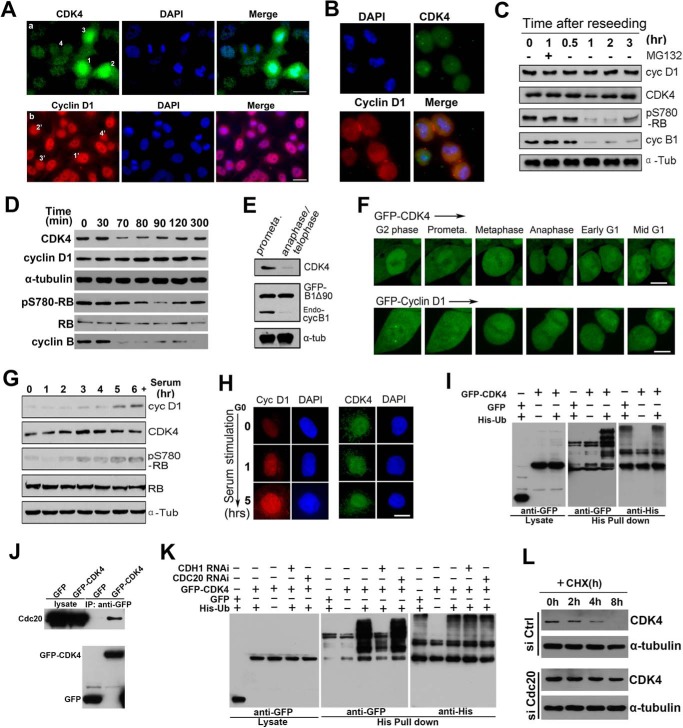
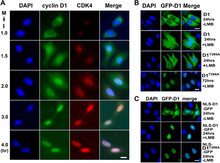
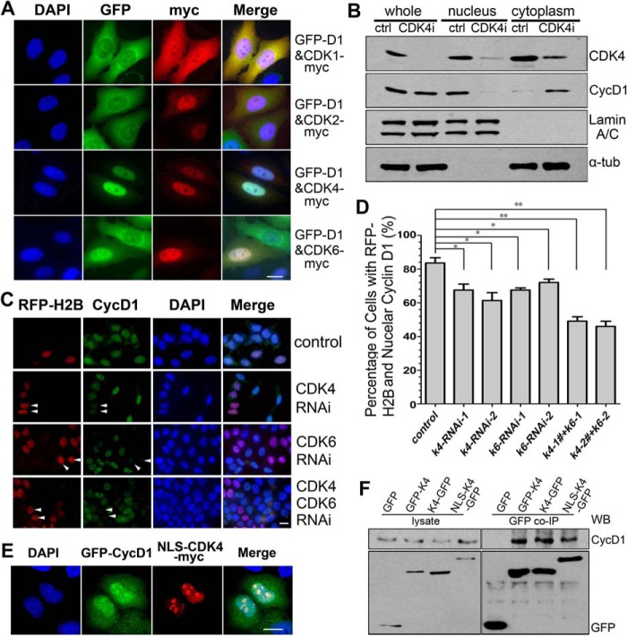
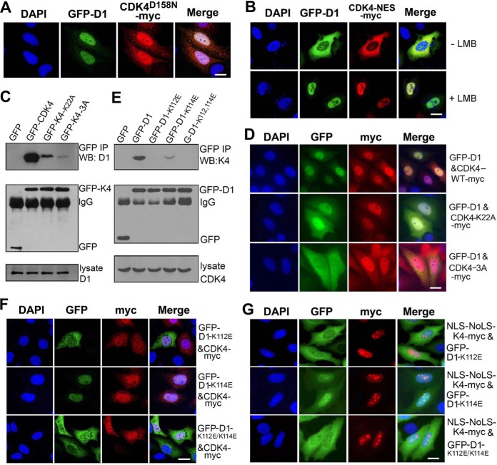
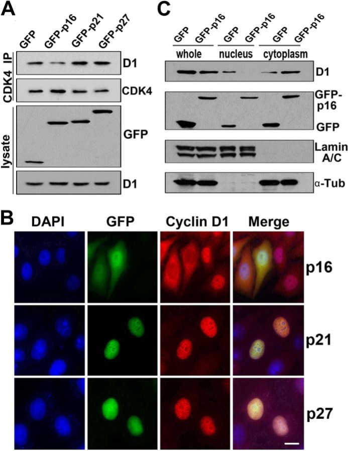
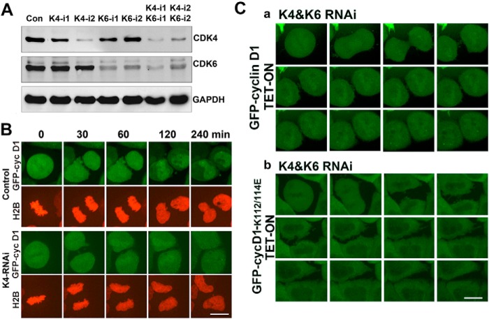
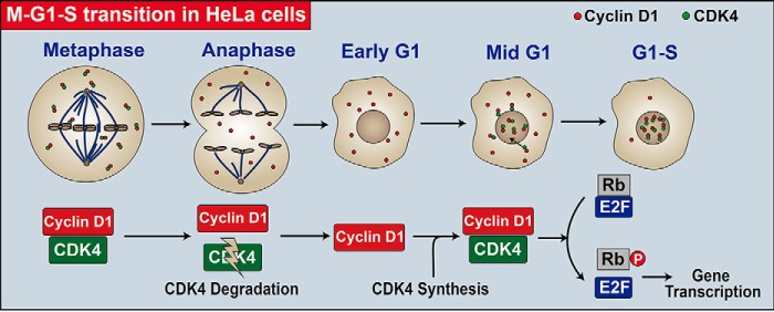
CDK4 regulates G1/S phase transition in the mammalian cell cycle by phosphorylating retinoblastoma family proteins. However, the mechanism underlying the regulation of CDK4 activity is not fully understood. Here, we show that CDK4 protein is degraded by anaphase-promoting complex/cyclosome (APC/C) during metaphase-anaphase transition in HeLa cells, whereas its main regulator, cyclin D1, remains intact but is sequestered in cytoplasm. CDK4 protein reaccumulates in the following G1 phase and shuttles between the nucleus and the cytoplasm to facilitate the nuclear import of cyclin D1. Without CDK4, cyclin D1 cannot enter the nucleus. Point mutations that disrupt CDK4 and cyclin D1 interaction impair the nuclear import of cyclin D1 and the activity of CDK4. RNAi knockdown of CDK4 also induces cytoplasmic retention of cyclin D1 and G0/G1 phase arrest of the cells. Collectively, our data demonstrate that CDK4 protein is degraded in late mitosis and reaccumulates in the following G1 phase to facilitate the nuclear import of cyclin D1 for activation of CKD4 to initiate a new cell cycle in HeLa cells.

Keywords: cell cycle; cyclin D1; cyclin-dependent kinase (CDK); protein degradation; retinoblastoma protein (pRb, RB).


Conflict of interest statement

The authors declare that they have no conflicts of interest with the contents of this article

Figures

Figure 1.
Figure 1.
CDK4 is degraded by APC/C-Cdc20 during M-A transition and reaccumulates from early to mid-G1 phase. A, HeLa cells were immunostained with anti CDK4 and cyclin D1 antibodies. The early prometaphase, late prometaphase, metaphase, and anaphase/telophase cells are labeled 1, 2, 3, and 4, respectively, in a, whereas the G2, metaphase, anaphase, and telophase cells are labeled with 1′, 2′, 3′, and 4′ in b. B, HeLa cells were synchronized at the G1/S transition and were then released into fresh culture medium for 10 h. Mitotic cells that were in prometaphase were shaken off, reseeded, and stained with DAPI. C, the cell lysate of the reseeded mitotic and released cells was subjected to Western blotting. D, HeLa cells were arrested at the G2/M border using RO3306 (9 μm) and released. The lysates from these cells were analyzed by Western blotting. E, HeLa cells transfected with GFP-cyclin B1Δ90 were arrested at prometaphase and anaphase/telophase and analyzed by Western blotting. F, HeLa cells expressing GFP-CDK4 and GFP-cyclin D1 were processed for time-lapse microscopy. More than 20 cells were filmed in each of three repeated experiments, and representative movie images are shown. G, HeLa cells arrested at the quiescent-like phase were released into G1 phase by serum at the indicated time points. The cell lysates were analyzed by Western blotting. H, quiescent-like phase to G1 phase cells were fixed and immunostained. DNA was stained by DAPI. 200 cells were randomly counted at each time point of the three repeated experiments. Representative images are shown. I, HeLa cells were co-transfected with His-ubiquitin and GFP or GFP-CDK4 or were transfected with GFP-CDK4 alone. Transfected cells were synchronized at the G2/M border using RO3306 and then released to M-A transition. Lysate of cells was subjected to a pull-down assay using nickel-Sepharose beads. The isolated proteins were analyzed by Western blotting. J, HeLa cells were transfected with GFP-CDK4, and the total cell extract was used for immunoprecipitation with a GFP antibody and probed with Cdc20 and GFP antibodies. K, HeLa cells were separately transfected with negative control (NC), Cdc20, or Cdh1 RNAi double-stranded RNA followed by co-transfection with His-ubiquitin and GFP-CDK4. As negative controls, HeLa cells transfected with NC RNAi double-stranded RNA were transfected with GFP-CDK4 or co-transfected with His-ubiquitin and GFP. Transfected cells were synchronized at the G2/M border using RO3306 and then released to M-A transition. Lysates of cells were subjected to a pull-down assay using nickel-Sepharose beads followed by Western blotting using anti-His and -GFP antibodies. L, HeLa cells were separately transfected with negative control or Cdc20 RNAi double-stranded RNA for 72 h. The cells were then treated with or without 30 μg/ml CHX for the indicated number of hours, lysed, and probed with CDK4 and α-tubulin antibodies. Scale bars, 10 μm.
Figure 2.
Figure 2.
Slow nuclear import and fast export retain cyclin D1 in the cytoplasm in the absence of CDK4. A, mitosis (M)-arrested HeLa cells were released to interphase (I) at the indicated time points. Cyclin D1 and CDK4 were immunostained. The DNA was stained with DAPI. 200 pairs of the cells were randomly counted at each time point of three repeated experiments. Representative images are shown. B, HeLa cells were transfected with the wild-type GFP-cyclin D1 (GFP-D1) or mutant GFP-cyclin D1T286A (GFP-D1T286A) for 24 or 72 h. The cells were then treated with or without 10 ng/ml LMB for 4 h. C, HeLa cells transfected with the wild-type NLS-cyclin D1-GFP (NLS-D1-GFP) or mutant (NLS-D1T286A-GFP) were treated with or without 10 ng/ml LMB for 4 h. More than 200 cells each of three experiments were randomly counted. Representative images are shown. Scale bars, 10 μm.
Figure 3.
Figure 3.
CDK4 shuttles between the nucleus and the cytoplasm and promotes the nuclear accumulation of cyclin D1. A, HeLa cells were co-transfected with GFP- or Myc-tagged cyclin D1 and CDK4 for 24 h. Myc-tagged protein was immunostained. 200 cells were randomly counted in each of three repeated experiments. Representative images are shown. B, HeLa cells were transfected with CDK4 siRNA for 72 h and fractionated into nuclear and cytoplasmic fractions, followed by Western blotting. C and D, asynchronous HeLa cells co-transfected with control, CDK4 or CDK6 siRNA, and RFP-H2B for 72 h were stained using an anti-cyclin D1 antibody and DAPI (C), and the cells with cyclin D1 in the nucleus were counted (D). *, p < 0.05; **, p < 0.01 (Student's t test). E and F, HeLa cells co-transfected with GFP-cyclin D1 (GFP-D1) and NLS-CDK4-Myc for 24 h were visualized by IFM (E; scale bars, 10 μm) or a co-immunoprecipitation assay (F).
Figure 4.
Figure 4.
Direct binding is required for CDK4-mediated nuclear import of cyclin D1. A, HeLa cells co-transfected with GFP-cyclin D1 (GFP-D1) and CDK4K158N-Myc plasmids for 24 h were visualized by IFM. B, asynchronous HeLa cells co-transfected with wild-type GFP-cyclin D1 (GFP-D1) and CDK4-NES-Myc plasmids for 24 h and treated without or with 10 ng/ml LMB for 4 h were visualized by IFM. C, HeLa cells were transfected with GFP, wild-type GFP-CDK4, and mutant plasmids for 22 h. The cell lysates were immunoprecipitated with a GFP antibody, and the co-immunoprecipitated cyclin D1 was detected. D, asynchronous HeLa cells co-transfected with GFP-cyclin D1 and CDK4-Myc mutant for 24 h were visualized by IFM. E, HeLa cells transfected with the wild-type GFP-cyclin D1 or mutant plasmid for 24 h followed by a co-immunoprecipitation assay using an anti-GFP antibody. The co-immunoprecipitated CDK4 with cyclin D1 or mutants was detected. F, HeLa cells co-transfected with CDK4-Myc and wild-type GFP-cyclin D1 or mutant cyclin D1 for 24 h were visualized by IFM. G, HeLa cells transfected with NLS-NoLS-CDK4-Myc and GFP-cyclin D1 mutant for 24 h were visualized by IFM. The DNA in all microscopy images was stained with DAPI. More than 200 cells in each of three repeated experiments were randomly counted. Representative images are shown. Scale bars, 10 μm.
Figure 5.
Figure 5.
Stable binding of D-type cyclins with CDK4 enhances its nuclear localization, and CDK4/CDK6 knockdown results in cell-cycle arrest at the G1 phase. A, HeLa cells were transfected with GFP, GFP-p16, GFP-p21, or GFP-p27 for 24 h. The cell lysates were immunoprecipitated with anti-CDK4 antibody. The co-immunoprecipitated cyclin D1 was detected by Western blotting. B, HeLa cells transfected with GFP-tagged p16, p21, or p27 for 24 h were visualized by IFM. The DNA was stained with DAPI. More than 200 cells in each of three repeated experiments were randomly counted. Representative images are shown. C, HeLa cells were transfected with GFP or GFP-tagged p16 for 24 h. The nuclear and cytoplasmic extracts were analyzed by Western blotting. Scale bars, 10 μm.
Figure 6.
Figure 6.
CDK4/CDK6 knockdown results in cell-cycle arrest at G1 phase. A, HeLa cells were transfected with specific siRNA for 72 h to knock down CDK4 and CDK6 individually or simultaneously. Cell lysates were analyzed by Western blotting. B, GFP-cyclin D1 tet-on cells were transfected with specific double-stranded CDK4-knockdown siRNA and RFP-H2B for 64 h, treated with tetracycline for 8 h to induce GFP-cyclin D1 expression, and viewed by time-lapse microscopy. C, GFP-cyclin D1 and GFP-cyclin D1K112E/K114E tet-on cell lines were transfected with specific CDK4/CDK6 knockdown siRNA for 64 h, treated with tetracycline for 8 h to induce GFP-cyclin D1 (a) and GFP-cyclin D1K112E/K114E (b) expression, and viewed by time-lapse microscopy. More than 20 cells were filmed in each of three repeated experiments (a and b). Representative movie images are shown. Scale bars, 10 μm.
Figure 7.
Figure 7.
A CDK4 reaccumulation-dependent model elucidating the regulation of a new cell-cycle initiation in HeLa cells. In HeLa cells, CDK4 is degraded during the M-A transition and reaccumulates in the following early G1 phase. The newly synthesized CDK4 shuttles between the nucleus and the cytoplasm and facilitates the nuclear transport of cyclin D1 to activate the kinase activity of CDK4. The activated nuclear CDK4 kinase then phosphorylates RB to release RB-bound E2F family transcription factors for the new cell-cycle initiation.

Similar articles

Cited by

References

    1. Murray A. W. (2004) Recycling the cell cycle: cyclins revisited. Cell 116, 221–234 - PubMed
    1. Nevins J. R. (2001) The Rb/E2F pathway and cancer. Hum. Mol. Genet. 10, 699–703 - PubMed
    1. Dyson N. (1998) The regulation of E2F by pRB-family proteins. Genes Dev. 12, 2245–2262 - PubMed
    1. Massagué J. (2004) G1 cell-cycle control and cancer. Nature 432, 298–306 - PubMed
    1. Masamha C. P., and Benbrook D. M. (2009) Cyclin D1 degradation is sufficient to induce G1 cell cycle arrest despite constitutive expression of cyclin E2 in ovarian cancer cells. Cancer Res. 69, 6565–6572 - PubMed

Publication types

MeSH terms

LinkOut - more resources