

Pacmanvirus, a New Giant Icosahedral Virus at the Crossroads between Asfarviridae and Faustoviruses
- PMID: 28446673
- PMCID: PMC5487549
- DOI: 10.1128/JVI.00212-17
Pacmanvirus, a New Giant Icosahedral Virus at the Crossroads between Asfarviridae and Faustoviruses
Abstract
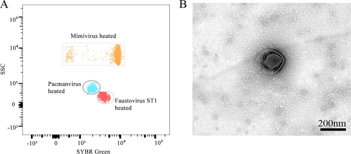
African swine fever virus, a double-stranded DNA virus that infects pigs, is the only known member of the Asfarviridae family. Nevertheless, during our isolation and sequencing of the complete genome of faustovirus, followed by the description of kaumoebavirus, carried out over the past 2 years, we observed the emergence of previously unknown related viruses within this group of viruses. Here we describe the isolation of pacmanvirus, a fourth member in this group, which is capable of infecting Acanthamoeba castellanii Pacmanvirus A23 has a linear compact genome of 395,405 bp, with a 33.62% G+C content. The pacmanvirus genome harbors 465 genes, with a high coding density. An analysis of reciprocal best hits shows that 31 genes are conserved between African swine fever virus, pacmanvirus, faustovirus, and kaumoebavirus. Moreover, the major capsid protein locus of pacmanvirus appears to be different from those of kaumoebavirus and faustovirus. Overall, comparative and genomic analyses reveal the emergence of a new group or cluster of viruses encompassing African swine fever virus, faustovirus, pacmanvirus, and kaumoebavirus.IMPORTANCE Pacmanvirus is a newly discovered icosahedral double-stranded DNA virus that was isolated from an environmental sample by amoeba coculture. We describe herein its structure and replicative cycle, along with genomic analysis and genomic comparisons with previously known viruses. This virus represents the third virus, after faustovirus and kaumoebavirus, that is most closely related to classical representatives of the Asfarviridae family. These results highlight the emergence of previously unknown double-stranded DNA viruses which delineate and extend the diversity of a group around the asfarvirus members.
Keywords: Acanthamoeba castellanii; African swine fever virus; Asfarviridae; NCLDV; faustovirus; giant viruses; kaumoebavirus; pacmanvirus.
Copyright © 2017 American Society for Microbiology.
Figures


References
-
- Philippe N, Legendre M, Doutre G, Couté Y, Poirot O, Lescot M, Arslan D, Seltzer V, Bertaux L, Bruley C, Garin J, Claverie J-M, Abergel C. 2013. Pandoraviruses: amoeba viruses with genomes up to 2.5 Mb reaching that of parasitic eukaryotes. Science 341:281–286. doi: 10.1126/science.1239181. - DOI - PubMed
-
- Legendre M, Bartoli J, Shmakova L, Jeudy S, Labadie K, Adrait A, Lescot M, Poirot O, Bertaux L, Bruley C, Couté Y, Rivkina E, Abergel C, Claverie J-M. 2014. Thirty-thousand-year-old distant relative of giant icosahedral DNA viruses with a pandoravirus morphology. Proc Natl Acad Sci U S A 111:4274–4279. doi: 10.1073/pnas.1320670111. - DOI - PMC - PubMed
-
- Boyer M, Yutin N, Pagnier I, Barrassi L, Fournous G, Espinosa L, Robert C, Azza S, Sun S, Rossmann MG, Suzan-Monti M, La Scola B, Koonin EV, Raoult D. 2009. Giant marseillevirus highlights the role of amoebae as a melting pot in emergence of chimeric microorganisms. Proc Natl Acad Sci U S A 106:21848–21853. doi: 10.1073/pnas.0911354106. - DOI - PMC - PubMed
-
- Legendre M, Lartigue A, Bertaux L, Jeudy S, Bartoli J, Lescot M, Alempic J-M, Ramus C, Bruley C, Labadie K, Shmakova L, Rivkina E, Couté Y, Abergel C, Claverie J-M. 2015. In-depth study of Mollivirus sibericum, a new 30,000-y-old giant virus infecting Acanthamoeba. Proc Natl Acad Sci U S A 112:E5327–E5335. doi: 10.1073/pnas.1510795112. - DOI - PMC - PubMed
Publication types
MeSH terms
Substances
Grants and funding
LinkOut - more resources
Full Text Sources
Other Literature Sources
Miscellaneous
