. 2017 Apr 26;9(387):eaal3986.
doi: 10.1126/scitranslmed.aal3986.

Transient CDK4/6 inhibition protects hematopoietic stem cells from chemotherapy-induced exhaustion

Affiliations

Transient CDK4/6 inhibition protects hematopoietic stem cells from chemotherapy-induced exhaustion

Shenghui He et al. Sci Transl Med. .

Abstract

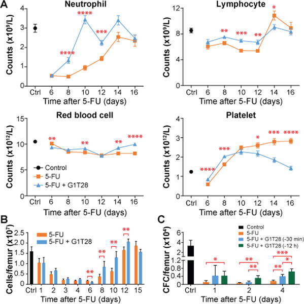
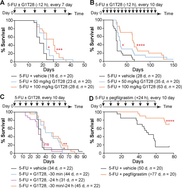
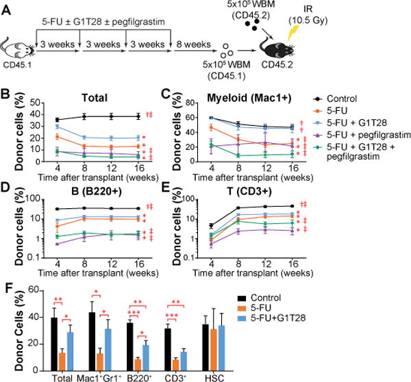
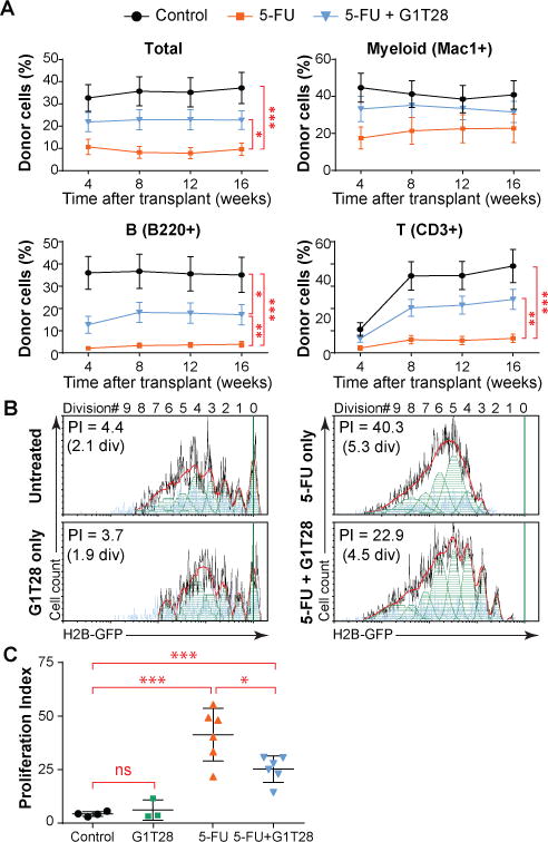
Conventional cytotoxic chemotherapy is highly effective in certain cancers but causes dose-limiting damage to normal proliferating cells, especially hematopoietic stem and progenitor cells (HSPCs). Serial exposure to cytotoxics causes a long-term hematopoietic compromise ("exhaustion"), which limits the use of chemotherapy and success of cancer therapy. We show that the coadministration of G1T28 (trilaciclib), which is a small-molecule inhibitor of cyclin-dependent kinases 4 and 6 (CDK4/6), contemporaneously with cytotoxic chemotherapy protects murine hematopoietic stem cells (HSCs) from chemotherapy-induced exhaustion in a serial 5-fluorouracil treatment model. Consistent with a cell-intrinsic effect, we show directly preserved HSC function resulting in a more rapid recovery of peripheral blood counts, enhanced serial transplantation capacity, and reduced myeloid skewing. When administered to healthy human volunteers, G1T28 demonstrated excellent in vivo pharmacology and transiently inhibited bone marrow (BM) HSPC proliferation. These findings suggest that the combination of CDK4/6 inhibitors with cytotoxic chemotherapy should provide a means to attenuate therapy-induced BM exhaustion in patients with cancer.


Conflict of interest statement

COMPETING INTERESTS:

N.E.S holds an equity interest in G1 Therapeutics Inc. P.J.R, J.A.S, H.S.W, J.E.B, K.M, J.C.S, and R.M were employees of G1 Therapeutics at the time the study was conducted and have an equity interest in the company; S.H, W.A.W, M.S, R.G.T, H.T, E.H report that they have no competing interests. Authors who are inventors on patents related to this work are:

  1. Roberts and Sharpless: Cyclin dependent kinase inhibitors and methods of use www.google.com/patents/US20120100100

  2. Strum, Bisi and Roberts: Transient protection of normal cells during chemotherapy www.google.com/patents/CA2906156A1?cl=en

  3. Strum, Bisi, Roberts and Sharpless: Transient Protection of Hematopoietic Stem and Progenitor Cells against ionizing radiation www.google.com/patents/US20140274896

  4. Strum, Bisi, Roberts and Sharpless: Hematopoietic protection against chemotherapeutic compounds using selective cyclin-dependent kinase 4/6 inhibitors www.google.com/patents/WO2010039997A3?cl=en

  5. Strum: CDK inhibitors www.google.com/patents/US8598186

Figures

Fig. 1
Fig. 1. G1T28 induces transient, reversible G1 cell cycle arrest in both murine and human BM HSPCs
(A) Rate of murine BM HSC proliferation 12 hours after a single IP injection of the indicated doses of G1T28 (n = 5 mice/dose analyzed in 5 independent experiments, same for B and C). (B) Median effective dose (ED50) of G1T28 to inhibit the in vivo proliferation of the indicated murine hematopoietic cell types with identification scheme shown in fig. S1 (LSK = LinSca-1+c-Kit+ cells, MP = myeloid progenitors, E = erythroid, M = myeloid, B = B lymphocytes, T = T lymphocytes). (C) Relative proliferation rate of each murine hematopoietic cell type at maximum CDK4/6 inhibition, compared to untreated mice. (D) Rate of murine BM HSC proliferation at various time points after a single IP injection of 50 or 100 mg/kg of G1T28 (n = 3-6 mice/dose/time point). (E) Mean plasma concentration curve of G1T28 after single dose IV administration in healthy human volunteers. (F, G) Frequency of total human BM cells (F) and HSC-enriched CD45dimCD34+CD38 BM cells (G) in S/G2/M phase of the cell cycle either before or at 24 and 32 hours after single dose of G1T28 treatment. Data were from Cohort 7 (BED) after single 192 mg/m2 dose of G1T28. Error bars represent SD in A-D and SEM in E-G. The statistical significance of differences between vehicle and G1T28 treated groups in A and D and between indicated groups in B, C, and F was assessed using unpaired, two-tailed Student’s t-tests (*P < 0.05, **P < 0.01, ***P < 0.001, ****P < 0.0001).
Fig. 2
Fig. 2. Transient G1 arrest by G1T28 pretreatment accelerates hematologic recovery after single dose 5FU treatment
(A, B) Complete blood cell counts (A) and BM cellularity (B) at various time points after a single dose of 5FU with or without G1T28 pretreatment (n = 5-18 mice/treatment/time point). (C) Number of colony-forming cells (CFC) per femur 1 to 4 days after a single treatment with 5FU ± G1T28 (n = 4-11 mice/treatment/time point). Data represent mean ± SEM in A and mean ± SD in B and C. The statistical significance of differences between indicated groups was assessed using unpaired, two-tailed Student’s t-tests (*P < 0.05, **P < 0.01, ***P < 0.001, ****P < 0.0001).
Fig. 3
Fig. 3. G1T28 pretreatment protects mice from repeated doses of 5FU challenge
(A–D) Survival curves of mice that were treated with 5FU plus vehicle, G1T28, or pegfilgrastim at 7 (A) or 10 (B-D) day intervals. G1T28 was given 12 hours before 5FU in A and B, and either 30 minutes before, 24 hours before, or both in C. Pegfilgrastim was given 24 hours after 5FU in (D). Numbers in parenthesis indicate median survival (d = day), and animal number (n) in each treatment group. Significance for the pairwise comparison to 5FU + vehicle was calculated using the log-rank test in A, B, and D and Gehan-Breslow-Wilcoxon test in C (*P < 0.05, **P < 0.01, ***P < 0.001, ****P < 0.0001, ns, not significant).
Fig. 4
Fig. 4. G1T28-induced PQ protects HSCs from proliferative exhaustion
(A) Treatment and BM transplantation schematic. WBM, whole bone marrow cells (B-E) The percentage of donor-derived cells (CD45.1+) in total PB leukocytes (B), or myeloid cell (Mac1+, C), B lymphocyte (B220+, D), and T lymphocyte (CD3+, E) fractions 1 to 4 months after primary BM transplantation. 3-8 donor mice/treatment, 5 recipients/donor. Statistical significance of differences between treatment groups at 16 weeks after transplantation was assessed using one-way ANOVA with correction for multiple comparisons. (*, P < 0.05 compared to control; †, P < 0.05 compared to 5FU + vehicle, ‡, P < 0.05 compared to 5FU + G1T28). (F) The percentages of donor-derived cells in total BM cells, HSCs, and major BM cell lineages 32 weeks after BM transplantation. 5 donor mice/treatment, 5 recipients/donor. Statistical significance was assessed by two-tailed Student’s t-test (*P < 0.05, **P < 0.01, ***P < 0.001). Average donor cell reconstitution in B-F was calculated by first averaging the value of the 5 recipients from each donor, followed by calculating the mean of all donors. Error bars represent SEM.
Fig. 5
Fig. 5. Transient CDK4/6 inhibition protects long-term HSC function against serial 5FU treatment by reducing overall proliferative burden
(A) The percentage of donor-derived cells (CD45.1+) in total PB leukocytes or myeloid cell (Mac1+), B lymphocyte (B220+), and T lymphocyte (CD3+) fractions 1 to 4 months after secondary BM transplantation. Data represent the mean ± SEM from 20-22 mice per group. (B, C) Cumulative HSC proliferation after a single dose of 5FU ± G1T28 treatment as determined by H2B-GFP label retention 4 weeks after 5FU administration. Representative H2B-GFP label dilution histograms (and curve fitting) are shown in (B), with quantification in (C). Proliferation index (PI) indicates the average number of daughter cells generated from a single, originally labeled HSC by the time of analysis, such that Log2 (PI) equals the average number of HSC divisions. Data represent mean ± SD from 3-6 mice in 3 experiments. Statistical significance was assessed by unpaired, two-tailed Student’s t-test (*P < 0.05, **P < 0.01, ***P < 0.001). div, division.

References

    1. Harris J, Sengar D, Stewart T, Hyslop D. The effect of immunosuppressive chemotherapy on immune function in patients with malignant disease. Cancer. 1976 Feb;37:1058–1069. - PubMed
    1. Gardner RV, Lerner C, Astle CM, Harrison DE. Assessing permanent damage to primitive hematopoietic stem cells after chemotherapy using the competitive repopulation assay. Cancer Chemother Pharmacol. 1993;32:450–454. - PubMed
    1. Mauch P, Constine L, Greenberger J, Knospe W, Sullivan J, Liesveld JL, Deeg HJ. Hematopoietic stem cell compartment: acute and late effects of radiation therapy and chemotherapy. Int J Radiat Oncol Biol Phys. 1995 Mar 30;31:1319–1339. - PubMed
    1. Gardner RV. Long term hematopoietic damage after chemotherapy and cytokine. Front Biosci. 1999 Jul 15;4:e47–57. - PubMed
    1. Wang Y, Probin V, Zhou D. Cancer therapy-induced residual bone marrow injury-Mechanisms of induction and implication for therapy. Curr Cancer Ther Rev. 2006 Aug 1;2:271–279. - PMC - PubMed

Publication types

MeSH terms