. 2017 Apr 26;7(1):1173.
doi: 10.1038/s41598-017-01214-4.

Expandable Mg-based Helical Stent Assessment using Static, Dynamic, and Porcine Ex Vivo Models

Affiliations

Expandable Mg-based Helical Stent Assessment using Static, Dynamic, and Porcine Ex Vivo Models

Youngmi Koo et al. Sci Rep. .

Abstract

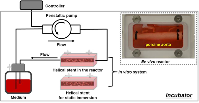
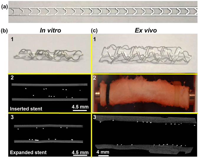
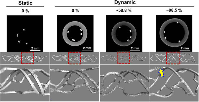
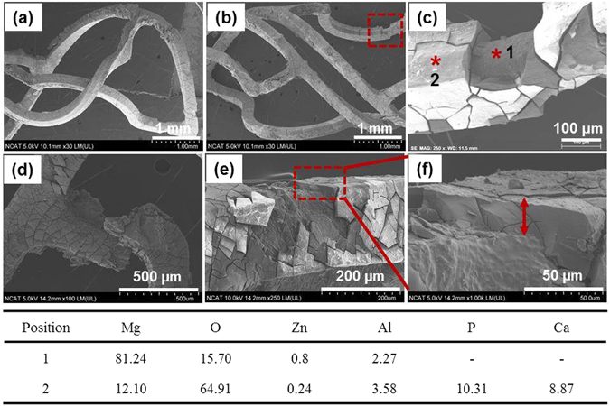
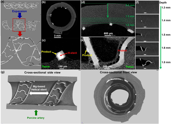
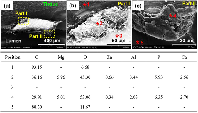
A bioresorbable metallic helical stent was explored as a new device opportunity (magnesium scaffold), which can be absorbed by the body without leaving a trace and simultaneously allowing restoration of vasoreactivity with the potential for vessel remodeling. In this study, developed Mg-based helical stent was inserted and expanded in vessels with subsequent degradation in various environments including static, dynamic, and porcine ex vivo models. By assessing stent degradation in three different environments, we observed: (1) stress- and flow-induced degradation; (2) a high degradation rate in the dynamic reactor; (3) production of intermediate products (MgO/Mg(OH)2 and Ca/P) during degradation; and (4) intermediate micro-gas pocket formation in the neighboring tissue ex vivo model. Overall, the expandable Mg-based helical stent employed as a scaffold performed well, with expansion rate (>100%) in porcine ex vivo model.


Conflict of interest statement

The authors declare that they have no competing interests.

Figures

Figure 1
Figure 1
Schematic illustrating the manufacturing steps in the photo-chemical etching approach for making Mg helical stents.
Figure 2
Figure 2
Bioreactor configuration for the in vitro and ex vivo simulation of the Mg-based helical stent.
Figure 3
Figure 3
Geometries and stenting of Mg-based helical stent for vascular bioreactor tests. (a) Optical image of 2D Mg-based ribbon before coiling to form the device, (b) In vitro stent testing: optical image of helical stent for in vitro test (b1), inserted stent in artificial vessel (b2), and expanded stent in vessel (b3), (c) Ex vivo stent testing: optical image of helical stent for ex vivo (c1), optical image of the inserted stent in porcine artery (c2), and expanded stent in artery (c3).
Figure 4
Figure 4
Cross-sectional 2D (before test) and 3D (after test) micro-CT structure images revealing corrosion products accumulated during the in vitro static and dynamic fluid flow simulations for 3 days in DMEM (10% FBS, 1% P/S) at 37 °C, 5% CO2. Red dot box images were enlarged. Yellow arrow indicates broken strut after in vitro dynamic fluid flow simulation.
Figure 5
Figure 5
SEM images of degraded areas on tested helical stents under flow induced shear stress, 0.68 Pa for 3 days in DMEM (10% FBS, 1% P/S) at 37 °C, 5% CO2. (ac) Represent the ~58.8% expanded helical stent, (df) represent the ~98.5% expanded helical stent. The Table displays the atomic percent (at %) detected in the two positions by EDX.
Figure 6
Figure 6
3D structure and cross-sectional 2D micro-CT images of expended helical stents in artery revealing the with corrosion products ex vivo dynamic simulation for 3 days in DMEM (10% FBS, 1% P/S) at 37 °C, 5% CO2. (a) Bare stent and extracted stent in artery, (b) 2D sliced image, (c) Enlarged one strut of (b), (d) Interface between implanted helical stent and tissue, (e) Enlarged images at a depth of 1.6 mm in (d,f) sliced images from outmost of artery to lumen (arrow direction in (d)), (g) Actual representative X-ray micro-CT 3D structures of expanded Mg-based helical stent scaffold at the porcine artery. Product: Ca/P complex, H-stent: Mg helical stent, Scale bars on (f): 1 mm.
Figure 7
Figure 7
SEM images and EDX analysis of corrosion products surrounding expanded helical stents under the flow induced wall shear stress value of 0.154 Pa for 3 days in DMEM (10% FBS, 1% P/S) at 37 °C, 5% CO2. aIndicates Mg-based helical stent has been implanted.

Similar articles

Cited by

References

    1. Kaul, S. Advisory Board. http://www.advisory.com/daily-briefing/2013/10/30/cardiologists-will-sto... (2013).
    1. Meier B, Bachmann D, Lüscher TF. 25 years of coronary angioplasty: almost a fairy tale. The Lancet. 2003;361:527. doi: 10.1016/S0140-6736(03)12470-1. - DOI - PubMed
    1. Grech G. Percutaneous coronary intervention. I: History and development. BMJ. 2003;326:1080–1082. doi: 10.1136/bmj.326.7398.1080. - DOI - PMC - PubMed
    1. Dangas GD, et al. In-stent restenosis in the drug-eluting stent era. JACC. 2010;56:1897–1907. doi: 10.1016/j.jacc.2010.07.028. - DOI - PubMed
    1. Moravej M, Mantoviani D. Biodegradable metals for cardiovascular stent application: interests and new opportunities. Int. J. Mol.Sci. 2011;12:4250–4270. doi: 10.3390/ijms12074250. - DOI - PMC - PubMed

Publication types