

UVB promotes the initiation of uveitic inflammatory injury in vivo and is attenuated by UV-blocking protection
- PMID: 28446860
- PMCID: PMC5390783
UVB promotes the initiation of uveitic inflammatory injury in vivo and is attenuated by UV-blocking protection
Abstract
Purpose: Uveitic inflammatory injury can cause irreversible visual loss; however, no single animal model recapitulates all the characteristics of human uveitis. Ultraviolet radiation (UVR) is one of the risk factors for uveitis, but the role of UVR in the pathogenesis of uveitic injury is unclear. The aim of this study was to elucidate whether UVB promotes the initiation of, and subsequently contributes to, uveitic inflammatory injury.
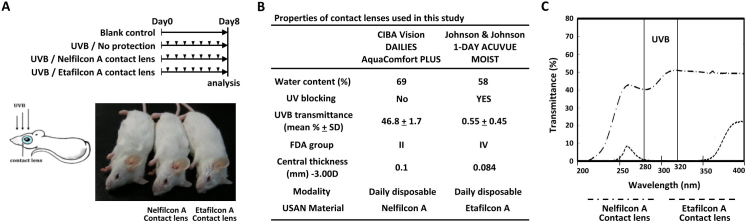
Methods: Mice were assigned to either a blank control group or one of three UVB treatment groups: no protection, protection with Nelfilcon A contact lens (Food and Drug Administration [FDA] class II, about 46.8% UVB transmittance), or protection with Etafilcon A contact lens (FDA class IV, about 0.55% UVB transmittance). The contact lenses acted as blocking barriers against UVR. After the application of UVR, pathologic injuries were determined with slit-lamp microscopy and histologic examination.
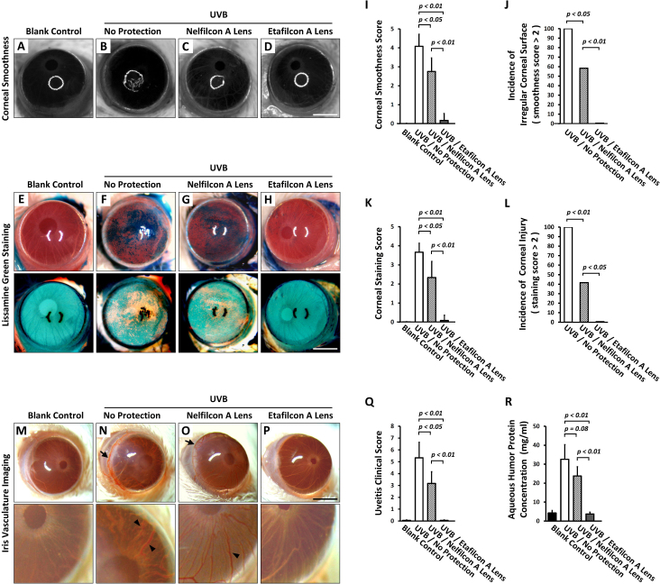
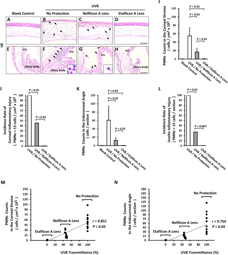
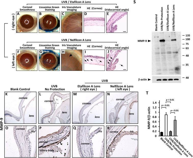
Results: Compared with the intact status of the controls, the anterior eyes of the UVB groups showed pathologic alterations in physiologic properties and tissue integrity. UVR promoted anterior uveitic inflammatory injury, with expansion of the hyperemic iris vessels, over-production of aqueous humor protein, disruption of the blood-aqueous barrier, and embedding of infiltrative leukocytes inside the iridocorneal angle. However, blockage of UVR in vivo retarded the progression of uveitic inflammatory injury. The highest level of UV protection in the Etafilcon A group resulted in greater inhibition of uveitic inflammatory injury than that in the Nelfilcon A group.
Conclusions: This study demonstrates that UVB initiated and promoted uveitic inflammatory injury. UV protection is needed for the clinical management of anterior uveitis. The Etafilcon A lenses provide better protection of the anterior segment of the eye against UVB damage compared with the Nelfilcon A lenses.
Figures
Similar articles
-
Prevention of UV-induced damage to the anterior segment using class I UV-absorbing hydrogel contact lenses.Invest Ophthalmol Vis Sci. 2010 Jan;51(1):172-8. doi: 10.1167/iovs.09-3996. Epub 2009 Aug 26. Invest Ophthalmol Vis Sci. 2010. PMID: 19710408
-
A class I (Senofilcon A) soft contact lens prevents UVB-induced ocular effects, including cataract, in the rabbit in vivo.Invest Ophthalmol Vis Sci. 2011 Jun 1;52(6):3667-75. doi: 10.1167/iovs.10-6885. Invest Ophthalmol Vis Sci. 2011. PMID: 21421866 Free PMC article.
-
Prevention of the adverse photic effects of peripheral light-focusing using UV-blocking contact lenses.Invest Ophthalmol Vis Sci. 2003 Apr;44(4):1501-7. doi: 10.1167/iovs.02-0380. Invest Ophthalmol Vis Sci. 2003. PMID: 12657585
-
Does the eye benefit from wearing ultraviolet-blocking contact lenses?Eye Contact Lens. 2011 Jul;37(4):267-72. doi: 10.1097/ICL.0b013e3182235777. Eye Contact Lens. 2011. PMID: 21670694 Review.
-
Ultraviolet absorption by contact lenses and the significance on the ocular anterior segment.Eye Contact Lens. 2011 Jul;37(4):259-66. doi: 10.1097/ICL.0b013e3182240945. Eye Contact Lens. 2011. PMID: 21646978 Review.
Cited by
-
P1G10, the Proteolytic Fraction from Vasconcellea cundinamarcensis, Stimulates Tissue Repair after Acute Exposure to Ultraviolet B Radiation.Int J Mol Sci. 2019 Sep 6;20(18):4373. doi: 10.3390/ijms20184373. Int J Mol Sci. 2019. PMID: 31489890 Free PMC article.
-
The protective effect of simvastatin against ultraviolet B-induced corneal endothelial cell death.Indian J Ophthalmol. 2018 Aug;66(8):1080-1083. doi: 10.4103/ijo.IJO_93_18. Indian J Ophthalmol. 2018. PMID: 30038146 Free PMC article.
-
UV Protection in the Cornea: Failure and Rescue.Biology (Basel). 2022 Feb 10;11(2):278. doi: 10.3390/biology11020278. Biology (Basel). 2022. PMID: 35205145 Free PMC article. Review.
-
The Impact of Corneal Oedema on UV Light Transmission: An Experimental Study in Porcine Eyes.J Clin Med. 2024 Nov 28;13(23):7228. doi: 10.3390/jcm13237228. J Clin Med. 2024. PMID: 39685687 Free PMC article.
References
-
- Taylor HR. The biological effects of UV-B on the eye. Photochem Photobiol. 1989;50:489–92. - PubMed
-
- DePry J, Brescoll J, Szczotka-Flynn L, Rambhatla P, Lim HW, Cooper K. Phototherapy-related ophthalmologic disorders. Clin Dermatol. 2015;33:247–55. - PubMed
-
- Willmann G. Ultraviolet Keratitis: From the Pathophysiological Basis to Prevention and Clinical Management. High Alt Med Biol. 2015;16:277–82. - PubMed
-
- Muresan S, Filip A, Muresan A, Simon V, Moldovan R, Gal AF, Miclaus V. Histological findings in the Wistar rat cornea following UVB irradiation. Romanian journal of morphology and embryology = Rev Roum Morphol Embryol. 2013;54:247–52. - PubMed
-
- Cejka C, Rosina J, Sirc J, Michalek J, Brunova B, Cejkova J. The reversibility of UV-B induced alterations in optical properties of the rabbit cornea depends on dose of UV irradiation. Photochem Photobiol. 2013;89:474–82. - PubMed
Publication types
MeSH terms
Substances
LinkOut - more resources
Full Text Sources
Medical
