

Endoscopic management of esophageal leaks
- PMID: 28446977
- PMCID: PMC5384896
- DOI: 10.21037/jtd.2017.03.100
Endoscopic management of esophageal leaks
Abstract
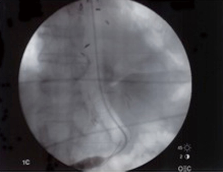
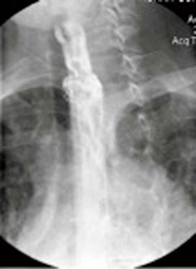
Traditionally, gold standard treatment for an acute esophageal perforation has been operative repair. Over the past two decades, there has been a paradigm shift towards the use of esophageal stents. Recent advances in biomaterial allowed a new generation of stents to be manufactured that combined (I) a non-permeable covering; (II) radial force sufficient to occlude a transmural esophageal injury and (III) improved ease of removability. The amalgamation of these developments set the stage for utilizing esophageal stents as part of the management algorithm of an acute esophageal perforation. This provides a safe and less invasive treatment route in lieu of direct primary repair and its well-documented significant failure rate. Esophageal stent placement for failed operative repair or esophageal leaks also had the potential to minimize the need for esophageal resection and diversion. When included in a multimodality hybrid treatment protocol, esophageal stents can optimize healing success rates and minimize the risks of adverse complications. This review summarizes the modern history of esophageal stent use in the treatment of esophageal perforation as well as the evidenced based recommendations for the use of esophageal stent placement in the treatment of acute esophageal perforation.
Keywords: Esophageal perforation; esophageal fistula; esophageal stent.
Conflict of interest statement
Conflicts of Interest: The authors have no conflicts of interest to declare.
Figures
References
-
- Guisez J. De l’intubation caouchoutee oesophienne. Presse Med 1914;22:85.
-
- Mousseau M, Le Forestier J, Barbin J. Role of permanent intubation in palliative treatment of esophageal cancer. Arch Mal Appar Dig Mal Nutr 1956;45:208-14. - PubMed
Publication types
LinkOut - more resources
Full Text Sources
Other Literature Sources
Miscellaneous