. 2017 Sep;22(5):687-697.
doi: 10.1007/s12192-017-0792-y. Epub 2017 Apr 26.

Interplay between recombinant Hsp70 and proteasomes: proteasome activity modulation and ubiquitin-independent cleavage of Hsp70

Affiliations

Interplay between recombinant Hsp70 and proteasomes: proteasome activity modulation and ubiquitin-independent cleavage of Hsp70

Alexey V Morozov et al. Cell Stress Chaperones. 2017 Sep.

Abstract

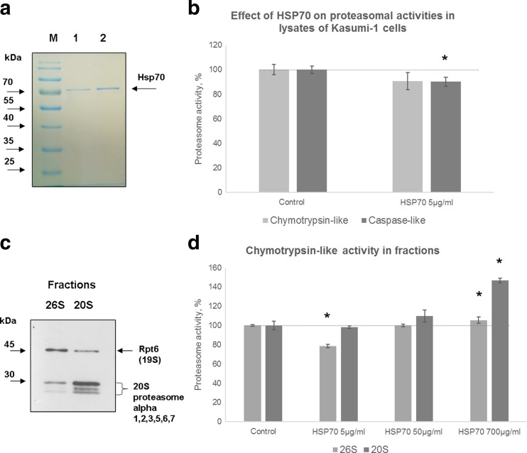
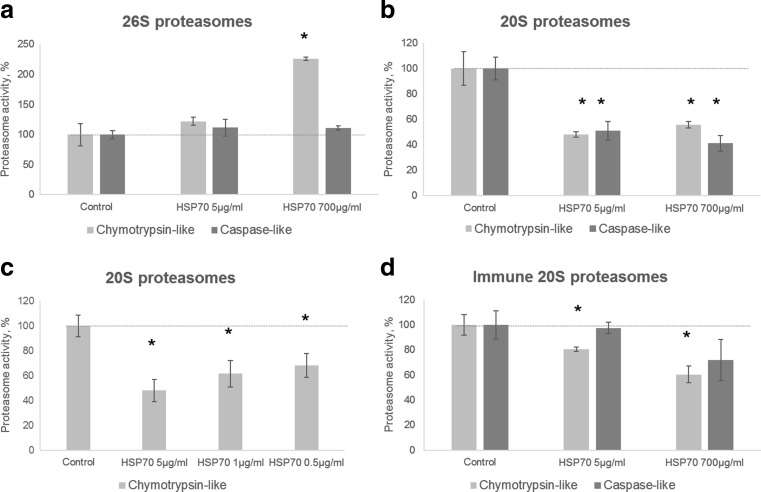
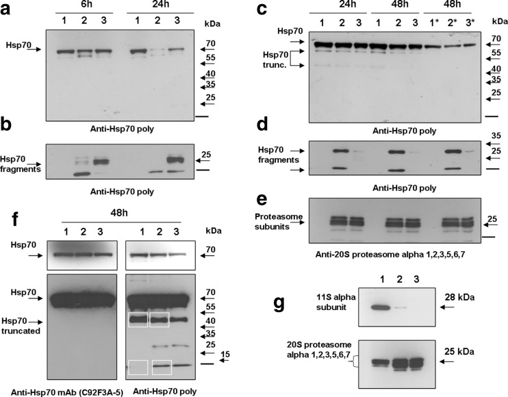
The heat shock protein 70 (Hsp70, human HSPA1A) plays indispensable roles in cellular stress responses and protein quality control (PQC). In the framework of PQC, it cooperates with the ubiquitin-proteasome system (UPS) to clear damaged and dysfunctional proteins in the cell. Moreover, Hsp70 itself is rapidly degraded following the recovery from stress. It was demonstrated that its fast turnover is mediated via ubiquitination and subsequent degradation by the 26S proteasome. At the same time, the effect of Hsp70 on the functional state of proteasomes has been insufficiently investigated. Here, we characterized the direct effect of recombinant Hsp70 on the activity of 20S and 26S proteasomes and studied Hsp70 degradation by the 20S proteasome in vitro. We have shown that the activity of purified 20S proteasomes is decreased following incubation with recombinant human Hsp70. On the other hand, high concentrations of Hsp70 activated 26S proteasomes. Finally, we obtained evidence that in addition to previously reported ubiquitin-dependent degradation, Hsp70 could be cleaved independent of ubiquitination by the 20S proteasome. The results obtained reveal novel aspects of the interplay between Hsp70 and proteasomes.

Keywords: Heat shock proteins; Hsp70; Proteasome; Proteasome regulators; Ubiquitin-independent degradation.


Conflict of interest statement

The investigation of proteasome activity in the culture cells and extracts was supported by Ministry of Education and Science of Russian Federation (agreement no. 14.Z50.31.0014) Russian Science Foundation grant (14-50-00060) and Russian President Foundation grant (МК-3613.2017.4) to A.M.

Figures

Fig. 1
Fig. 1
Hsp70 influences proteasome activity in lysates of Kasumi-1 cells and in the proteasome-enriched fractions. a Obtained recombinant Hsp70 is as pure as commercial protein. Of commercial (track 1) and obtained Hsp70 (track 2), 0.2 μg was separated in SDS-PAGE followed by staining with Roti blue quick. Prestained protein ladder (Fermentas, Lithuania) (M). b Chymotrypsin-like and caspase-like activity in crude Kasumi-1 cellular lysates incubated with 5 μg/ml of Hsp70 for 40 min. Chymotrypsin-like activity is shown in gray and caspase-like activity in dark gray. Proteolytic activity in untreated lysates was set as control. The data are mean values from three experiments ± SD. Statistically significant differences versus control are indicated by asterisks: *p < 0.05, calculated by two-tailed Student’s t test. Dotted line indicates proteasome activity in control samples. c Western blot of 26S and 20S proteasome-enriched protein fractions, obtained from Kasumi-1 cells. α-Subunits (1, 2, 3, 5, 6, 7 (∑α)) of the 20S proteasomes and Rpt6 subunits of 19S regulators were revealed in both fractions and indicated by arrowhead and a curly brace. The prevalence of the 26S proteasomes in the 26S fraction and domination of 20S proteasomes in the 20S fraction were confirmed: 26S fraction contained 5 times less α-subunits but 2.27 times more of Rpt6 subunits than the 20S fraction (estimated using ImageJ software). d Chymotrypsin-like proteasome activity in 26S proteasome-enriched (gray) and 20S proteasome-enriched (dark gray) fractions from Kasumi-1 cells after incubation with Hsp70. Aliquots of fractions (~8 μg of total protein) were incubated with three different concentrations of Hsp70: 5 μg/ml (0.07 μM), 50 μg/ml (0.7 μM), and 700 μg/ml (10 μM). Proteolytic activity in untreated fractions was set as control. The data are mean values from three experiments ± SD. Statistically significant differences versus control are indicated by asterisks: *p < 0.05, calculated by two-tailed Student’s t test. Dotted line indicates proteasome activity in control samples
Fig. 2
Fig. 2
The effect of Hsp70 on proteolytic activity of highly purified proteasomes. The effect of Hsp70 on proteolytic activity of 26S (a), constitutive 20S (b, c), and immune 20S (d) proteasomes. Proteasomal activity was measured after treatment with 5 and 700 μg/ml of the Hsp70 (a, b, d). Chymotrypsin-like activity is shown in gray and caspase-like activity in dark gray. c Dose-dependent effect of Hsp70 on chymotrypsin-like activity of 20S proteasomes. The activity was measured in samples incubated with 5, 1, and 0.5 μg/ml of Hsp70. Proteolytic activity in protein-free samples was set as control. The data are mean values from three experiments ± SD. Statistically significant differences versus control are indicated by asterisks *p < 0.05, calculated by two-tailed Student’s t test. Dotted line indicates proteasome activity in control samples
Fig. 3
Fig. 3
Hsp70 is cleaved without ubiquitination by the 20S proteasomes. a Western blot analysis of Hsp70 and SDS-activated constitutive 20S proteasome mixtures. Samples in order: 1 μg of Hsp70 (1), 1 μg of Hsp70 and 1 μg of 20S proteasome (2), 1 μg of Hsp70, 1 μg of 20S proteasome, and 10 μM of MG132 (3). Samples were incubated 6 (left panel) or 24 (right panel) h. Membrane was stained with polyclonal anti-Hsp70 antibodies. In samples containing activated 20S proteasomes, 47 and 93% of the Hsp70 were degraded following 6 and 24 h, respectively. Addition of chymotrypsin-like activity inhibitor MG132 partially attenuated the degradation. Optical density of the protein bands was quantified using ImageJ software. Proteins and molecular weight markers are indicated by arrowheads. Bar represents electrophoresis front. b Lower part of the X-ray film a obtained at longer exposure. c Western blot analysis of Hsp70 and constitutive 20S proteasome mixtures incubated without SDS. Samples in order: 1 μg of Hsp70 (1), 1 μg of Hsp70 and 1 μg of 20S proteasome (2), 1 μg of Hsp70, 1 μg of 20S proteasome, and 1 mM of MG132 (3). Asterisks indicate that in the reaction mixtures, the Hsp70 128 was used. Membrane was stained with anti-Hsp70 antibodies. Samples were incubated 24 or 48 h. Samples, containing 20S proteasomes approximately from 10 and 20–40% (depending on Hsp70 used) of the Hsp70, were degraded following 24 and 48 h, respectively. Addition of chymotrypsin-like activity inhibitor MG132 partially attenuated the degradation. Optical density of the protein bands was quantified using ImageJ software. Proteins and molecular weight markers are indicated by arrowheads. Bar represents electrophoresis front. d Lower part of the X-ray film c obtained at longer exposure. e The membrane depicted in c and d stripped and stained with antibodies to 20S proteasome alpha subunits α 1, 2, 3, 5, 6, and 7. f Western blot analysis of Hsp70 and mixtures of Hsp70 with constitutive and immune 20S proteasomes incubated in SDS-free conditions. Samples in order: 1 μg of Hsp70 (1), 1 μg of Hsp70 and 1 μg of 20S proteasome (2), 1 μg of Hsp70 and 1 μg of immune 20S proteasome (3). Samples were incubated 48 h. Reactions were performed in triplicates. Three gels were run. Two were used for blotting and subsequent staining with antibodies. One membrane was stained with mouse monoclonal (C92F3A-5) and another with rabbit polyclonal anti-Hsp70 antibodies. Upper part of the image represents short, while lower a longer exposure of the film. Third gel was stained with Coomassie brilliant blue, and the white frames indicate corresponding gel slices that were excised and analyzed by mass spectrometry (ESM File 1 Fig. 4, ESM Files 2–4). Proteins and molecular weight markers are indicated by arrowheads. Bar represents electrophoresis front. g Comparative analysis of 20S fraction obtained from Kasumi-1 cells and commercial highly pure 20S and immune 20S proteasome preparations. Samples were analyzed by SDS-PAGE in the 12% gel. Ten micrograms of 20S fraction (track 1) and 1 μg of commercial 20S (track 2) and immune 20S (track 3) proteasomes were loaded onto the gel. Western blot with antibodies to the α-subunit of 11S regulator and α-subunits (1, 2, 3, 5, 6, 7 (∑α)) of the 20S proteasomes was performed

References

    1. Abramova EB, Astakhova TM, Erokhov PA, Sharova NP (2004) Multiple forms of the proteasomes and some approaches to their separation Izvestiia Akademii nauk Seriia biologicheskaia / Rossiiskaia akademiia nauk:150–156 - PubMed
    1. Abramova EB, Sharova NP, Karpov VL. The proteasome: destroy to live. Mol Biol. 2002;36:761–776. - PubMed
    1. Alberti S, Esser C, Hohfeld J. BAG-1—a nucleotide exchange factor of Hsc70 with multiple cellular functions. Cell Stress Chaperones. 2003;8:225–231. doi: 10.1379/1466-1268(2003)008<0225:BNEFOH>2.0.CO;2. - DOI - PMC - PubMed
    1. Alexandrova A, Petrov L, Georgieva A, Kirkova M, Kukan M. Effects of proteasome inhibitor, MG132, on proteasome activity and oxidative status of rat liver. Cell Biochem Funct. 2008;26:392–398. doi: 10.1002/cbf.1459. - DOI - PubMed
    1. Arlt A, et al. Increased proteasome subunit protein expression and proteasome activity in colon cancer relate to an enhanced activation of nuclear factor E2-related factor 2 (Nrf2) Oncogene. 2009;28:3983–3996. doi: 10.1038/onc.2009.264. - DOI - PubMed

MeSH terms

LinkOut - more resources