. 2017 Dec;55(1):1679-1687.
doi: 10.1080/13880209.2017.1319867.

Neuroprotective effects of ellagic acid on cuprizone-induced acute demyelination through limitation of microgliosis, adjustment of CXCL12/IL-17/IL-11 axis and restriction of mature oligodendrocytes apoptosis

Affiliations

Neuroprotective effects of ellagic acid on cuprizone-induced acute demyelination through limitation of microgliosis, adjustment of CXCL12/IL-17/IL-11 axis and restriction of mature oligodendrocytes apoptosis

Nima Sanadgol et al. Pharm Biol. 2017 Dec.

Retraction in

Abstract

Context: Ellagic acid (EA) is a natural phenol antioxidant with various therapeutic activities. However, the efficacy of EA has not been examined in neuropathologic conditions.

Objective: In vivo neuroprotective effects of EA on cuprizone (cup)-induced demyelination were evaluated.

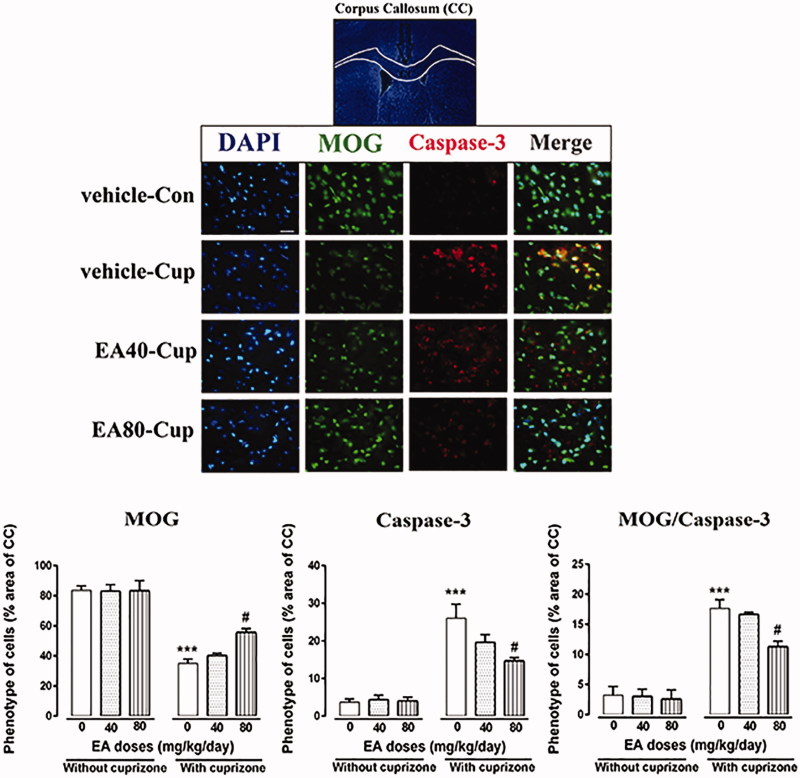
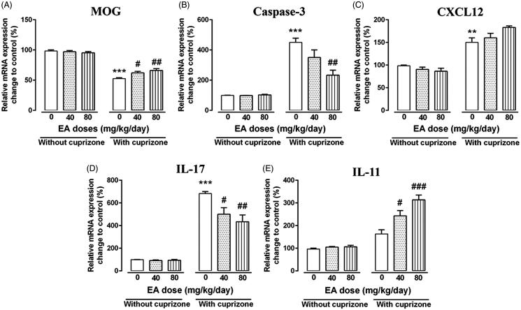
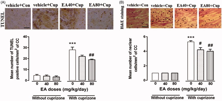
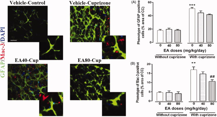
Material and methods: C57BL/6 J mice were fed with chow containing 0.2% cup for 4 weeks to induce oligodendrocytes (OLGs) depletion predominantly in the corpus callosum (CC). EA was administered at different doses (40 or 80 mg/kg body weight/day/i.p.) from the first day of cup diet. Oligodendrocytes apoptosis [TUNEL assay and myelin oligodendrocyte glycoprotein (MOG+)/caspase-3+ cells), gliosis (H&E staining, glial fibrillary acidic protein (GFAP+) and macrophage-3 (Mac-3+) cells) and inflammatory markers (interleukin 17 (IL-17), interleukin 11 (IL-11) and stromal cell-derived factor 1 α (SDF-1α) or CXCL12] during cup intoxication were examined.

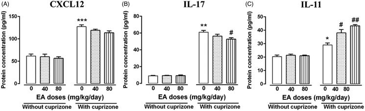
Results: High dose of EA (EA-80) increased mature oligodendrocytes population (MOG+ cells, p < 0.001), and decreased apoptosis (p < 0.05) compared with the cup mice. Treatment with both EA doses did not show any considerable effects on the expression of CXCL12, but significantly down-regulated the expression of IL-17 and up-regulated the expression of IL-11 in mRNA levels compared with the cup mice. Only treatment with EA-80 significantly decreased the population of active macrophage (MAC-3+ cells, p < 0.001) but not reactive astrocytes (GFAP+ cells) compared with the cup mice.

Discussion and conclusion: In this model, EA-80 effectively reduces lesions via reduction of neuroinflammation and toxic effects of cup on mature OLGs. EA is a suitable therapeutic agent for moderate brain damage in neurodegenerative diseases such as multiple sclerosis.

Keywords: Antioxidant; multiple sclerosis; neurodegeneration; neuroinflammation; toxic demyelination.


Figures

Figure 1.
Figure 1.
Effects of EA treatment on mature OLGs population (MOG + cells) and apoptosis (caspase-3 + cells). IHC of coronal sections through the CC showing labeling with a monoclonal antibody that is specific to the mature OLGs marker (MOG), and apoptosis marker (caspase-3) along with DAPI nuclear stain. MOG staining showed significantly decrease in immunoreactivity after 4 weeks of cup treatment that is significantly increased throughout 4-week co-treatment with EA-80. MOG and caspase-3 double-positive cells significantly increased after cup treatment and decreased throughout TP treatment. Scale bar =25 μm, original magnification ×100. Vehicle + con: mice on a regular diet and injected with vehicle for 4 weeks (n = 3), vehicle + cup: cuprizone plus vehicle injection for 4 weeks (n = 3), EA-40 + cup: cuprizone mice were injected with 40 mg/kg of EA for 4 weeks (n = 3), EA80 + cup: cuprizone mice were injected with 80 mg/kg of EA for 4 weeks (n = 3). Data are expressed as means ± SEM. *Compared with control mice, #compared with cuprizone (#p < 0.05, ##p < 0.01 and ***p < 0.001).
Figure 2.
Figure 2.
Analysis of immune, apoptosis and OLGs-related transcripts after EA treatment. Using quantitative PCR technique, the effects of EA on MOG (A), caspase-3 (B), CXCL12 (C), IL-17 (D) and IL-11(E) were tested in the CC region of mice after 4 weeks treatment. Quantitative RT-PCR was conducted and results were normalized to β-actin and reported as % changes to the control group. Data are presented as means ± SEM, analyzed using two-way ANOVA. *Compared with control mice, #compared with cuprizone (#p < 0.05, **, ##p < 0.01 and ***, ###p < 0.001).
Figure 3.
Figure 3.
Evaluation of gliosis and apoptosis during EA treatment. Haematoxylin and eosin (H&E) staining was performed to study effect of different doses of EA treatments on cup-induced reactive gliosis and trans-endothelial migration of immune cells in the CC region. TUNEL assay confirmed that high dose of EA significantly reduced population of apoptotic cells in CC (A). Moreover, quantification of H&E indicate significantly lower amount of cell infiltration after EA treatments (B). Vehicle + con: mice on a regular diet and injected with vehicle for 4 weeks (n = 3), vehicle + cup: cuprizone plus vehicle injection for 4 weeks (n = 3), EA-40 + cup: cuprizone mice were injected with 40 mg/kg of EA for 4 weeks (n = 3), EA80 + cup: cuprizone mice were injected with 80 mg/kg of EA for 4 weeks (n = 3). Scale bar =75 μm, original magnification ×40. Data are expressed as means ± SEM. *Compared with control mice, #compared with cuprizone (#p < 0.05, ##p < 0.01 and ***p < 0.001).
Figure 4.
Figure 4.
Effects of EA treatment on astrogliosis (GFAP + cells) and microgliosis (Mac-3 + cells). IHC of coronal sections through the CC showing labeling with a monoclonal antibody that is specific to the activated astrocytes marker (GFAP), and microglial marker (Mac-3) along with DAPI nuclear stain. GFAP staining showed significant increase in immunoreactivity after 4 weeks of cup treatment compared with control mice. EA treatments have no significant effects on GFAP population. Mac-3-positive cells significantly increased after cup treatment and decreased throughout EA treatments. Scale bar =25 μm, original magnification ×100. Vehicle + con: mice on a regular diet and injected with vehicle for 4 weeks (n = 3), vehicle + cup: cuprizone plus vehicle injection for 4 weeks (n = 3), EA-40 + cup: cuprizone mice were injected with 40 mg/kg of EA for 4 weeks (n = 3), EA80 + cup: cuprizone mice were injected with 80 mg/kg of EA for 4 weeks (n = 3). Data are expressed as means ± SEM. *Compared with control mice, #compared with cuprizone (##p < 0.05, **p < 0.01 and ***p < 0.001).
Figure 5.
Figure 5.
Evaluation of protein levels of immune mediators in brain after EA treatment. Using quantitative ELISA technique, the effects of EA on CXCL12 (A), IL-17 (B) and IL-11(C) were tested in the CC region of mice after 4 weeks treatment. Data are presented as means ± SEM, analyzed using two-way ANOVA. *Compared with control mice, #compared with cuprizone (*, #p < 0.05, **, ##p < 0.01 and ***p < 0.001).

Similar articles

Cited by

References

    1. Abakumova TO, Kuz'kina AA, Zharova ME, Pozdeeva DA, Gubskii IL, Shepeleva II, Antonova OM, Nukolova NV, Kekelidze ZI, Chekhonin VP.. 2015. Cuprizone model as a tool for preclinical studies of the efficacy of multiple sclerosis diagnosis and therapy. Bull Exp Biol Med. 159:111–115. - PubMed
    1. Abdul-Wahab RH, Waleed MAM, Janakat S, Sawsan AO.. 2009. Bioavailability of ellagic acid after single dose administration using HPLC. Pakistan J Nutr. 8:1661–1664.
    1. Amakura Y, Okada M, Tsuji A, Tonogai Y.. 2000. High-performance liquid chromatography determination with photodiode array detection of ellagic acid in fresh and processed fruits. J Chromatog B. 896:87–93. - PubMed
    1. Baeeri M, Momtaz S, Navaei-Nigjeh M, Niaz K, Rahimifard M, Ghasemi-Niri SF, Sanadgol N, Hodjat M, Sharifzadeh M, Abdollahi M.. 2017. Molecular evidence on the protective effect of ellagic acid on phosalone-induced senescence in rat embryonic fibroblast cells. Food Chem Toxicol. 100:8–23. - PubMed
    1. Baxi EG, DeBruin J, Tosi DM, Grishkan IV, Smith MD, Kirby LA, Strasburger HJ, Fairchild AN, Calabresi PA, Gocke AR.. 2015. Transfer of myelin-reactive Th17 cells impairs endogenous remyelination in the central nervous system of cuprizone-fed mice. J Neurosci. 35:8626–8639. - PMC - PubMed

Publication types

MeSH terms