. 2017 Apr 27:8:15110.
doi: 10.1038/ncomms15110.

PARP3 is a promoter of chromosomal rearrangements and limits G4 DNA

Affiliations

PARP3 is a promoter of chromosomal rearrangements and limits G4 DNA

Tovah A Day et al. Nat Commun. .

Erratum in

Abstract

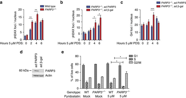
Chromosomal rearrangements are essential events in the pathogenesis of both malignant and nonmalignant disorders, yet the factors affecting their formation are incompletely understood. Here we develop a zinc-finger nuclease translocation reporter and screen for factors that modulate rearrangements in human cells. We identify UBC9 and RAD50 as suppressors and 53BP1, DDB1 and poly(ADP)ribose polymerase 3 (PARP3) as promoters of chromosomal rearrangements across human cell types. We focus on PARP3 as it is dispensable for murine viability and has druggable catalytic activity. We find that PARP3 regulates G quadruplex (G4) DNA in response to DNA damage, which suppresses repair by nonhomologous end-joining and homologous recombination. Chemical stabilization of G4 DNA in PARP3-/- cells leads to widespread DNA double-strand breaks and synthetic lethality. We propose a model in which PARP3 suppresses G4 DNA and facilitates DNA repair by multiple pathways.


Conflict of interest statement

The authors declare no competing financial interests.

Figures

Figure 1
Figure 1. Identification of promoters and suppressors of chromosomal rearrangements in human cells.
(a) Flow cytometry-based assay for chromosomal rearrangements. Scissors, AAVS1 zinc-finger nucleases (ZFNs). Dotted lines, targeted cutting at AAVS1 recognition sequence upstream of either GFP or CD4. (b) Design of shRNA rearrangement screen. A549 cells harbouring a randomly integrated GFP or CD4 reporter were transduced clonally with 1 of 966 shRNA, pooled and 2.5 × 10⁷ shRNA-expressing cells were infected with AAVS1 ZFN adenovirus, and flow-sorted by transgene expression. (c) Representation of different pathways in the shRNA library of 169 genes. (d) Representation of different protein functions in the shRNA library of 169 genes. (e) Gene hits identified in the shRNA screen. Average of log2 reads in transgene-positive population/average log2 reads in transgene-negative population for GFP replicates (x axis) and CD4 replicates (y axis).
Figure 2
Figure 2. Validation of factors that modulate chromosomal rearrangements in human cells.
Normalized frequency of rearrangements, immunoblots and correlation of rearrangement frequency with the degree of knockdown in HeLa ZITR cells with shRNA-mediated knockdown of RAD50 (a), UBC9 (b), 53BP1 (c), DDB1 (d) and PARP3 (e). Data in bar graphs are presented as mean±s.e. of n=3. Immunoblots were quantified using ImageJ software and normalized to β-actin, tubulin or loading control. The dagger in c indicates that this data point (shRNA3) was excluded from the R2 calculation as it resulted in 53BP1 overexpression. The shRNAs that scored in the screen are marked by asterisks. To compare rearrangement frequency with the degree of knockdown, we performed linear regression (R2). P values for R2 calculations are as follows: RAD50, P=0.2206; UBC9, P=0.0861; 53BP1, P=0.2841; DDB1, P=0.0048; PARP3, P=0.0311.
Figure 3
Figure 3. PARP3 promotes chromosomal rearrangements in several human cell types.
(ac) Normalized frequency of rearrangements (a) compared to control (Ct), PARP3 transcript levels (b) and cleavage by the AAVS1 ZFNs measured by quantitative PCR across the targeted site (c) in HeLa cells transduced with shRNA targeting PARP3. P3, PARP3. (d,e) Normalized frequency of rearrangements (d) and qRT–PCR (e) in HeLa ZITR cells transduced with lentivirus expressing control (Ct) shRNA or shRNA targeting PARP3 3′-untranslated region (P3) and expressing control (Ct) cDNA or PARP3 cDNA. (f,g) Normalized frequency of rearrangements (f) and immunoblots (g) in PARP3−/− A549 cells with adenovirus-mediated re-expression of wild-type (WT) PARP3. (hj) Schematic of CRITR assay for translocations between CD71 and CD4 loci (h), flow cytometry (i) and metaphase fluorescence in situ hybridization (j) of CD4+ 293T cells 48 h after transfection of CAS9 and gRNAs. Green probe, RP11-436M6, CD71. Red probe, RP11-277E18, CD4. Scale bar, 10 μm. (ko) Normalized frequency of rearrangements using CRITR assay in wild-type (WT), 53BP1−/−, PARP3−/− and LIG4−/− 293T cells (k) with qRT–PCR of PARP3 (l), immunoblots of indicated proteins (m,n) and intact locus after expression of CAS9 and gRNA measured by quantitative PCR across the targeted site (o). HA-PARP3 occupancy at CD4 18 h after transient expression of CAS9 and gRNA by ChIP, represented as ratio of CAS9 with CD4-directed gRNA to CAS9 with empty vector (EV) (p). bp, base pairs. Data are presented as mean±s.e. of n=3. P values were calculated using unpaired Student’s t-test. *P<0.05, **P<0.01, ***P<0.001.
Figure 4
Figure 4. PARP3−/− cells are sensitive to the stabilization of G4 DNA
(ad) Colony survival assays using wild-type and PARP3−/− A549 cells with the indicated doses of ionizing radiation (a), etoposide (b), bleomycin (c) and pyridostatin (d). (e) Colony survival assay in wild-type A549 cells with siRNA for control (siCont), PARP1, PARP2 or PARP3 with corresponding immunoblots. (f) Colony survival assay in wild-type A549 cells treated with vehicle, 3.0 μM ME0328 or 500 nM KU58948 and pyridostatin. (g) Schematic of sequences predicted by QGRS (purple) and Quadbase2 (blue) and experimentally observed to form G4 DNA by Chambers et al. (green) in a 2,000 bp window surrounding the CD4 CRISPR-CAS9 gRNA. Scissors, CRISPR-CAS9 cut site. (h) BLM occupancy at the CD4 locus measured by ChIP 18 h after transient expression of CAS9 alone or with gRNA targeting CD4. (i) Schematic of sequences G4 DNA sequences depicted as in g in a 2,000 bp window surrounding ESR1 CRISPR-CAS9 gRNA. Scissors, CRISPR-CAS9 cut site. (j) BLM occupancy at the ESR1 locus measured by ChIP 18 h after transient expression of CAS9 alone or with gRNA targeting CD4. (k) G4 DNA content measured by immunoprecipitation (IP) with hf2 antibody following 24 h treatment with vehicle or pyridostatin (PDS) at the telomere (TEL), MYC promoter (MYC), ESR1 enhancer, chr8 or chr22 loci. (l) Quantification of G4 DNA content in the indicated genotypes at the CD4 locus measured by IP with hf2 antibody 24 h after pyridostatin (PDS) treatment and 18 h after transfection with CRISPR-CAS9 with or without gRNA for CD4. bp, base pairs; PDS, pyridostatin. Data are mean±s.e. of n=3. P values calculated using unpaired Student’s t-test. *P<0.05, **P<0.01, ***P<0.001. NS, not significant.
Figure 5
Figure 5. PARP3−/− cells are more susceptible to pyridostatin-induced DSBs.
(a) Quantification of immunofluorescence (IF) for γH2AX at the indicated time points following treatment with 5 μM pyridostatin (PDS) in wild-type and PARP3−/− A549 cells. (b,c) Quantification of IF for γH2AX (b) and G4 DNA using the 1H6 antibody (c) at the indicated time points, and immunoblots (d) in PARP3−/− A549 cells infected with adenovirus-expressing PARP3 (ad PARP3) or β-galactosidase (ad β-gal). (e) Cell cycle dynamics for wild-type (WT) or PARP3−/− A549 cells mock treated or treated with 5 μM pyridostatin. Data are presented as mean±s.e. of n=3. P values calculated using unpaired Student’s t-test. A two-way analysis of variance (alpha set at 0.05) gave the following P values for cell cycle phases: G1, P=0.5955, S, P=0.0152, and G2, P=0.0111. *P<0.05, **P<0.01, ***P<0.001.
Figure 6
Figure 6. PARP3 promotes deposition of CtIP and RPA at DNA DSBs.
(a) CAS9-mediated cleavage at CD4 measured by quantitative PCR across the targeted site 18 h after transient expression of CAS9 and gRNA targeting CD4 (CD4) or CAS9 with empty vector (EV). (b–d) CtIP (b), RPA (c) and γH2AX (d) occupancy 18 h after transient expression of CAS9 alone or with gRNA targeting CD4. bp, base pairs. Data are represented as mean±s.e. of n≥2 biological replicates. P values calculated using unpaired Student’s t-test. *P<0.05, **P<0.01, ***P<0.001. (e,f) Immunofluorescence for γH2AX and RPA (e), and quantification of γH2AX and RPA double-positive cells (f) at 2 h following 10 Gy IR. Scale bars, 10 μm. (g) Quantification of γH2AX and RPA double-positive cells at 24 h after release from serum starvation and 2 h after 10 Gy IR. Data are represented as per cent nuclei per high-power field (HPF). Two-sided t-test was calculated on three biological replicates with a representative replicate shown.
Figure 7
Figure 7. PARP3 and BLM cooperate to repair DNA DSBs.
(ac) Quantification of IF for 53BP1 (a) and γH2AX (b) at the indicated time points in wild-type (WT) or PARP3−/− A549 cells transfected with siRNA targeting control (siCont) or BLM (siBLM). Data are represented as mean±s.e. of three biological replicates. P values calculated using unpaired Student’s t-test. *P<0.05, **P<0.01, ***P<0.001. (c) immunoblot after transfection with siRNA targeting control (Ct) or BLM.
Figure 8
Figure 8. Model of PARP3 regulation of G4 DNA at DNA DSBs.
PARP3 negatively regulates G4 DNA after DNA DSBs to facilitate repair of the damage (left). In the absence of PARP3, G4 DNA accumulates at sites of damage and delays repair of DSBs by preventing deposition of repair factors.

Similar articles

Cited by

References

    1. Kong F. et al. dbCRID: a database of chromosomal rearrangements in human diseases. Nucleic Acids Res. 39, D895–D900 (2011). - PMC - PubMed
    1. Piomboni P., Stendardi A. & Gambera L. Chromosomal aberrations and aneuploidies of spermatozoa. Adv. Exp. Med. Biol. 791, 27–52 (2014). - PubMed
    1. Tabet A. C. et al. Complex nature of apparently balanced chromosomal rearrangements in patients with autism spectrum disorder. Mol. Autism 6, 19 (2015). - PMC - PubMed
    1. Shou Y. et al. Diverse karyotypic abnormalities of the c-myc locus associated with c-myc dysregulation and tumor progression in multiple myeloma. Proc. Natl Acad. Sci. USA 97, 228–233 (2000). - PMC - PubMed
    1. Bochtler T., Frohling S. & Kramer A. Role of chromosomal aberrations in clonal diversity and progression of acute myeloid leukemia. Leukemia 29, 1243–1252 (2015). - PubMed

Publication types

MeSH terms