

Diamide Inhibitors of the Bacillus subtilis N-Acetylglucosaminidase LytG That Exhibit Antibacterial Activity
- PMID: 28448118
- PMCID: PMC5789777
- DOI: 10.1021/acsinfecdis.7b00005
Diamide Inhibitors of the Bacillus subtilis N-Acetylglucosaminidase LytG That Exhibit Antibacterial Activity
Abstract
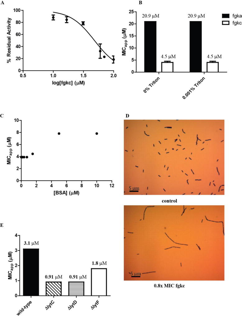
N-Acetylglucosaminidases (GlcNAcases) play an important role in the remodeling and recycling of bacterial peptidoglycan by degrading the polysaccharide backbone. Genetic deletions of autolysins can impair cell division and growth, suggesting an opportunity for using small molecule autolysin inhibitors both as tools for studying the chemical biology of autolysins and also as antibacterial agents. We report here the synthesis and evaluation of a panel of diamides that inhibit the growth of Bacillus subtilis. Two compounds, fgkc (21) and fgka (5), were found to be potent inhibitors (MIC 3.8 ± 1.0 and 21.3 ± 0.1 μM, respectively). These compounds inhibit the B. subtilis family 73 glycosyl hydrolase LytG, an exo GlcNAcase. Phenotypic analysis of fgkc (21)-treated cells demonstrates a propensity for cells to form linked chains, suggesting impaired cell growth and division.
Keywords: N-acetylglucosaminidase; autolysin; diamide; inhibitor; peptidoglycan.
Figures


Similar articles
-
LytG of Bacillus subtilis is a novel peptidoglycan hydrolase: the major active glucosaminidase.Biochemistry. 2003 Jan 21;42(2):257-64. doi: 10.1021/bi020498c. Biochemistry. 2003. PMID: 12525152
-
A mechanism-based GlcNAc-inspired cyclophellitol inactivator of the peptidoglycan recycling enzyme NagZ reverses resistance to β-lactams in Pseudomonas aeruginosa.Chem Commun (Camb). 2018 Sep 25;54(75):10630-10633. doi: 10.1039/c8cc05281f. Epub 2018 Sep 4. Chem Commun (Camb). 2018. PMID: 30178799
-
Identification of novel inhibitors of phospho-MurNAc-pentapeptide translocase MraY from library screening: Isoquinoline alkaloid michellamine B and xanthene dye phloxine B.Bioorg Med Chem. 2014 Sep 1;22(17):4566-71. doi: 10.1016/j.bmc.2014.07.035. Epub 2014 Jul 27. Bioorg Med Chem. 2014. PMID: 25127465
-
The characteristics of autolysins associated with cell separation in Bacillus subtilis.J Bacteriol. 2024 Aug 22;206(8):e0013324. doi: 10.1128/jb.00133-24. Epub 2024 Jul 16. J Bacteriol. 2024. PMID: 39012109 Free PMC article.
-
The functions of autolysins in the growth and division of Bacillus subtilis.Crit Rev Microbiol. 1987;15(2):169-222. doi: 10.3109/10408418709104457. Crit Rev Microbiol. 1987. PMID: 3123142 Review.
Cited by
-
Endogenous cell wall degrading enzyme LytD is important for the biocontrol activity of Bacillus subtilis.Front Plant Sci. 2024 Apr 10;15:1381018. doi: 10.3389/fpls.2024.1381018. eCollection 2024. Front Plant Sci. 2024. PMID: 38660441 Free PMC article.
-
Synthesis and Cytotoxic Analysis of Novel Myrtenyl Grafted Pseudo-Peptides Revealed Potential Candidates for Anticancer Therapy.Molecules. 2020 Apr 21;25(8):1911. doi: 10.3390/molecules25081911. Molecules. 2020. PMID: 32326138 Free PMC article.
-
Inhibition of Streptococcus pneumoniae growth by masarimycin.Microbiology (Reading). 2022 Apr;168(4):001182. doi: 10.1099/mic.0.001182. Microbiology (Reading). 2022. PMID: 35467499 Free PMC article.
-
Metabolic inhibitors of bacterial glycan biosynthesis.Chem Sci. 2020 Jan 8;11(7):1761-1774. doi: 10.1039/c9sc05955e. Chem Sci. 2020. PMID: 34123271 Free PMC article.
References
Publication types
MeSH terms
Substances
Grants and funding
LinkOut - more resources
Full Text Sources
Other Literature Sources
Medical
Molecular Biology Databases
