. 2017 Apr 27;13(4):e1006748.
doi: 10.1371/journal.pgen.1006748. eCollection 2017 Apr.

Dysregulation of INF2-mediated mitochondrial fission in SPOP-mutated prostate cancer

Affiliations

Dysregulation of INF2-mediated mitochondrial fission in SPOP-mutated prostate cancer

Xiaofeng Jin et al. PLoS Genet. .

Abstract

Next-generation sequencing of the exome and genome of prostate cancers has identified numerous genetic alternations. SPOP (Speckle-type POZ Protein) was one of the most frequently mutated genes in primary prostate cancer, suggesting SPOP is a potential driver of prostate cancer development and progression. However, how SPOP mutations contribute to prostate cancer pathogenesis remains poorly understood. SPOP acts as an adaptor protein of the CUL3-RBX1 E3 ubiquitin ligase complex that generally recruits substrates for ubiquitination and subsequent degradation. ER-localized isoform of the formin protein inverted formin 2 (INF2) mediates actin polymerization at ER-mitochondria intersections and facilitates DRP1 recruitment to mitochondria, which is a critical step in mitochondrial fission. Here, we revealed that SPOP recognizes a Ser/Thr (S/T)-rich motif in the C-terminal region of INF2 and triggers atypical polyubiquitination of INF2. These ubiquitination modifications do not lead to INF2 instability, but rather reduces INF2 localization in ER and mitochondrially associated DRP1 puncta formation, therefore abrogates its ability to facilitate mitochondrial fission. INF2 mutant escaping from SPOP-mediated ubiquitination is more potent in prompting mitochondrial fission. Moreover, prostate cancer-associated SPOP mutants increase INF2 localization in ER and promote mitochondrial fission, probably through a dominant-negative effect to inhibit endogenous SPOP. Moreover, INF2 is important for SPOP inactivation-induced prostate cancer cell migration and invasion. These findings reveal novel molecular events underlying the regulation of INF2 function and localization, and provided insights in understanding the relationship between SPOP mutations and dysregulation of mitochondrial dynamics in prostate cancer.


Conflict of interest statement

The authors have declared that no competing interests exist.

Figures

Fig 1
Fig 1. SPOP interacts with INF2 protein in cells.
(A) Western blot of whole cell lysates (WCL) and co-IP samples of anti-FLAG antibody obtained from 293T cells transfected with indicated plasmids. (B) Western blot of WCL and co-IP samples of anti-Myc antibody obtained from 293T cells transfected with indicated plasmids. (C) Western blot of WCL and co-IP samples of anti-FLAG antibody obtained from LNCaP cells infected with lentivirus expressing FLAG-SPOP or control. The cells were treated with 20 μM MG132 for 8 h before harvesting. (D) Western blot of co-IP samples of IgG or anti-INF2 antibodies obtained from cell lysates of LNCaP cells. (E) Schematic representation of SPOP deletion mutants. Binding capacity of SPOP to INF2 is indicated with the symbol. (F) Western blot of WCL and co-IP samples of anti-FLAG antibody obtained from 293T cells transfected with indicated plasmids.
Fig 2
Fig 2. SPOP promotes INF2 protein ubiquitination but not degradation.
(A) Western blot of WCL from 293T cells transfected with indicated plasmids. (B) Western blot of WCL from LNCaP or DU145 cells infected with lentivirus expressing FLAG-SPOP or control. (C) Western blot of WCL of LNCaP or DU145 cells infected with lentivirus expressing SPOP-specific shRNA or scramble control. (D) Western blot of the products of in vivo ubiquitination assay performed using cell lysates form 293T cells transfected with indicated plasmids. (E) Western blot of the products of in vivo ubiquitination assay performed using cell lysates from LNCaP cells transfected with HA-Ub and indicated shRNAs. (F) Western blot of the products of in vivo ubiquitination assay performed using cell lysates from 293T cells transfected with indicated plasmids. (G) Western blot of the products of in vivo ubiquitination assay performed using cell lysates from 293T cells transfected with FLAG-INF2, Myc-SPOP and HA-Ub WT or mutants carrying a single K/R substitution, as indicated. (H) In vivo ubiquitination assay was performed as in (G) by using the K-only set of HA-Ub. (i) Identification of ubiquitin attachment sites on INF2 (see Materials and methods for details). (J) Schematic representation of INF2 domain architecture and its deletion mutants. Red asterisk indicates ubiquitin attachment sites. (K) Western blot of WCL and co-IP samples of anti-FLAG antibody obtained from 293T cells transfected with indicated plasmids. (I) Western blot of the products of in vivo ubiquitination assay performed using cell lysates from 293T cells transfected with indicated plasmids.
Fig 3
Fig 3. The SBC motif in INF2 is recognized by SPOP.
(A) Diagram showing a putative SBC motif in INF2. It also shows the INF2 fragments identified in Y2H screening. (B) Alignment of the SBC motif in INF2 with other known SPOP substrates. (C) Western blot of WCL and co-IP samples of anti-FLAG antibody obtained from 293T cells transfected with indicated plasmids. (D) Western blot of the products of in vivo ubiquitination assay performed using cell lysates from 293T cells transfected with indicated plasmids.
Fig 4
Fig 4. Prostate cancer-associated SPOP mutants cannot bind to and promote INF2 ubiquitination.
(A) Distribution of the point mutations on the SPOP gene found in prostate cancer samples. (B) Western blot of WCL and co-IP samples of anti-FLAG antibody obtained from 293T cells transfected with indicated plasmids. (C) Western blot of the products of in vivo ubiquitination assay of 293T cells transfected with indicated plasmids. (D) Western blot of WCL and co-IP samples of anti-FLAG antibody obtained from 293T cells transfected with indicated plasmids. (E) Western blot of the products of in vivo ubiquitination assay of 293T cells transfected with indicated plasmids.
Fig 5
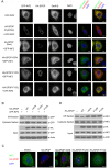
Fig 5. SPOP promotes INF2 disassociation from ER.
(A) Representative images of HeLa cells transfected with indicated plasmids, stained with SPOP(HA)and DAPI. Scale bar, 20 μm. (B) HeLa cells were transfected with indicated plasmids. Cytosol and purified ER Fractions were isolated and GFP-INF2 was detected by Western Blot. (C) Representative images of HeLa cells transfected with indicated plasmids, stained with SPOP(HA), INF2 and DAPI. (D) HeLa cells were infected with lentivirus expressing HA-SPOP (WT or mutants) or control. Cytosol and purified ER Fractions were isolated and endogenous INF2 was detected by Western Blot.
Fig 6
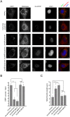
Fig 6. Wild-type SPOP increases, and the prostate cancer-associated SPOP mutants decrease, mitochondria average length.
(A) Representative images of DU145 cells infected with lentivirus expressing HA-SPOP (WT or mutants) or control. stained with INF2, SPOP(HA), Mitotracker Red and DAPI. Scale bar, 20 μm. (B) Quantification of mitochondria lengths in (A). n = 40 to 50 cells. Error bars, ± SD for triplicate. (C) DU145 cells were transfected with indicated with indicated plasmids. The mitochondria lengths were quantified similar as (A, B). (D) DU145 cells were infected with lentivirus expressing indicated shRNAs, and the mitochondrial lengths were quantified similar as (A, B).
Fig 7
Fig 7. SPOP-ΔNLS mutant is more potent in suppressing mitochondrial fission than SPOP-WT.
(A) Representative images of DU145 cells infected with lentivirus expressing HA-SPOP (WT or mutants) or control, stained with DRP1, SPOP(HA), Mitotracker Red and DAPI. (B) Quantification of DRP1 puncta per mitochondrial length in (A). (C) Quantification of mitochondria lengths in (A).
Fig 8
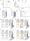
Fig 8. SPOP suppresses cell invasion and migration partially dependent on INF2.
(A) MitoSOX Red was added to DU145 cells expressing shRNAs targeting INF2 or scramble control and fluorescence was measured by flow cytometry. (B) Oxygen consumption rate was measured using an XF24 extracellular flux analyzer in DU145 cells expressing shRNA targeting INF2 or scramble control. Oligomycin, FCCP, Rotenone and Antimycin A were added at the indicated timepoints (arrows). Spare respiratory capacity is measured as the difference between basal oxygen consumption rate and the FCCP uncoupled oxygen consumption rate. (C) JC-1 fluorescent dye was added to DU145 cells expressing shRNA targeting INF2 or scramble control. For quantification, the green fluorescence intensity (representing the degree of decreased ΔΨm) was measured by flow cytometry. Data represent three replicates. n.s, not statistically significant. (D) Cell cycle analysis of DU145 cells infected with lentivirus expressing indicated shRNAs. (E) Cell growth analysis of DU145 cells infected with lentivirus expressing indicated shRNAs. (F) DU145 cells were infected with lentivirus expressing indicated shRNAs. Cell migration assay was shown on the left panel, and the quantitative analysis is shown on the right panel. All data shown are mean values ± SD (error bar) from three replicates. *p < 0.01. (G) DU145 cells were infected with lentivirus expressing indicated shRNAs for cell invasion assay. (H) DU145 cells were infected with lentivirus expressing indicated shRNAs and treated with Mdivi-1 (1 μM)for cell migration assay. (I) DU145 cells were infected with lentivirus expressing indicated shRNAs and treated with Mdivi-1 (1 μM)for cell invasion assay.
Fig 9
Fig 9. A proposed model how SPOP is a regulator of mitochondrial fission and dysregulated by prostate cancer-associated mutations.

Similar articles

Cited by

References

    1. Berger MF, Lawrence MS, Demichelis F, Drier Y, Cibulskis K, Sivachenko AY et al. The genomic complexity of primary human prostate cancer. Nature 2011; 470: 214–220. 10.1038/nature09744 - DOI - PMC - PubMed
    1. Barbieri CE, Baca SC, Lawrence MS, Demichelis F, Blattner M, Theurillat JP et al. Exome sequencing identifies recurrent SPOP, FOXA1 and MED12 mutations in prostate cancer. Nat Genet 2012; 44: 685–689. 10.1038/ng.2279 - DOI - PMC - PubMed
    1. Grasso CS, Wu YM, Robinson DR, Cao X, Dhanasekaran SM, Khan AP et al. The mutational landscape of lethal castration-resistant prostate cancer. Nature 2012; 487: 239–243. 10.1038/nature11125 - DOI - PMC - PubMed
    1. Cancer Genome Atlas Research N. The Molecular Taxonomy of Primary Prostate Cancer. Cell 2015; 163: 1011–1025. 10.1016/j.cell.2015.10.025 - DOI - PMC - PubMed
    1. Errington WJ, Khan MQ, Bueler SA, Rubinstein JL, Chakrabartty A, Privé GG. Adaptor protein self-assembly drives the control of a cullin-RING ubiquitin ligase. Structure. 2012; 20: 1141–53. 10.1016/j.str.2012.04.009 - DOI - PubMed

MeSH terms