. 2017 Apr 27;12(4):e0176371.
doi: 10.1371/journal.pone.0176371. eCollection 2017.

Characterization of physiological defects in adult SIRT6-/- mice

Affiliations

Characterization of physiological defects in adult SIRT6-/- mice

Victoria Peshti et al. PLoS One. .

Abstract

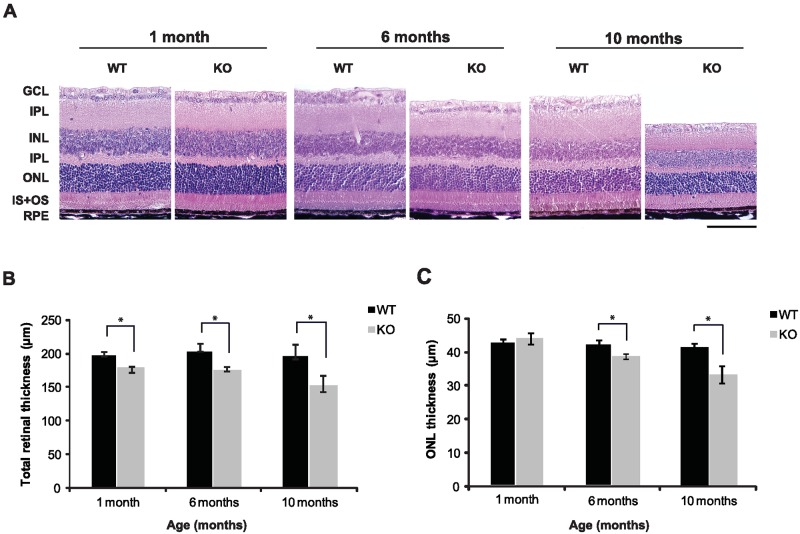
The NAD+-dependent SIRT6 deacetylase was shown to be a major regulator of lifespan and healthspan. Mice deficient for SIRT6 develop a premature aging phenotype and metabolic defects, and die before four weeks of age. Thus, the effect of SIRT6 deficiency in adult mice is unknown. Here we show that SIRT6-/- mice in mixed 129/SvJ/BALB/c background reach adulthood, allowing examination of SIRT6-related metabolic and developmental phenotypes in adult mice. In this mixed background, at 200 days of age, more than 80% of the female knock-out mice were alive whereas only 10% of male knock-out mice survived. In comparison to their wild-type littermates, SIRT6 deficient mice have reduced body weight, increased glucose uptake and exhibit an age-dependent progressive impairment of retinal function accompanied by thinning of retinal layers. Together, these results demonstrate a role for SIRT6 in metabolism and age-related ocular changes in adult mice and suggest a gender specific regulation of lifespan by SIRT6.


Conflict of interest statement

Competing Interests: The authors have declared that no competing interests exist.

Figures

Fig 1
Fig 1. Gender specific effect of SIRT6 deficiency on mouse survival.
Kaplan—Meier survival curves of WT, HET and KO male (left panel) and female (right panel) mice in 129/SvJ/BALB/c background. 90% of KO male mice died before 200 days of age and more than 75% of KO female mice survived over 300 days of age. Males, n = 23–39 per group; Females, n = 14–40 per group.
Fig 2
Fig 2. Effect of SIRT6 deficiency on body weight and adiposity.
(A) Average body weight of WT and KO male (left panel) and female (right panel) mice. In both genders, the weight of SIRT6 KO mice was significantly lower as compared with age-matched WT animals. (B) KO mice showed a trend towards lower percentage of total body fat in comparison to their WT littermates. The percentage of total body fat was measured at 6–8 months of age. Results are presented as mean ± SEM. n = 3–14 per group.
Fig 3
Fig 3. Increased glucose uptake in SIRT6 deficient mice.
Glucose tolerance test (GTT) in WT and KO male (A) and female (B) mice. Area under curve (AUC) is shown on the right for each GTT. Both male and female KO mice show increased glucose uptake in comparison to WT mice. (C) Insulin tolerance test in male (left panel) and female (right panel) mice showed no significant difference between genotypes. (D) Insulin blood levels after glucose stimulation as determined by ELISA. All assays were done at 3–4 months old. Results are presented as mean ± SEM. n = 5–14 per group. * P < 0.05, ** P < 0.01
Fig 4
Fig 4. Effect of SIRT6 deficiency on GLUT1 expression and serum IGF-1 levels.
(A) Relative expression of GLUT1 in MEF's, liver and muscle of WT and KO male mice as determined by quantitative real-time PCR. (B) Serum IGF-1 levels of WT and KO male and female mice. All measurements were done at 6–8 months of age. Results are presented as mean ± SEM. n = 5–14 per group. * P < 0.05.
Fig 5
Fig 5. Corneal changes in SIRT6 deficient mice over time.
(A-B) Photographs and histological sections of corneas in 10-month-old WT and KO mice. Corneas of normal WT mice are fully transparent, with no ingrowth of blood vessels (A, left panel). In contrast, from 6 months of age, corneal scarring and vascularization were often present in KO mice eyes (A, right panel). Histological section in KO mice (B, lower panel) shows corneal thickening, stromal edema, inflammation and scarring that are not present in WT mice of the same age (B, upper panel). B: Paraffin embedded sections stained with hematoxylin and eosin; C- Cornea; L-Lens; original magnification x10; Scale bar = 200μm.
Fig 6
Fig 6. Retinal function in SIRT6 deficient mice.
Scotopic (A-F) and photopic (G, H) full-field electroretinography (ERG) recordings may suggest accelerated aging in KO eyes. At one month of age, dark-adapted ERG responses to a series of increasing white flash intensities showed preserved a-wave amplitudes in KO as compared to WT and HET eyes (A), while a trend towards lower amplitudes was observed in the scotopic b-wave amplitudes, attaining statistical significance at 3 high intensities (B). At the age of 6 months, scotopic a-wave responses at higher intensities showed a trend towards lower amplitudes (C), and b-wave amplitudes were markedly reduced (D). At 10 months, both a- and b-wave amplitudes in KO mice were markedly reduced as compared to WT and HET mice (E, F). Light adapted 16Hz flicker responses were also markedly reduced in KO mice at 6 and 10 months (G, H). ERG responses were similar in WT and HET mice at all ages. Results are presented as mean ± SEM; n = 6–9 for WT, n = 7–8 for HET, and n = 4–5 for KO mice. * P < 0.05.
Fig 7
Fig 7. Retinal structural changes well correlated with the ERG findings.
(A) Representative retinal sections stained with hematoxylin and eosin demonstrate marked decrease of retinal thickness in KO eyes by the age of 6 months with further thinning by 10 months. Original magnification x20; Scale bar = 100μm. (B) Throughout the experiment, total retinal thickness was reduced in KO eyes as compared with WT littermates. (C) Thickness of the outer nuclear layer (ONL, containing the photoreceptor nuclei) did not differ between WT and KO mice at the age of 1 month, but by the age of 6 and 10 months, the ONL was significantly thinner in animals lacking SIRT6 activity. Results are presented as mean ± SEM; n = 5–7 for WT, n = 4–5 for KO mice. * P < 0.05.

References

    1. Tissenbaum HA, Guarente L. Increased dosage of a sir-2 gene extends lifespan in Caenorhabditis elegans. Nature. 2001;410(6825):227–30. 10.1038/35065638 - DOI - PubMed
    1. Rogina B, Helfand SL. Sir2 mediates longevity in the fly through a pathway related to calorie restriction. Proc Natl Acad Sci U S A. 2004;101(45):15998–6003. 10.1073/pnas.0404184101 - DOI - PMC - PubMed
    1. Kaeberlein M, Kirkland KT, Fields S, Kennedy BK. Sir2-independent life span extension by calorie restriction in yeast. PLoS Biol. 2004;2(9):E296 Epub 2004/08/26. 10.1371/journal.pbio.0020296 - DOI - PMC - PubMed
    1. Herranz D, Muñoz-Martin M, Cañamero M, Mulero F, Martinez-Pastor B, Fernandez-Capetillo O, et al. Sirt1 improves healthy ageing and protects from metabolic syndrome-associated cancer. Nat Commun. 2010;1:3 10.1038/ncomms1001 - DOI - PMC - PubMed
    1. Satoh A, Brace CS, Rensing N, Cliften P, Wozniak DF, Herzog ED, et al. Sirt1 extends life span and delays aging in mice through the regulation of Nk2 homeobox 1 in the DMH and LH. Cell Metab. 2013;18(3):416–30. 10.1016/j.cmet.2013.07.013 - DOI - PMC - PubMed