. 2017 Aug:12:843-853.
doi: 10.1016/j.redox.2017.04.024. Epub 2017 Apr 19.

Oral administration of methysticin improves cognitive deficits in a mouse model of Alzheimer's disease

Affiliations

Oral administration of methysticin improves cognitive deficits in a mouse model of Alzheimer's disease

Athanassios Fragoulis et al. Redox Biol. 2017 Aug.

Abstract

Introduction: There is increasing evidence for the involvement of chronic inflammation and oxidative stress in the pathogenesis of Alzheimer's disease (AD). Nuclear factor erythroid 2-related factor 2 (Nrf2) is an anti-inflammatory transcription factor that regulates the oxidative stress defense. Our previous experiments demonstrated that kavalactones protect neuronal cells against Amyloid β (Aβ)-induced oxidative stress in vitro by Nrf2 pathway activation. Here, we tested an in vivo kavalactone treatment in a mouse model of AD.

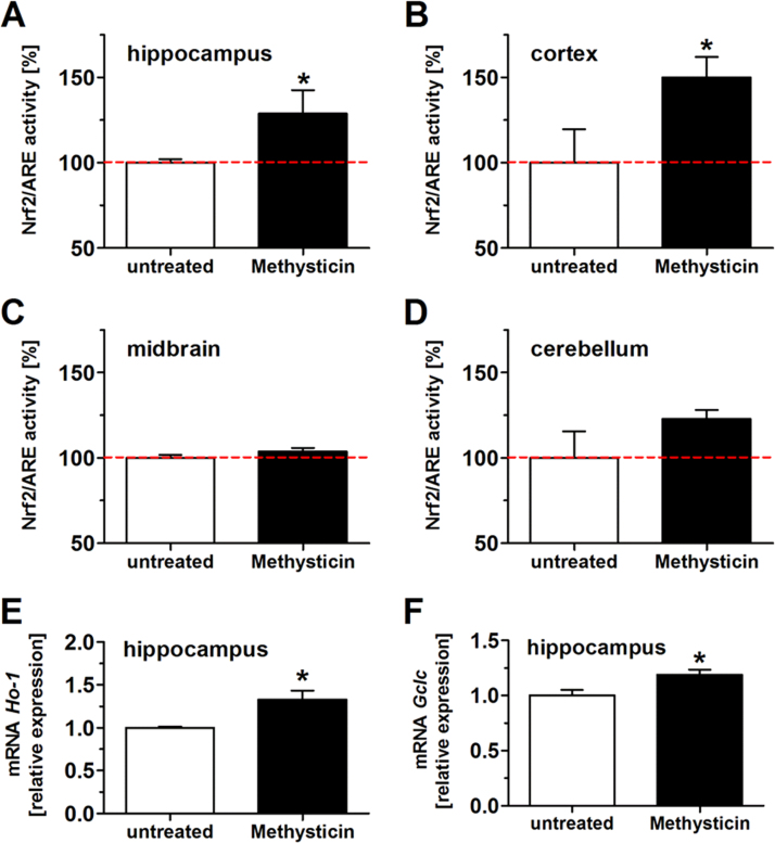
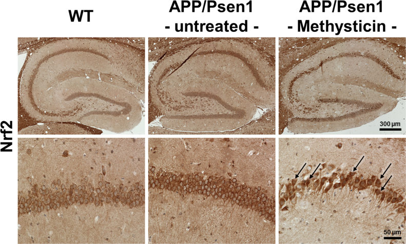
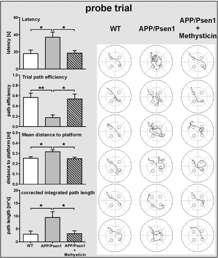
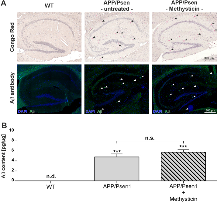
Methods: The kavalactone methysticin was administered once a week for a period of 6 months to 6 month old transgenic APP/Psen1 mice by oral gavage. Nrf2 pathway activation was measured by methysticin treatment of ARE-luciferase mice, by qPCR of Nrf2-target genes and immunohistochemical detection of Nrf2. Aβ burden was analyzed by CongoRed staining, immunofluorescent detection and ELISA. Neuroinflammation was assessed by immunohistochemical stainings for microglia and astrocytes. Pro-inflammatory cytokines in the hippocampus was determined by Luminex multi-plex assays. The hippocampal oxidative damage was detected by oxyblot technique and immunohistochemical staining against DT3 and 4-HNE. The cognitive ability of mice was evaluated using Morris water maze.

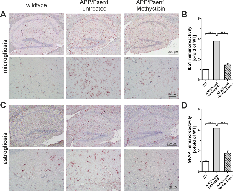
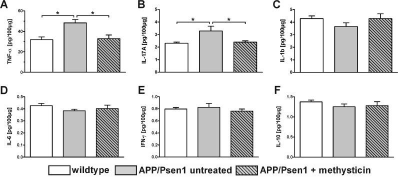
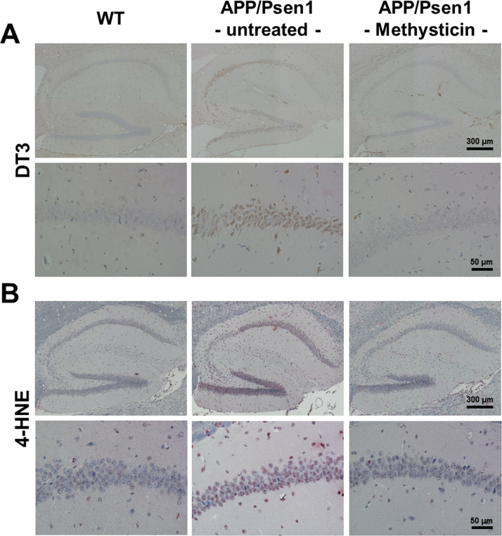
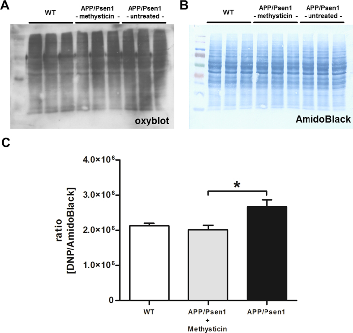
Results: Methysticin treatment activated the Nrf2 pathway in the hippocampus and cortex of mice. The Aβ deposition in brains of methysticin-treated APP/Psen1 mice was not altered compared to untreated mice. However, methysticin treatment significantly reduced microgliosis, astrogliosis and secretion of the pro-inflammatory cytokines TNF-α and IL-17A. In addition, the oxidative damage of hippocampi from APP/Psen1 mice was reduced by methysticin treatment. Most importantly, methysticin treatment significantly attenuated the long-term memory decline of APP/Psen1 mice.

Conclusion: In summary, these findings show that methysticin administration activates the Nrf2 pathway and reduces neuroinflammation, hippocampal oxidative damage and memory loss in a mouse model of AD. Therefore, kavalactones might be suitable candidates to serve as lead compounds for the development of a new class of neuroprotective drugs.

Keywords: Alzheimer's disease; Astrogliosis; Kava kava; Kavalactone; Methysticin; Neuroinflammation; Nrf2; Oxidative stress.


Figures

Fig. 1
Fig. 1
Methysticin application induced Nrf2 in the hippocampus and cortex of ARE-luciferase reporter gene mice. Nrf2 activation due to methysticin application was investigated in ARE-luciferase reporter gene mice. Animals were treated with methysticin once by oral gavage. Brains were prepared as described in the method section. Hippocampus, cortex, midbrain and cerebellum were separated and homogenized. The homogenates were used in a luminometer assay and for qRT-PCR. Methysticin significantly induced Nrf2/ARE activity in the hippocampus (A) and cortex (B) of treated animals. There was no effect on Nrf2/ARE activation in midbrain (C) and cerebellum (D). mRNA expressions of the Nrf2 target genes Ho-1 (E) and Gclc (F) were significantly increased after methysticin treatment (2-tailed unpaired t-test). Data represent mean+SEM; n =6; * p<0.05 vs. untreated.
Fig. 2
Fig. 2
Methysticin induces Nrf2 nuclear translocation in hippocampal neurons. Nrf2 activity and its cellular localization in the hippocampus was determined by IHC staining. Nrf2 was predominantly found in the cytosol of hippocampal neurons in WT and untreated APP/Psen1 mice. Methysticin treatment led to nuclear translocation of Nrf2 and thereby induced its signaling. Overview of hippocampi (upper row) and detailed depiction of CA1 region (lower row) of representative stainings are shown. n=6.
Fig. 3
Fig. 3
Schematic illustration of the Morris-Water Maze experimental design. To analyze long-term memory we performed a Morris water maze test. The apparatus measured 120 cm in diameter. To test long-term memory, latency, path efficiency, mean distance to the platform and the corrected integrated path length during the probe trial were analyzed. Track plot of each individual animal from the probe trial on day 5 are shown. APP/Psen1 mice showed a significant decline in long-term memory performance compared to age-matched wild-type littermates. Methysticin treatment rescued this effect significantly. (Statistics probe trial: one-way ANOVAs with Tukey's multiple comparisons post hoc test). Data represent mean+SEM; n=6; * p<0.05, ** p<0.01 as indicated.
Fig. 4
Fig. 4
Methysticin treatment did not significantly prevent amyloid-beta deposition in APP/Psen1 hippocampus. To visualize and quantify amyloid-beta deposition, Congo Red and immunohistochemical staining with a specific antibody against Aβ1-42 were conducted. The analyses were focused on the hippocampus. (A) Amyloid-beta plaque density in this area was analyzed by scanning the hippocampus at 20× magnification. Single pictures were merged using the BZ-II Analyzer Software (Keyence, Neu-Isenburg, Germany). Representative pictures (n=6) of wild type (1st column), untreated APP/Psen1 (2nd column) and methysticin treated (3rd column) hippocampi are shown. The first row represents the pictures of Congo Red staining and the second row the immunohistochemical staining against Aβ1-42. (B) Aβ content in the hippocampus of investigated animals was determined by ELISA. Hippocampal tissue was processed as described in the methods section and data are expressed as pg/µg total protein. (one-way ANOVA with Tukey's post hoc test). Data represent mean+SEM; n=6; *** p<0.001 vs. WT, n.d. not detectable.
Fig. 5
Fig. 5
Neuroinflammation in hippocampi of APP/Psen1 mice is reduced by methysticin treatment. (A & C) Microglia infiltration and astrogliosis was analyzed by immunohistochemical staining against Iba1and GFAP respectively (n=6). Pictures were generated by scanning the hippocampus at 20× magnification and subsequent merging of these pictures to a single picture. The upper lanes in A and C show representative pictures of wild type (1st column), untreated APP/Psen1 (2nd column) and methysticin-treated APP/Psen1 (3rd column) mouse hippocampi. The lower lanes in A and C provide detailed pictures at 40× magnification of the same groups. Scale bars: upper lanes=300 µm, lower lanes =50 µm. (B & D) Quantification of immunoreactivity of 3 consecutive sections of each hippocampus. Untreated APP/Psen1 mice showed prominent microglia infiltration and astrogliosis that was extensively elevated compared to wild type (WT) mice. Methysticin application led to a significant reduction of microglia infiltration as well as astrogliosis in these animals. (one-way ANOVA with Tukey's multiple comparison post hoc test). Data represent mean+SEM; n=6; *** p<0.001 as indicated in the chart.
Fig. 6
Fig. 6
The secretion of pro-inflammatory cytokines in hippocampi of APP/Psen1 mice is reduced by methysticin treatment. Cytokine secretion in the hippocampus was analyzed by Luminex Multi-plex assay. Hippocampal tissue was processed and the assay was conducted as described in the methods section. Cytokine secretion is expressed as pg/100 µg total protein. The pro-inflammatory cytokines TNF-α (A) and IL-17A (B) were significantly increased in untreated APP/Psen1 animals compared to the age-matched wild type control animals. Methysticin treatment in turn prevented this effect. The other investigated inflammatory mediators (IL-1b, IL-6, IFN-γ and IL-10) did not differ between the groups (C – F). (one-way ANOVA with Tukey's multiple comparison post hoc test). Data represent mean+SEM; n=6; * p<0.05 as indicated.
Fig. 7
Fig. 7
Methysticin reduces oxidative stress mediated damage. Oxidative damage to amino acids was analyzed by immunohistochemistry using antibodies against DT3 (A) and 4-HNE (B). The elevated oxidative protein modification induced by the APP/Psen1-transgene was significantly reduced by methysticin treatment. n=6.
Fig. 8
Fig. 8
Methysticin reduces oxidative stress mediated damage. Oxidative damage to amino acids was additionally analyzed by oxyblot technique. The oxyblot using an DNP-specific antibody (A) and the corresponding loading control by AmidoBlack staining (B) is shown. Densitometry revealed increased protein carbonylation in untreated APP/Psen1 mice compared to WT littermates. Methysticin treatment reduced protein carbonylation to WT levels. (one-way ANOVA with Tukey's multiple comparison post hoc test). Data represent mean+SEM; n=6; * p<0.05 as indicated.

Similar articles

Cited by

References

    1. Rodriguez J.J., Verkhratsky A. Neuroglial roots of neurodegenerative diseases? Mol. Neurobiol. 2011;43:87–96. - PubMed
    1. Mandrekar-Colucci S., Landreth G.E. Microglia and inflammation in Alzheimer's disease. CNS Neurol. Disord. Drug Targets. 2010;9:156–167. - PMC - PubMed
    1. Wruck C.J., Gotz M.E., Herdegen T., Varoga D., Brandenburg L.O., Pufe T. Kavalactones protect neural cells against amyloid beta peptide-induced neurotoxicity via extracellular signal-regulated kinase 1/2-dependent nuclear factor erythroid 2-related factor 2 activation. Mol. Pharmacol. 2008;73:1785–1795. - PubMed
    1. Joshi G., Johnson J.A. The Nrf2-ARE pathway: a valuable therapeutic target for the treatment of neurodegenerative diseases. Recent Pat. CNS Drug Discov. 2012;7:218–229. - PMC - PubMed
    1. Wruck C.J., Claussen M., Fuhrmann G., Romer L., Schulz A., Pufe T., Waetzig V., Peipp M., Herdegen T., Gotz M.E. Luteolin protects rat PC12 and C6 cells against MPP+ induced toxicity via an ERK dependent Keap1-Nrf2-ARE pathway. J. Neural Transm. Suppl. 2007:57–67. - PubMed

Publication types

MeSH terms

LinkOut - more resources