. 2018 Jun 1;28(6):1946-1958.
doi: 10.1093/cercor/bhx101.

Secretagogin is Expressed by Developing Neocortical GABAergic Neurons in Humans but not Mice and Increases Neurite Arbor Size and Complexity

Affiliations

Secretagogin is Expressed by Developing Neocortical GABAergic Neurons in Humans but not Mice and Increases Neurite Arbor Size and Complexity

Chandrasekhar S Raju et al. Cereb Cortex. .

Abstract

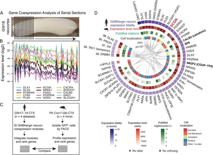
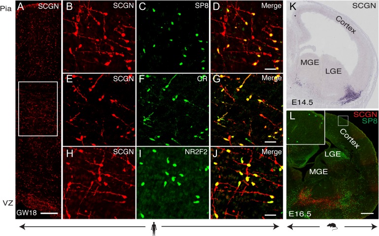
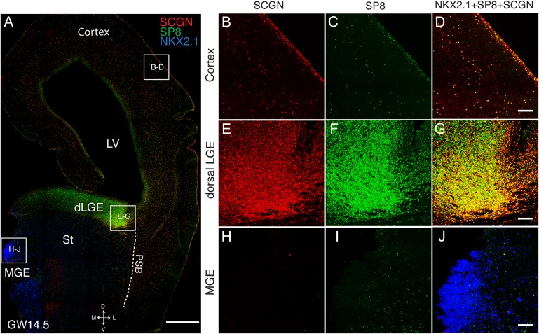
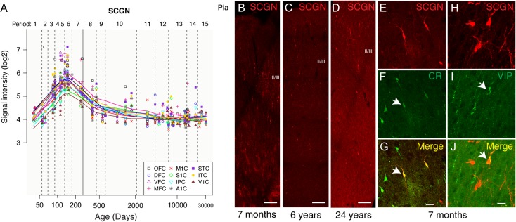
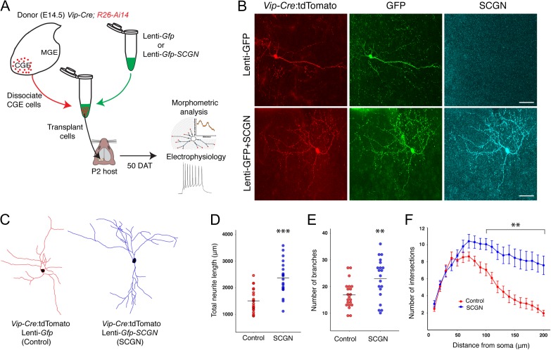
The neocortex of primates, including humans, contains more abundant and diverse inhibitory neurons compared with rodents, but the molecular foundations of these observations are unknown. Through integrative gene coexpression analysis, we determined a consensus transcriptional profile of GABAergic neurons in mid-gestation human neocortex. By comparing this profile to genes expressed in GABAergic neurons purified from neonatal mouse neocortex, we identified conserved and distinct aspects of gene expression in these cells between the species. We show here that the calcium-binding protein secretagogin (SCGN) is robustly expressed by neocortical GABAergic neurons derived from caudal ganglionic eminences (CGE) and lateral ganglionic eminences during human but not mouse brain development. Through electrophysiological and morphometric analyses, we examined the effects of SCGN expression on GABAergic neuron function and form. Forced expression of SCGN in CGE-derived mouse GABAergic neurons significantly increased total neurite length and arbor complexity following transplantation into mouse neocortex, revealing a molecular pathway that contributes to morphological differences in these cells between rodents and primates.


Figures

Figure 1.
Figure 1.
GCASS identifies a transcriptional signature of GABAergic neurons in prenatal human neocortex. (A) A GW18 human neocortical specimen was analyzed by GCASS (n = 87 sections). (B) Expression patterns of the top 15 members of the green coexpression module over all sections. (C) Schematic of strategy for comparing gene expression in GABAergic neurons between humans and mice. (D) Top genes ranked by consensus expression fidelity for GABAergic neurons in GW17-19 human neocortex (blue track). Red track denotes expression level rank in purified GABAergic neurons from P0 Cxcr7-Gfp mice. PubMed citations were obtained by queries with gene symbol and “GABAergic” or “interneuron”. Cellular localizations are from COMPARTMENTS and PPI are from STRING (Methods).
Figure 2.
Figure 2.
Neocortical GABAergic neurons express SCGN during human but not mouse brain development. (A) Immunostaining for SCGN in GW18 human neocortex. (BJ) Magnified images of boxed area in (A) (see Supplementary Fig. 2F,I,L for more detailed localization). (K and L) In situ hybridization for Scgn (K; http://www.eurexpress.org/ee/) and immunostaining (L) for SCGN and SP8 in embryonic mouse brain. Scale bars: 200 μm (A, K, L); 50 μm (BJ).
Figure 3.
Figure 3.
SCGN+ GABAergic neurons are highly concentrated in the dLGE and excluded from the MGE in prenatal human brain. (A) Coronal section of GW14.5 human telencephalon immunostained for SCGN, SP8, and NKX2.1. LV, lateral ventricle; St, striatum; PSB, pallial–subpallial boundary. (B–J) Magnified images of boxed areas in (A). Scale bars: 1 mm (A); 50 μm (BJ).
Figure 4.
Figure 4.
Expression of SCGN peaks before birth and preferentially labels interneurons that populate superficial layers of human neocortex. (A) Expression patterns in 11 neocortical areas across the human lifespan (Kang et al. 2011) suggest a role for SCGN during prenatal brain development (black vertical line denotes birth). (BD) Immunostaining for SCGN in human prefrontal cortex (7 months), superior frontal gyrus (6 years), and entorhinal cortex (24 years). (EJ) SCGN+ neurons in human prefrontal cortex (7 months) acquire bipolar/bitufted morphology and coexpress CR and VIP. Scale bars: 150 μm (BD); 50 μm (EG); 30 μm (HJ).
Figure 5.
Figure 5.
Forced expression of SCGN increases neurite length and arbor complexity in CGE-derived mouse GABAergic neurons. (A) Schematic of transplantation experiment. CGE cells from donor mice expressing tdTomato exclusively in VIP+ neurons (Vip-Cre; R26-Ai14) were harvested at E13.5, transduced with lentivirus, and transplanted into P2 recipient visual cortex. (B) Cortical sections from recipient mice were immunostained for GFP and SCGN 50 days after transplantation to confirm SCGN expression in transplanted cells. (C) Representative Neurolucida reconstructions of transplanted neurons expressing SCGN or empty vector (control). (DF) Comparison of total neurite length (D), number of branches (E), and number of intersections with concentric spheres drawn at 10 μm radius intervals from the soma via Sholl analysis (F). P-values are from Student’s t-test (D, E) and Wilcoxon rank-sum test (F; performed separately for each distance): **P < 0.01; ***P < 0.001. Neurolucida tracings are from n = 21 (control) and n = 20 (SCGN) cells (4 animals per condition, minimum 5 cells per animal). Error bars: one SEM. Scale bars: 50 μm (B).

References

    1. Al-Jaberi N, Lindsay S, Sarma S, Bayatti N, Clowry GJ. 2015. The early fetal development of human neocortical GABAergic interneurons. Cereb Cortex. 25:631–645. - PMC - PubMed
    1. Alpar A, Attems J, Mulder J, Hokfelt T, Harkany T. 2012. The renaissance of Ca2+-binding proteins in the nervous system: secretagogin takes center stage. Cell Signal. 24:378–387. - PMC - PubMed
    1. Barbosa-Morais NL, Dunning MJ, Samarajiwa SA, Darot JF, Ritchie ME, Lynch AG, Tavare S. 2010. A re-annotation pipeline for Illumina BeadArrays: improving the interpretation of gene expression data. Nucleic Acids Res. 38:e17. - PMC - PubMed
    1. Barinka F, Magloczky Z, Zecevic N. 2015. Editorial: at the top of the interneuronal pyramid-calretinin expressing cortical interneurons. Front Neuroanat. 9:108. - PMC - PubMed
    1. Bayatti N, Moss JA, Sun L, Ambrose P, Ward JF, Lindsay S, Clowry GJ. 2008. A molecular neuroanatomical study of the developing human neocortex from 8 to 17 postconceptional weeks revealing the early differentiation of the subplate and subventricular zone. Cereb Cortex. 18:1536–1548. - PMC - PubMed

Publication types