. 2017 Jul 1;26(13):2541-2550.
doi: 10.1093/hmg/ddx152.

A homozygous missense mutation in ERAL1, encoding a mitochondrial rRNA chaperone, causes Perrault syndrome

Affiliations

A homozygous missense mutation in ERAL1, encoding a mitochondrial rRNA chaperone, causes Perrault syndrome

Iliana A Chatzispyrou et al. Hum Mol Genet. .

Abstract

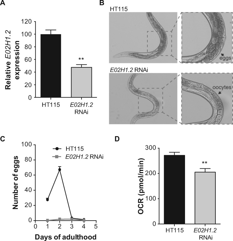
Perrault syndrome (PS) is a rare recessive disorder characterized by ovarian dysgenesis and sensorineural deafness. It is clinically and genetically heterogeneous, and previously mutations have been described in different genes, mostly related to mitochondrial proteostasis. We diagnosed three unrelated females with PS and set out to identify the underlying genetic cause using exome sequencing. We excluded mutations in the known PS genes, but identified a single homozygous mutation in the ERAL1 gene (c.707A > T; p.Asn236Ile). Since ERAL1 protein binds to the mitochondrial 12S rRNA and is involved in the assembly of the small mitochondrial ribosomal subunit, the identified variant represented a likely candidate. In silico analysis of a 3D model for ERAL1 suggested that the mutated residue hinders protein-substrate interactions, potentially affecting its function. On a molecular basis, PS skin fibroblasts had reduced ERAL1 protein levels. Complexome profiling of the cells showed an overall decrease in the levels of assembled small ribosomal subunit, indicating that the ERAL1 variant affects mitochondrial ribosome assembly. Moreover, levels of the 12S rRNA were reduced in the patients, and were rescued by lentiviral expression of wild type ERAL1. At the physiological level, mitochondrial respiration was markedly decreased in PS fibroblasts, confirming disturbed mitochondrial function. Finally, knockdown of the C. elegans ERAL1 homologue E02H1.2 almost completely blocked egg production in worms, mimicking the compromised fertility in PS-affected women. Our cross-species data in patient cells and worms support the hypothesis that mutations in ERAL1 can cause PS and are associated with changes in mitochondrial metabolism.


Figures

Figure 1
Figure 1
Three individuals diagnosed with PS, carry the same homozygous missense mutation in the ERAL1 gene. (A) The mutated residue is highly conserved as evidenced by sequence alignment using the Alamut software. (B) Pedigrees of the two initial PS patients (indicated by P1 and P2, as annotated in the remainder of the paper) in whom WES was performed and skin fibroblasts were analyzed. Individuals that were sequenced for the mutation are indicated with asterisks, and the results are annotated (wt: wildtype ERAL1, mut: mutated ERAL1). (C) Pedigree (left panel) and sequencing results (right panel) of a third PS patient that was found homozygous for the same mutation. (D) Left panel: Structural Alignment of modelled human ERAL1 (gray) and crystallized Aquifex aeolicus ERA (dark blue), showing the high similarity between the two structures. Ligands of ERA are shown in yellow. Middle panel: Active center of modelled wild type ERAL1 in complex with a non-hydrolizable GTP analog (GNP, phosphoaminophosphonic acid-guanylate ester) (yellow sticks) and magnesium (yellow sphere). Hydrogen bonds between asparagine 236 (magenta sticks), GNP and alanines 124 and 310 (cyan) are shown as red dashes. Right panel: Active center of modelled N236I mutated ERAL1 in complex with GNP (yellow sticks) and magnesium (yellow sphere), showing that no interactions can be made between the mutated amino acid, the ligand and the two flanking alanines (cyan). (E) Western blot of skin fibroblasts from PS patients and controls; both PS patients present with decreased ERAL1 protein levels. P1: PS patient 1, P2: PS patient 2. (F) Bar graph depicting the levels of ERAL1 in patient and control fibroblasts normalized to tubulin, as quantified from the blot in panel E.
Figure 2
Figure 2
Cells from PS patients show defected assembly of the small 28S mitochondrial ribosomal subunit. (A-B) Heat map representation of migration profiles in blue native gels of proteins of the small 28S (A) and large 39S (B) mitochondrial ribosomal subunits isolated from PS patient and control skin fibroblasts. (C-D) Graphs depicting the average normalized relative abundance of proteins of the 28S (C) and 39S (D) mitochondrial ribosomal subunits spanning the blue native gel. Protein abundance was determined by label-free quantitation using the composite iBAQ intensity values determined by MaxQuant (31) and normalized as in (3030) considering multiple migration profiles of individual proteins, that is taking into account iBAQ values from all 180 gel slices (60 slices per sample). Both patients show decreased levels of assembled small mitochondrial ribosomal subunit (A, C), while the levels of assembled large subunit remain unaffected (B, D). P1: patient 1, P2: patient 2. (E) Western blot of skin fibroblasts from PS patients and controls; both PS patients present with decreased MRPS22 protein levels, while MRPL54 levels are unaffected. (F) Bar graphs depicting the levels of MRPS22 (left) and MRPL54 (right) in patient and control fibroblasts normalized to tubulin, as quantified from the blot in panel E. Data are represented as the mean±SEM. *p<0.05 as calculated by a 2-tailed student’s t-test.
Figure 3
Figure 3
12S rRNA levels are decreased in PS cells and are rescued after ectopic ERAL1 expression. (A) The 12S to 16S rRNA ratio in control and PS fibroblasts as measured by qPCR. Both patients present with significantly lower 12S/16S rRNA ratio compared to controls. Data are represented as the mean±SEM of six biological replicates. **p<0.01, ***p<0.001 as calculated by a one-way ANOVA Tukey's multiple comparisons test. (B) Western blot from PS fibroblasts infected by GFP or ERAL1 overexpressing lentivirus. Patient cells infected with ERAL1 overexpressing lentivirus stably express ERAL1 protein compared to those infected by a GFP overexpressing virus. Quantification of the levels of ERAL1 normalized to tubulin are given below the blot. (C) 12S to 16S rRNA ratio in PS fibroblasts stably expressing ERAL1 or GFP. Patient cells stably expressing ERAL1 present a significantly increased 12S/16S rRNA ratio compared to those expressing GFP, that almost reaches the levels of healthy cells (panel A). Data are represented as the mean±SEM of six biological replicates. *p<0.05, **p<0.01 as calculated by a 2-tailed student’s t-test.
Figure 4
Figure 4
Cells from PS patients show impaired mitochondrial function. (A) Western blot from PS patient and control skin fibroblasts; both patients present with decreased MT-CO1 protein levels, while SDHA levels are unaffected. P1: PS patient 1, P2: PS patient 2. (B) Histogram depicting the MT-CO1 to SDHA ratio, as quantified from a western blot with samples depicted in panel A, in biological duplicates. Data are represented as the mean±SEM. **p<0.01 as calculated by a 2-tailed student’s t-test. (C) Seahorse respirometry on PS patient and control skin fibroblasts; both PS patients have reduced basal and maximal respiration. Injections of compounds during measurement are indicated with arrows. OCR: oxygen consumption rate, P1: PS patient 1, P2: PS patient 2.
Figure 5
Figure 5
Knockdown of the ERAL1 worm homologue E02H1.2 compromises fecundity and reduces respiration in C. elegans. (A) mRNA levels of knockdown efficiency of E02H1.2 in worms fed with control HT115 and E02H1.2 RNAi as measured by qPCR. Worms fed with E02H1.2 RNAi present with 50% reduction of E02H1.2 gene expression. Data are represented as the mean±SEM of biological triplicates. **p<0.01 as calculated by a 2-tailed student’s t-test. (B) Wide-field images of adult (day 1) worms fed with control HT115 (upper panels) and E02H1.2 (lower panels) RNAi; worms with E02H1.2 knockdown do not carry any eggs, in contrast to the controls. (C) Graph showing the number of eggs laid over the first four days of adulthood. Worms with E02H1.2 knockdown laid almost no eggs compared to the controls. Egg-laying of 30 individual worms was followed. Error bars correspond to SEM. (D) Basal respiration measured on worms fed with control (HT115) or E02H1.2 (ERAL1) RNAi. Worms with E02H1.2 knockdown have significantly (p < 0.01) decreased basal respiration compared to the controls. n = 8. Error bars correspond to SEM.

Similar articles

Cited by

References

    1. Perrault M., Klotz B., Housset E. (1951) [Two cases of Turner syndrome with deaf-mutism in two sisters]. Bull. Mem. Soc. Med. Hop. Paris, 67, 79–84. - PubMed
    1. Nishi Y., Hamamoto K., Kajiyama M., Kawamura I. (1988) The Perrault syndrome: clinical report and review. Am. J. Med. Genet., 31, 623–629. - PubMed
    1. Gottschalk M.E., Coker S.B., Fox L.A. (1996) Neurologic anomalies of Perrault syndrome. Am. J. Med. Genet., 65, 274–276. - PubMed
    1. Fiumara A., Sorge G., Toscano A., Parano E., Pavone L., Opitz J.M. (2004) Perrault syndrome: evidence for progressive nervous system involvement. Am. J. Med. Genet. A, 128A, 246–249. - PubMed
    1. Pierce S.B., Walsh T., Chisholm K.M., Lee M.K., Thornton A.M., Fiumara A., Opitz J.M., Levy-Lahad E., Klevit R.E., King M.C. (2010) Mutations in the DBP-deficiency protein HSD17B4 cause ovarian dysgenesis, hearing loss, and ataxia of Perrault Syndrome. Am. J. Hum. Genet., 87, 282–288. - PMC - PubMed

Publication types

MeSH terms

Supplementary concepts