Review
. 2017 Jul 31;75(5):ftx046.
doi: 10.1093/femspd/ftx046.

Functions of galectins as 'self/non-self'-recognition and effector factors

Affiliations
Review

Functions of galectins as 'self/non-self'-recognition and effector factors

Gerardo R Vasta et al. Pathog Dis. .

Abstract

Carbohydrate structures on the cell surface encode complex information that through specific recognition by carbohydrate-binding proteins (lectins) modulates interactions between cells, cells and the extracellular matrix, or mediates recognition of potential microbial pathogens. Galectins are a family of ß-galactoside-binding lectins, which are evolutionary conserved and have been identified in most organisms, from fungi to invertebrates and vertebrates, including mammals. Since their discovery in the 1970s, their biological roles, initially understood as limited to recognition of endogenous carbohydrate ligands in embryogenesis and development, have expanded in recent years by the discovery of their roles in tissue repair and regulation of immune homeostasis. More recently, evidence has accumulated to support the notion that galectins can also bind glycans on the surface of potentially pathogenic microbes, and function as recognition and effector factors in innate immunity, thus establishing a new paradigm. Furthermore, some parasites 'subvert' the recognition roles of the vector/host galectins for successful attachment or invasion. These recent findings have revealed a striking functional diversification in this structurally conserved lectin family.

Keywords: carbohydrate recognition domain: glycans; chimera; function; galectins, ß-galactoside; pattern recognition receptors; proto-type; structure; tandem repeat.


Figures

Figure 1.
Figure 1.
Structural aspects of mammalian galectins. (A) Schematic display of three types of mammalian galectins. (B) Structure of the galectin-1 in complex with LacNAc (indicated by the white box) at 1.9 Å resolution. (C) Carbohydrate-binding site of galectin-1. Three continuous concave strands (S4–S6) contain all residues involved in direct interactions with LacNAc, plus additional interactions with His52, Asp54 and Arg73 bridged by a water molecule. (D) Schematic representation of interactions between amino acid residues of galectin-1 and LacNAc. (Adapted from Liao et al. and Bianchet et al.2000).
Figure 2.
Figure 2.
Multiple functions of galectins. Galectins are synthesized in the cytosol (A), and some can be translocated into the nucleus and interact with RNP particles (B). Most galectins are transported and secreted to the extracellular space (C), where they interact with glycans in the ECM (D), bridge the cell surface ligands (E) or function as PRRs by recognizing microbial pathogens (F).
Figure 3.
Figure 3.
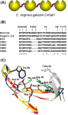
Structural aspects of galectins from invertebrates. (A) Schematic representation of the CRD organization of the galectin of CvGal1 from eastern oyster (C. virginica). (B) Sequence alignment of the carbohydrate-binding regions from bovine galectin-1, zebrafish Drgal1-L2, C. elegans N16 and the CRDs-1 to -4 of CvGal1. (C) Homology modeling of the CRDs from CvGal1: bovine galectin-1(white); CvGal1: CRD-1, -2, -3 and -4 are shown in blue, yellow, red and green, respectively. Numbering of amino acid residues is based on bovine galectin-1. The solid arrow shows loop 4 of CvGal1 CRD-2, 3 and 4, whereas a dashed arrow shows loop 4 of CRD-1. (Adapted from Tasumi and Vasta and Feng et al. 2013).
Figure 4.
Figure 4.
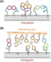
Recognition of ‘self’ and ‘non-self’ glycans by galectins. (A) In the extracellular space, galectins form multivalent oligomers that cross-link cell surface glycoproteins and glycolipids, form microdomains and activate signaling pathways. (B) Proto, chimera and tandem-repeat galectins can function as PRRs and establish transinteractions with the host cell surface and microbial glycans.
Figure 5.
Figure 5.
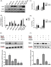
Galectins and microbial neuraminidases modulate bacterial adhesion and expression of SOCS1 and RIG1. (A and B) Mice were challenged with a sublethal dose of influenza PR8 strain (PR8) followed by a S. pneumoniae (Sp3) challenge 14 days afterwards. (A) Transcript levels of galectin-1 (Gal1) and galectin-3 (Gal3) were assessed by RT-PCR in broncho-alveolar lavage collected from control (unchallenged) or challenged groups of mice at indicated time points. (B) Bar graphs represent fold change of galectin transcription compared to control mice after normalized to β-actin. (C) A549 cells were treated with neuraminidases from Arthrobacter ureafaciens and Clostridium perfringens (Neu K) or S. pneumoniae neuraminidase (Neu Sp), and subsequently incubated with S. pneumoniae type 3 (Sp3, MOI 10) in medium (Sp) or with either 15 μg/ml of exogenous rhGal1 (Sp + rhGal1) or rhGal3 (Sp + rhGal3). The bound bacteria were released in water and quantified after 24 h incubation on 5% sheep's blood agar plates by counting colony-forming units (CFU). The fold changes of CFU from galectin-mediated Sp adhesion compared to that without exogenous galectin are shown. (D) A549 cells were exposed to PR8 (MOI 5) for 72 h (A549+PR8) or medium only (A549), and then incubated with Sp3 (MOI 10) in medium (Sp) or with either 15 μg/ml of exogenous rhGal1 (Sp + rhGal1) or rhGal3 (Sp + rhGal3) for bacterial adhesion, which was determined and shown as described above. (E and F) Total RNA was extracted from A549 control (Ctrl) or neuraminidase (A. ureafaciens and C. perfringens) treated cells (NeuK) incubated in presence or absence of 15 μg/ml exogenous rhGal1 or rhGal3 for 1 h. (E) SOCS1 transcript level was analyzed by RT-PCR. (F) RIG1 transcript levels were analyzed by RT-PCR. Bar graphs show the fold change in mRNA expression levels in neuraminidase-treated cells as well as galectin-treated cells in comparison with control cell without neuraminidase and galectin treatment (Ctrl) after normalized to β-actin. In all studies, representative data from at least three independent experiments are shown. *P < 0.05; **P < 0.001, non-paired Student's t test. (Adapted from Nita-Lazar et al. 2015a,b).

Similar articles

Cited by

References

    1. Ahmed H. Promoter methylation in prostate cancer and its application for the early detection of prostate cancer using serum and urine samples. Biomark Cancer 2010;2:17–33. - PMC - PubMed
    1. Ahmed H, Banerjee PP, Vasta GR. Differential expression of galectins in normal, benign and malignant prostate epithelial cells: silencing of galectin-3 expression in prostate cancer by its promoter methylation. Biochem Bioph Res Co 2007;358:241–6. - PubMed
    1. Ahmed H, Bianchet MA, Amzel LM et al. . Novel carbohydrate specificity of the 16-kDa galectin from Caenorhabditis elegans: binding to blood group precursor oligosaccharides (type 1, type 2, Talpha, and Tbeta) and gangliosides. Glycobiology 2002;12:451–61. - PubMed
    1. Ahmed H, Cappello F, Rodolico V et al. . Evidence of heavy methylation in the galectin 3 promoter in early stages of prostate adenocarcinoma: development and validation of a methylated marker for early diagnosis of prostate cancer. Transl Oncol 2009;2:146–56. - PMC - PubMed
    1. Ahmed H, Du S-J, O’Leary N et al. . Biochemical and molecular characterization of galectins from zebrafish (Danio rerio): notochord-specific expression of a prototype galectin during early embryogenesis. Glycobiology 2004;14:219–32. - PubMed

Publication types

LinkOut - more resources