. 2017 Jul 7;45(12):7382-7400.
doi: 10.1093/nar/gkx303.

Two ribosome recruitment sites direct multiple translation events within HIV1 Gag open reading frame

Affiliations

Two ribosome recruitment sites direct multiple translation events within HIV1 Gag open reading frame

Jules Deforges et al. Nucleic Acids Res. .

Erratum in

Abstract

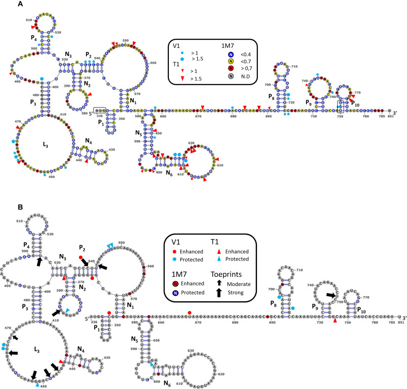
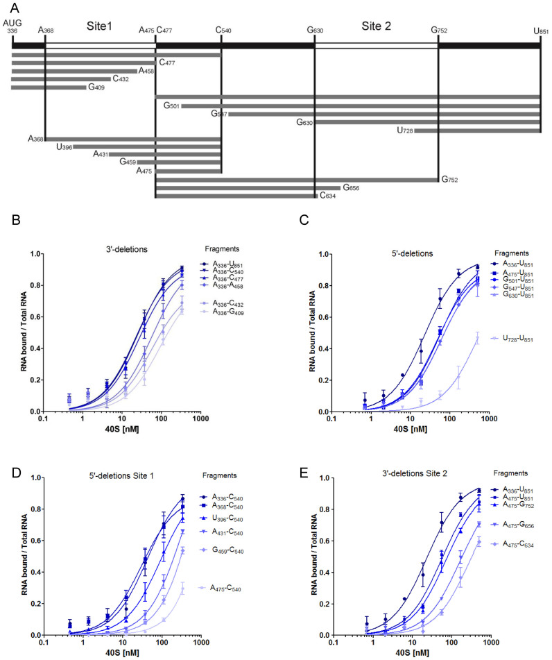
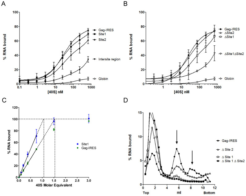
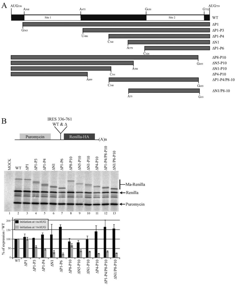
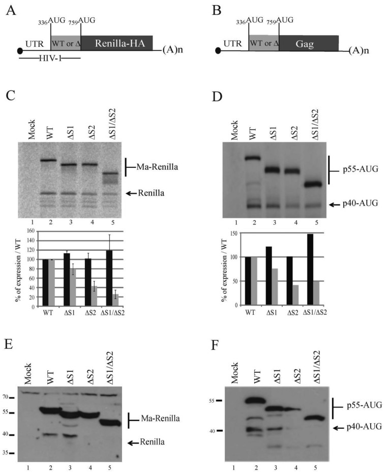
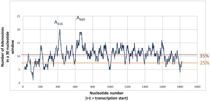
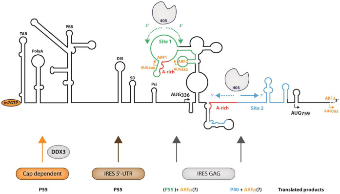
In the late phase of the HIV virus cycle, the unspliced genomic RNA is exported to the cytoplasm for the necessary translation of the Gag and Gag-pol polyproteins. Three distinct translation initiation mechanisms ensuring Gag production have been described with little rationale for their multiplicity. The Gag-IRES has the singularity to be located within Gag ORF and to directly interact with ribosomal 40S. Aiming at elucidating the specificity and the relevance of this interaction, we probed HIV-1 Gag-IRES structure and developed an innovative integrative modelling strategy to take into account all the gathered information. We propose a novel Gag-IRES secondary structure strongly supported by all experimental data. We further demonstrate the presence of two regions within Gag-IRES that independently and directly interact with the ribosome. Importantly, these binding sites are functionally relevant to Gag translation both in vitro and ex vivo. This work provides insight into the Gag-IRES molecular mechanism and gives compelling evidence for its physiological importance. It allows us to propose original hypotheses about the IRES physiological role and conservation among primate lentiviruses.


Figures

Figure 1.
Figure 1.
RNA secondary structure model of the HIV1 Gag-IRES and 40S ribosome subunit footprints and toeprints. (A) Schematic representation of the secondary structure model of Gag-IRES. Nucleotides are colored according to their reactivity toward 1M7 as indicated in the box. Red triangles and blue dots represent RNAse T1 and V1 cleavages. Pairings numbering (Pn and Nn) are as described in the text. Nucleotide numbering is from the +1 of transcription (First nucleotide of TAR). Experimental values result from the mean of three independent experiments (see material and methods and supplementary material). (B) Footprints and toeprints of the 40S ribosomal subunit on Gag-IRES. As indicated in the box, the colors and signs compare the reactivities without and with saturating concentration of 40S ribosomal subunit. Nucleotides in red are more reactive to 1M7, while nucleotides in blue are less. Red and Blue triangles indicate nucleotides exposed or protected to RNAse T1 respectively. Red and Blue dots indicate nucleotides exposed or protected to RNAse V1 respectively. Black Arrows indicate premature RT stops observed in the presence of 40S ribosomal subunits. Small and big arrows are respectively moderate and strong toeprints. Experimental values result from the mean of three independent experiments (see material and methods and supplementary material).
Figure 2.
Figure 2.
Mapping the 40S ribosomal subunit on HIV1 Gag-IRES using truncated fragments of the IRES. (A) Schematic representation of the fragments used in this study. Nucleotides numbering is from the +1 of transcription (first nucleotide of Tar). (B–E) Binding curves of 32P-labeled fragments to purified 40S ribosomal subunits as measured by filter binding assays (see Materials and Methods). Fragments were obtained by successive deletions from the 3΄ (B) and the 5΄ (C) end of Gag-IRES, or from the 5΄ of A336–C540 fragment (D) or by the 3΄ of the C477–U851 fragment (E). The results are the mean of at least three independent experiments ± s.e.m.
Figure 3.
Figure 3.
Two sites within the Gag IRES can independently recruit the 40S ribosomal subunit. (A) Binding curves of 32P-labeled Site 1 (▪, G367–U486), Site 2 (▴, G630–A751), full length HIV-1 Gag-IRES (•, A336–U851), the intersite region (▾), and the globin transcript (○) with purified 40S ribosomal subunits. (B) Binding curves for HIV-1 Gag-IRES deleted of site 1 (Δ Site 1: A336–U851Δ363–485), of site 2 (Δ Site 2: A336-U851Δ632–756), or of both sites (Δ Site 1 Δ Site 2: A336–U851Δ363–485Δ632–756) as compared to the full length HIV-1 Gag-IRES (A336–U851), and to the globin transcript. (C) Determination of the stoichiometry of the 40S ribosomal subunit and the Gag-IRES RNA. A constant concentration of RNA corresponding to the whole IRES (▪) or to the isolated site 1 (G367–U486 •), tenfold over the Kd of the Gag-IRES/40S complex (250 nM) was incubated with increasing concentration of 40S ribosome. The proportion of RNA in complex in the mix was determined by filter binding assay. The % of complex was plotted as a function of [RNA] equivalent of 40S, the linear part of the curve was calculated drawn, and extrapolated to 100% binding. The values are the mean of at least three independent experiments ± standard deviation. The two curves are statistically different (P < 10-4). (D) Analysis of the complexes formed by the full length HIV-1 Gag-IRES deleted of site 1 (Δ Site 1: A336–U851Δ363–485), of site 2 (Δ Site 2: A336–U851Δ632–756), or of both sites (Δ Site 1 Δ Site 2: A336–U851Δ363–485Δ632–756) and the full length HIV-1 Gag-IRES (A336–U851) with the 40S ribosomal subunit. 32P-labeled RNA transcripts were separated on 10–30% sucrose gradients. Peaks were identified by comparison with UV profiles obtained with purified 40S ribosomal subunits.
Figure 4.
Figure 4.
The presence of the ribosome binding sites influence IRES driven HIV-1 Gag translation in vitro. (A) Schematic representation of the fragments of the Gag coding region IRES used as intergenic region in the in vitro bicistronic translation assay (nucleotides numbering are from the +1 of transcription). (B) Schematic representation of the bicistronic construct. The first gene encodes for the puromycin N-acetyl transferase. In the intergenic region are cloned the Gag IRES fragments with 759AUG fused to the HA-tagged Renilla luciferase open reading frame (upper panel). Translation of the second gene from 336AUG yields a protein which is a fusion of the Gag Matrix sequence with the Renilla Luciferase (Ma-Renilla), while initiation from 759AUG yields the Renilla luciferase from. RRL was programmed for 45 min at 30°C with bicistronic mRNAs that contain in the intergenic region the full length Gag IRES (nt.331 to nt.761) or the different truncated fragments described in panel A (lanes 2–13), or with water (lane 1). Protein products were resolved by 12% SDS-PAGE and quantified using with a PhosphorImager. Expression initiated at both 336AUG and 759AUG codons was normalized to wild type expression and to the puromycin; Black and grey squares of the histograms represent the quantification of translation initiated at 336AUG (p55) and 759AUG (p40) codons respectively. Data are representative of three independent experiments, error bars are ± the S.E.M.
Figure 5.
Figure 5.
The presence of the ribosome binding sites influences the translation of HIV1 Gag short isoform both in vitro and ex vivo. Schematic representation of the monocitronic mRNA encoding for (A) the HA-tagged renilla luciferase reporter gene under controlled of the HIV-1 sequence from the +1 of transcription up to the 759AUG codon, or (B) the HIV-1 Gag mRNA starting from the +1 of transcription and ending at the stop codon of Gag open reading frame. Both initiation codons are indicated. HIV-1-Renilla (C) or UTR-GAG (D) mRNAs (200 fmol) containing either the Full length Gag IRES (lane 2), or fragments of the Gag IRES (Δsite 1 (Δ363–485), Δsite 2 (Δ632–756) and Δsite 1/Δ site 2 (Δ363–485Δ632–756); lanes 3–5) were translated in RRL during 45 min at 30°C. Protein products were analyzed and quantified as previously (Figure 4). Jurkat T-cells were transfected with mRNAs coding with the HA-tagged renilla luciferase constructs (lanes 2–5) (E) or the constructs carrying HIV-1 5’UTR and Gag ORF (lanes 2–5) (F), or water (lane 1) (E and F). Cells were then incubated for 90 min at 37°C. The cellular extracts were sonicated and equal amount of proteins were loaded on 12% SDS-PAGE. Proteins of interest were revealed by western blotting. Data are representative of three independent experiments, error bars are ± the S.E.M.
Figure 6.
Figure 6.
Adenosine distribution in HIV-1 Gag open reading frame. For each nucleotide within the 5’ UTR and the whole Gag ORF of the PNL4.3, the number of Adenosine within the 20 surrounding positions (9 in 5΄ and 10 in 3΄) was plotted. Two dotted lines indicate the level of 25% adenosine (as expected in a random distribution) and 35% (as the mean observed in Gag ORF).The nucleotides corresponding to the two major peaks (A416 and A669) are indicated.
Figure 7.
Figure 7.
Summary of the translation initiation events on HIV-1 genomic RNA. The secondary structure of HIV-1 gRNA is schematically represented with the names of the structural elements in the 5΄UTR: TAR (TARget of Tat), PolyA stem loop, PBS (Primer Binding Site), DIS (Dimerization Initiation Site), SD (Splicing Donor), Psi (Encapsidation). AUG336 and AUG759 responsible for the production of Gag p55 and p40 are represented. AUG440, AUG380 and AUG795 represented in orange are non in frame potential initiation triplets on which could be initiated the translation of ARFp (Alternative Reading Frame peptide). The ribosome binding sites 1 and 2 as defined in this work are represented in green and blue respectively, they both include an ‘A-rich region (in red). The arrows under the representation of the 40S ribosomal subunits represent the potential ribosome scanning, dotted arrows suggest that the progression of the ribosome might be difficult due to the presence of stable structures. At the bottom of the figure are schematized the different translation initiation pathways and the proteins yielded from each individual pathway. Under the IRES gag label p55 is in green and between parentheses because its translation from site 1 is probably only marginal.

Similar articles

Cited by

References

    1. Jackson R.J., Hellen C.U., Pestova T.V.. The mechanism of eukaryotic translation initiation and principles of its regulation. Nat. Rev. Mol. Cell. Biol. 2009; 11:113–127. - PMC - PubMed
    1. Jang S.K., Krausslich H.G., Nicklin M.J., Duke G.M., Palmenberg A.C., Wimmer E.. A segment of the 5΄ nontranslated region of encephalomyocarditis virus RNA directs internal entry of ribosomes during in vitro translation. J. Virol. 1988; 62:2636–2643. - PMC - PubMed
    1. Pelletier J., Sonenberg N.. Internal initiation of translation of eukaryotic mRNA directed by a sequence derived from poliovirus RNA. Nature. 1988; 334:320–325. - PubMed
    1. Kolupaeva V.G., Lomakin I.B., Pestova T.V., Hellen C.U.. Eukaryotic initiation factors 4G and 4A mediate conformational changes downstream of the initiation codon of the encephalomyocarditis virus internal ribosomal entry site. Mol. Cell. Biol. 2003; 23:687–698. - PMC - PubMed
    1. Lomakin I.B., Hellen C.U., Pestova T.V.. Physical association of eukaryotic initiation factor 4G (eIF4G) with eIF4A strongly enhances binding of eIF4G to the internal ribosomal entry site of encephalomyocarditis virus and is required for internal initiation of translation. Mol. Cell. Biol. 2000; 20:6019–6029. - PMC - PubMed

MeSH terms

Substances