. 2017 Mar;9(3):598-609.
doi: 10.21037/jtd.2017.02.89.

Pulmonary inflammatory myofibroblastic tumor versus IgG4-related inflammatory pseudotumor: differential diagnosis based on a case series

Affiliations

Pulmonary inflammatory myofibroblastic tumor versus IgG4-related inflammatory pseudotumor: differential diagnosis based on a case series

Longfei Zhu et al. J Thorac Dis. 2017 Mar.

Abstract

Background: Pulmonary inflammatory myofibroblastic tumor (IMT) has been considered as a synonym for inflammatory pseudotumor (IPT) for a long time. Recent studies have indicated that IMT and IgG4-related IPT are distinct diseases. However, no consensus criteria have been recommended. Here we propose a set of criteria for the differential diagnosis.

Methods: Twenty-six archived IMT and IgG4-related IPT samples were examined for histological characteristics and the expression of IgG, IgG4, SMA and ALK-1. Based on our proposed criteria, we reclassified the cases into either IMT or IgG4-related IPT group and compared the clinicopathological features, laboratory findings, overall survivals (OS) and disease-free survivals between groups to validate the effectiveness and dependability of the diagnostic criteria.

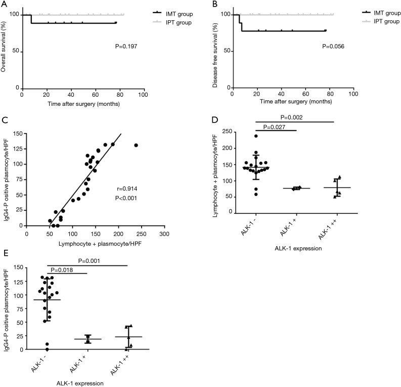
Results: The average age of IgG4-related IPT group was higher than IMTs (48.82 vs. 39.22 years, P=0.031). In IMT group, tumors were characterized by bigger tumor sizes (3.47 vs. 2.22 cm, P=0.007), diffuse and total destroyed alveoli (88.89% vs. 17.65%, P=0.002), fewer lymphoid follicles (1.6/HPF vs. 3.0/HPF, P=0.045) and lower expression of IgG (74.7/HPF vs. 149.1/HPF; P<0.001). As an exclusion criterion of IgG4-related IPT, ALK-positivity was correlated with the higher cytological atypia (mean 3.7/HPF, P<0.001) and lesser lymphoid follicles (mean 1.2/HPF, P=0.021). And it's the first study to show the liner positive correlation between the lymphocytes + plasma cells count and IgG4-positive plasma cells count in these lesions (r=0.914, P<0.001). The negative correlation between the IgG4-positive plasma cells count and the expression of ALK-1 are reported for the first time as well (rs=-0.632, P=0.001). However, despite two patients with recurrence or metastasis were divided into IMT group, only borderline values were detected in the survival analysis (OS 88.89% vs. 100%, P=0.197, DFS 77.78% vs. 100.00%; P=0.056).

Conclusions: The significant differences of clinicopathological characteristics between the IMTs and IgG4-related IPTs indicated that a combination of lymphocytes + plasma cells count, cytological atypia, IgG4 and ALK-1 staining will be helpful in differential diagnosis.

Keywords: Inflammatory myofibroblastic tumor (IMT); differential diagnosis; inflammatory pseudotumor (IPT).


Conflict of interest statement

Conflicts of Interest: The authors have no conflicts of interest to declare.

Figures

Figure 1
Figure 1
Computed tomography (CT) and macroscopic imaging data of the patient with intracranial metastasis (Case 2) (A,B). CT examinations of the patient before the resection. A solid tumor (Marked by the white arrows) in the right lower lobe of the lung with deep lobulated sign, pleural indentation and markedly inhomogeneous enhancement showed on CT imaging; (C) 5 months after surgery, magnetic resonance imaging (MRI) showed a few of subcortical nodules with obvious peripheral edema (Marked by the green arrows) which were considered as intracranial metastasis; (D) a macroscopic image of the lobectomy specimen showed that the tumor was circumscribed, and whitish-yellow in color on the cut-surface.
Figure 2
Figure 2
Hematoxylin and eosin staining in IMT and IgG4-related IPT. (A) Original alveolar structures were totally destroyed by intersecting fascicles of spindle tumor cells in IMTs; (B) tumor cells were admixed with numerous lymphocytes, plasma cells and lymphoid follicles (red arrows) in IgG4-related IPTs; (C) a large number of inflammatory cells were infiltrative and involves the small bronchi (green arrow) in IPTs; (D) inflammatory infiltration of vessel walls or obstructive phlebitis (yellow arrow) were only observed in the tumors diagnosed as IgG4-related IPT.
Figure 3
Figure 3
Immunohistochemical (IHC) staining of IMT and IgG4-related IPT. (A) Smooth muscle actin (SMA) was focally positive in spindle-shaped cells in inflammatory myofibroblastic tumors (IMT); (B) in IgG4-related IPTs, IgG expressions were patchy and cytoplasmic, arrows, IgG-positive plasma cells; (C) IgG4 immunostain showed numerous IgG4-positive plasma cells which marked by red arrows in the lesions of IgG4-related IPTs; (D) spindle-shaped tumor cells presented diffuse and strong reactivity for ALK-1 immunostain in the IMT, red arrows, ALK-1-positive spindle cells.
Figure 4
Figure 4
The results of statistical analysis based on the clinicopathological and immunohistochemical (IHC) findings. (A) Overall survival (OS) between the IMT and IgG4-related IPT group were found no statistically significant difference (P=0.197); (B) a borderline value was found in disease-free survival (DFS) between the two group (P=0.056); (C) Pearson correlation analysis showed that the lymphocytes + plasmocytes level had a possible linear positive correlation with the number of IgG4-positive plasma cells (r=0.914, P<0.001); (D) the significant differences were found in the average number of L + P between the ALK-negative and weakly or strongly positive lesions respectively (P=0.027 and P=0.002). And a negative correlation was found between the ALK-1 expression and L + P count as well (rs=−0.663, P<0.001); (E) similar results were also found between the ALK-1 expression and the number of IgG4-positive plasmocyte (P=0.018, P=0.001, rs=−0.632 and P=0.001). ALK-1 weakly focally positive reaction was represented by ALK-1 + in the graph.

References

    1. Travis WD, Brambilla E, Muller-Hermelink HK, et al. Pathology and Genetics of Tumours of the Lung, Pleura,Thymus and Heart. IARC Press, International Agency for Research on Cancer. 2004.
    1. Coffin CM, Hornick JL, Fletcher CD. Inflammatory myofibroblastic tumor: comparison of clinicopathologic, histologic, and immunohistochemical features including ALK expression in atypical and aggressive cases. Am J Surg Pathol 2007;31:509-20. 10.1097/01.pas.0000213393.57322.c7 - DOI - PubMed
    1. Chavez C, Hoffman MA. Complete remission of ALK-negative plasma cell granuloma (inflammatory myofibroblastic tumor) of the lung induced by celecoxib: A case report and review of the literature. Oncol Lett 2013;5:1672-6. - PMC - PubMed
    1. Griffin CA, Hawkins AL, Dvorak C, et al. Recurrent involvement of 2p23 in inflammatory myofibroblastic tumors. Cancer Res 1999;59:2776-80. - PubMed
    1. Zen Y, Kitagawa S, Minato H, et al. IgG4-positive plasma cells in inflammatory pseudotumor (plasma cell granuloma) of the lung. Hum Pathol 2005;36:710-7. 10.1016/j.humpath.2005.05.011 - DOI - PubMed