Case Reports
. 2017 Mar;9(3):E202-E209.
doi: 10.21037/jtd.2017.02.71.

Malignant pheochromocytoma in the anterior mediastinum with sternal invasion: a case report

Affiliations
Case Reports

Malignant pheochromocytoma in the anterior mediastinum with sternal invasion: a case report

Mingzhi Song et al. J Thorac Dis. 2017 Mar.

Abstract

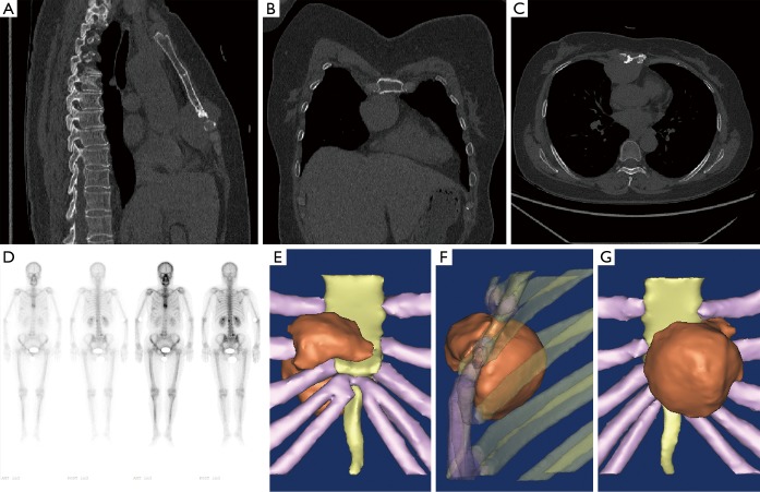
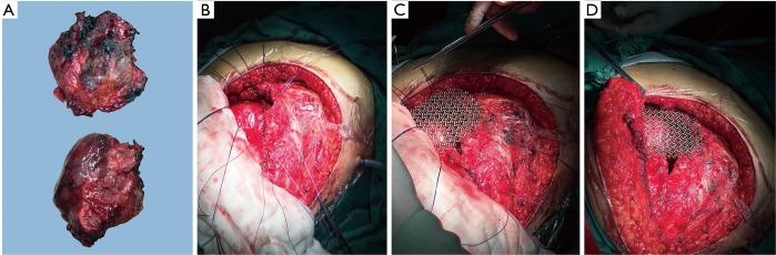
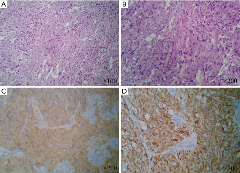
Pheochromocytomas are catecholamine-producing neuroendocrine tumors that usually occur in the adrenal medulla or sympathetic paraganglia. Anterior mediastinum involvement with pheochromocytoma is rare and may not present with typical symptoms. Its clinical manifestation may be unclear and a high index of suspicion is required for accurate diagnosis. We report a rare case of pheochromocytoma of the anterior mediastinum in a 51-year-old female. A painful hard mass on the sternum was the only clinical manifestation. Imageological examination indicated that there might be a malignant mass on the anterior mediastinum and thoracic wall. The patient accepted surgical curettage and thoracic wall reconstruction. Based on pathological results and WHO definition, the final diagnosis was malignant pheochromocytoma. After six months follow-up, the patient had no recurrence or any symptom. Malignant pheochromocytoma in the anterior mediastinum invading the sternum is rare. A local painful mass may be the only clinical manifestation without special laboratory results. Surgery remains as the first choice for these patients. For this rare case, 3D reconstruction by special software may be a good method to realize individualized treatment. The final decision of the diagnosis should be based on pathological results, past medical history and WHO definition. Long-term follow-up is necessary, while other suspicious lesions should also be given sufficient attention.

Keywords: Pheochromocytoma; sternum; surgery; thoracic wall; three-dimensional (3D) reconstruction.


Conflict of interest statement

Conflicts of Interest: The authors have no conflicts of interest to declare.

Figures

Figure 1
Figure 1
Lesion on the CT, bone scintigraphy and 3D reconstruction: (A,B,C) a soft tissue mass (approximately 3.7 cm × 4.7 cm × 5.8 cm) located in the anterior mediastinum and anterior thoracic wall. The mass was likely to be malignant thymoma. The adjacent sternum was destroyed by the tumor; (D) bone scintigraphy revealed a nuclide focus on the distal mesosternum, first lumbar and fifth lumbar. Furthermore, these abnormal locations were suspected to be bone metastases; (E,F,G) 3D reconstruction displayed the certain appearance and location of the tumor. An overall demonstration was recorded in the short video (Figure 2).
Figure 2
Figure 2
The dynamic display of the reconstructed model of malignant pheochromocytoma (12). Available online: http://www.asvide.com/articles/1427
Figure 3
Figure 3
Gross specimen of the tumor and surgery procedures. (A) The resected tumor (anterior and lateral view) was spherical and hard with a smooth surface. Its size was approximately 5.0 cm × 6.0 cm × 8.5 cm; (B,C,D) the right breast was upturned to expose the lesion during the surgical process. Following the complete resection of the tumor and adjacent tissue, a titanium mesh was selected and used to repair the defect of the thoracic wall.
Figure 4
Figure 4
Photomicrograph of the resected tumor. (A,B) Nest-like pattern of polygonal neoplastic cells with atypical nucleolus were round or ovate, possessing abundant cytoplasm (hematoxylin and eosin stain); (C,D) immunohistochemistry results demonstrated that NSE was positive in (C) and CHGA was positive in (D).

References

    1. Lenders JW, Eisenhofer G, Mannelli M, et al. Phaeochromocytoma. Lancet 2005. Aug;366:665-75. 10.1016/S0140-6736(05)67139-5 - DOI - PubMed
    1. Lenders JW, Pacak K, Walther MM, et al. Biochemical diagnosis of pheochromocytoma: which test is best? JAMA 2002;287:1427-34. 10.1001/jama.287.11.1427 - DOI - PubMed
    1. Eisenhofer G, Lenders JW, Pacak K. Biochemical diagnosis of pheochromocytoma. Front Horm Res 2004;31:76-106. 10.1159/000074659 - DOI - PubMed
    1. Elder EE, Elder G, Larsson C. Pheochromocytoma and functional paraganglioma syndrome: no longer the 10% tumor. J Surg Oncol 2005;89:193-201. 10.1002/jso.20177 - DOI - PubMed
    1. Fishbein L. Pheochromocytoma and Paraganglioma: Genetics, Diagnosis, and Treatment. Hematol Oncol Clin North Am 2016;30:135-50. 10.1016/j.hoc.2015.09.006 - DOI - PubMed

Publication types

LinkOut - more resources