Review
. 2017 Apr 27;17(1):59.
doi: 10.1186/s12886-017-0455-x.

CD34 + tumours of the orbit including solitary fibrous tumours: a six-case series

Affiliations
Review

CD34 + tumours of the orbit including solitary fibrous tumours: a six-case series

Su Kyung Jung et al. BMC Ophthalmol. .

Abstract

Background: To report six cases of CD34+ fibroblastic mesenchymal tumours, which are uncommon neoplasms in the orbit.

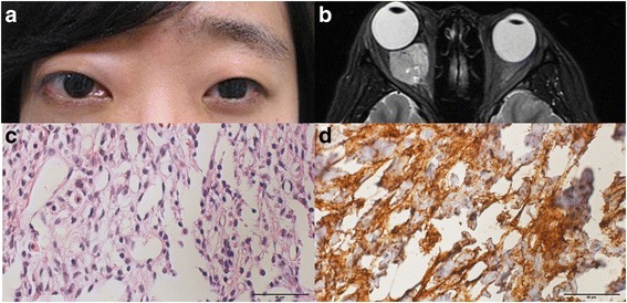
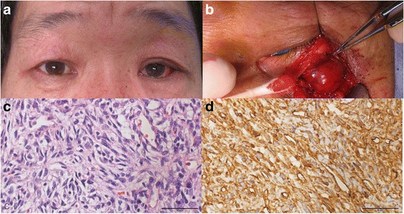
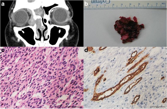
Case presentation: Six patients presenting with proptosis and palpable mass who were later diagnosed with fibrous solitary tumours, fibrous histocytoma or haemangiopericytoma in the orbit were included. All patients received radiologic examinations and surgical excision for histopathology and immunohistochemistry examinations. Five patients had no recurrence after a minimum follow-up of 12 months. One patient (case 6) experienced recurrence twice, and had debulking surgeries each time. At present, the patient still has remnant tumour in the orbit, but no growth has been detected during the past two years. The tumour size will be closely monitored.

Conclusions: Even though fibroblastic tumours are rarely found in the orbit, they can present as a palpable mass with proptosis. Complete surgical excision is important for long-term prognosis, and immunohistochemical study is helpful for confirming pathologic diagnosis.

Keywords: CD34; Fibrous histiocytoma; Haemangiopericytoma; Mesenchymal tumour; Solitary fibrous tumour.


Figures

Fig. 1
Fig. 1
a A clinical photograph shows the proptosis and conjunctival chemosis of the right eye. b Axial T2-weighted orbital MRI showed a strong enhancing mass at the retrobulbar region of the right orbit. The biopsy specimen showed spindle cells with the myxoid features (H-E stain, x400) c and intense reactivity for CD34 (CD34 stain, x 400), d
Fig. 2
Fig. 2
a A clinical photograph shows the proptosis of the left eye. b Contrast enhanced coronal CT scan shows the heterogeneous enhancing lesion in the left superomedial extraconal space. Histopathologic examination shows spindle cells with atypical vascular structures (H-E stain, x400) c and intense reactivity for CD34 (CD34 stain, x 400), d
Fig. 3
Fig. 3
a A clinical photograph shows a periorbital swelling with palpable mass in the left eye. b The surgically removed mass as a reddish surface and hard elastic consistency. The specimen has spindle-shaped cells (H-E stain, x400) without mitosis or nuclear atypism c and strong reactivity for CD34 (CD34 stain, x 400), d
Fig. 4
Fig. 4
a A preoperative photograph shows the proptosis of the right eye. b A 2.6 cm enhanced lesion was detected in the right retrobulbar space based on the axial contrast enhanced CT scans. The biopsy specimen shows spindle cells with the eosinophilic cytoplasm (H-E stain, x400) c and intense reactivity for CD34 (CD34 stain, x 400), d
Fig. 5
Fig. 5
a A movable mass is shown in the inferomedial side of the right eye. b T1-weighted orbital MRI shows 3 cm mass in the extraconal space of the right eye. The storiform pattern of fibroblasts is shown (H-E stain, x400) c and the positive reactivity for CD34 was detected in histopathologic finding (CD34 stain, x 400), d
Fig. 6
Fig. 6
a The coronal contrast enhanced orbit CT shows much progression of the mass involving the right ethmoid sinus, frontal sinus, orbit, and some portions of the nasal cavity. b The removed tumour has a cystic and fragile structure. The tightly packed spindle-shaped cells (H-E stain, x400) c are shown with positive reactivity for CD34 (CD34 stain, x 400), d

Similar articles

Cited by

References

    1. Furusato E, Valenzuela IA, Fanburg-Smith JC, Auerbach A, Furusato B, Cameron JD, Rushing EJ. Orbital solitary fibrous tumor: encompassing terminology for hemangiopericytoma, giant cell angiofibroma, and fibrous histiocytoma of the orbit: reappraisal of 41 cases. Hum Pathol. 2011;42(1):120–128. doi: 10.1016/j.humpath.2010.05.021. - DOI - PubMed
    1. Hsu CH, Wei YH, Peng Y, Liao SL. Orbital hemangiopericytoma in an Asian population. J Formos Med Assoc. 2014;113(6):356–363. doi: 10.1016/j.jfma.2012.08.004. - DOI - PubMed
    1. Brunnemann RB, Ro JY, Ordonez NG, Mooney J, El-Naggar AK, Ayala AG. Extrapleural solitary fibrous tumor: a clinicopathologic study of 24 cases. Mod Pathol. 1999;12(11):1034–1042. - PubMed
    1. Le CP, Jones S, Valenzuela AA. Orbital solitary fibrous tumor: a case series with review of the literature. Orbit. 2014;33(2):145–151. doi: 10.3109/01676830.2013.853806. - DOI - PubMed
    1. Kim HJ, Kim HJ, Kim YD, Yim YJ, Kim ST, Jeon P, Kim KH, Byun HS, Song HJ. Solitary fibrous tumor of the orbit: CT and MR imaging findings. Am J Neuroradiol. 2008;29(5):857–862. doi: 10.3174/ajnr.A0961. - DOI - PMC - PubMed

MeSH terms

LinkOut - more resources