

Arthroscopic treatment of focal osteochondral lesions of the first metatarsophalangeal joint
- PMID: 28449701
- PMCID: PMC5406985
- DOI: 10.1186/s13018-017-0562-7
Arthroscopic treatment of focal osteochondral lesions of the first metatarsophalangeal joint
Erratum in
-
Correction to: Arthroscopic treatment of focal osteochondral lesions of the first metatarsophalangeal joint.J Orthop Surg Res. 2019 Dec 23;14(1):460. doi: 10.1186/s13018-019-1456-7. J Orthop Surg Res. 2019. PMID: 31870392 Free PMC article.
Abstract
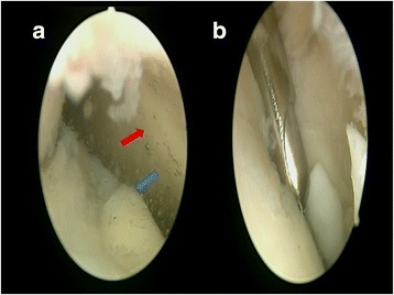
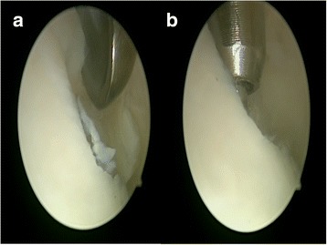
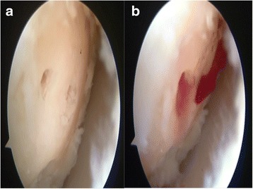
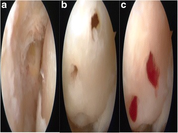
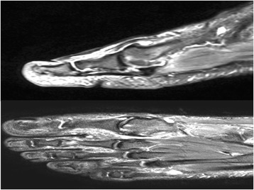
Background: Although arthroscopic surgical treatment of the first metatarsophalangeal (MTP) joint involves painful sesamoid excision, synovectomy, debridement, and partial cheilectomy, no gold standard treatment technique has been defined in the literature for hallux rigidus and focal osteochondral lesions. This study aimed to assess the arthroscopic treatment for early grade focal osteochondral lesions of the first MTP joint and to determine the impact of arthroscopic microhole drill surgery on foot function and activities of daily living in a group of patients who failed conservative treatment.
Methods: This prospective study included 14 patients with hallux rigidus and focal osteochondral lesions of the first MTP joint who underwent surgery in 2014 and were followed on a regular basis thereafter.
Results: The patients had mean preoperative VPS (visual pain score) and AOFAS (American Orthopedic Foot and ankle Society)-Hallux scores of 8.14 ± 0.86 SD and 48.64 ± 4.27, respectively; the corresponding postoperative values of both scores were 1.86 ± 0.66 SD and 87.00 ± 3.70. Both VPS and AOFAS-Hallux scores changed significantly.
Discussion: In this prospective study, we explored the impact of arthroscopic microhole drill surgery on foot function and activities of daily living in patients with focal osteochondral lesions of the first MTP joint. Our results showed significant improvements in VPS and AOFAS scores with this treatment.
Conclusions: An arthroscopic microhole drill technique can be used with impressive functional scores and without any complications in patients who failed conservative therapy for hallux rigidus with focal chondral injury.
Keywords: Arthroscopy; Hallux rigidus; Metatarsophalangeal joint.
Figures
Similar articles
-
Arthroscopic treatment of focal osteochondral lesions of the first metatarsophalangeal joint.J Orthop Surg Res. 2017 Jun 21;12(1):95. doi: 10.1186/s13018-017-0569-0. J Orthop Surg Res. 2017. PMID: 28637481 Free PMC article.
-
Hallux metatarsophalangeal (MTP) joint arthroscopy for hallux rigidus.Foot Ankle Int. 2015 Jan;36(1):113-9. doi: 10.1177/1071100714559728. Foot Ankle Int. 2015. PMID: 25550495
-
First Metatarsophalangeal Joint Arthroscopy of 36 Consecutive Cases.Acta Chir Orthop Traumatol Cech. 2021;88(3):211-216. Acta Chir Orthop Traumatol Cech. 2021. PMID: 34228617 English.
-
[Minimally invasive arthroscopic-assisted arthrodesis of the first metatarsophalangeal joint].Oper Orthop Traumatol. 2021 Dec;33(6):465-470. doi: 10.1007/s00064-021-00743-6. Epub 2021 Oct 28. Oper Orthop Traumatol. 2021. PMID: 34709415 Review. German.
-
First Metatarsophalangeal Joint Degeneration: Arthroscopic Treatment.Foot Ankle Clin. 2015 Sep;20(3):413-20. doi: 10.1016/j.fcl.2015.04.004. Epub 2015 Jun 6. Foot Ankle Clin. 2015. PMID: 26320556 Review.
Cited by
-
Correction to: Arthroscopic treatment of focal osteochondral lesions of the first metatarsophalangeal joint.J Orthop Surg Res. 2019 Dec 23;14(1):460. doi: 10.1186/s13018-019-1456-7. J Orthop Surg Res. 2019. PMID: 31870392 Free PMC article.
-
A comprehensive and narrative review of historical aspects and management of low-grade hallux rigidus: conservative and surgical possibilities.Musculoskelet Surg. 2018 Dec;102(3):201-211. doi: 10.1007/s12306-018-0530-3. Epub 2018 Feb 1. Musculoskelet Surg. 2018. PMID: 29392615 Review.
-
Metatarsophalangeal Joint Reconstruction Using Talar Osteochondral Allograft following a Failed Dorsal Cheilectomy.Case Rep Orthop. 2022 Sep 19;2022:6359108. doi: 10.1155/2022/6359108. eCollection 2022. Case Rep Orthop. 2022. PMID: 36171795 Free PMC article.
References
-
- Watanabe M. Selfoc-Arthroscope (Watanabe no. 24 arthroscope). Monograph. Tokyo: Teishin Hospital; 1972. pp. 46–53.
MeSH terms
LinkOut - more resources
Full Text Sources
Other Literature Sources
