. 2017 Apr 27;5(1):35.
doi: 10.1186/s40478-017-0431-y.

Altered Ca2+ homeostasis induces Calpain-Cathepsin axis activation in sporadic Creutzfeldt-Jakob disease

Affiliations

Altered Ca2+ homeostasis induces Calpain-Cathepsin axis activation in sporadic Creutzfeldt-Jakob disease

Franc Llorens et al. Acta Neuropathol Commun. .

Abstract

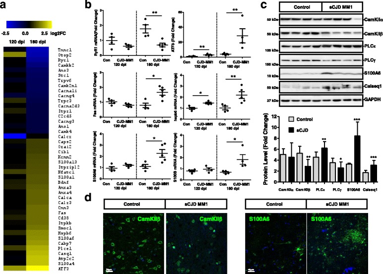
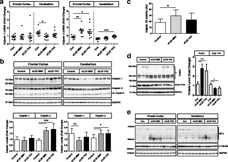
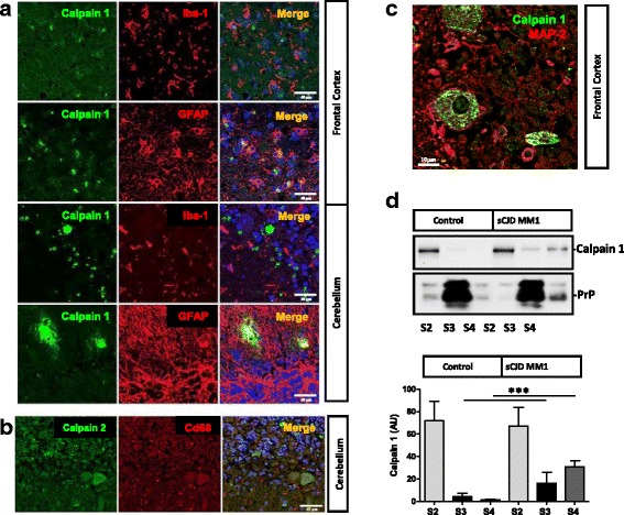
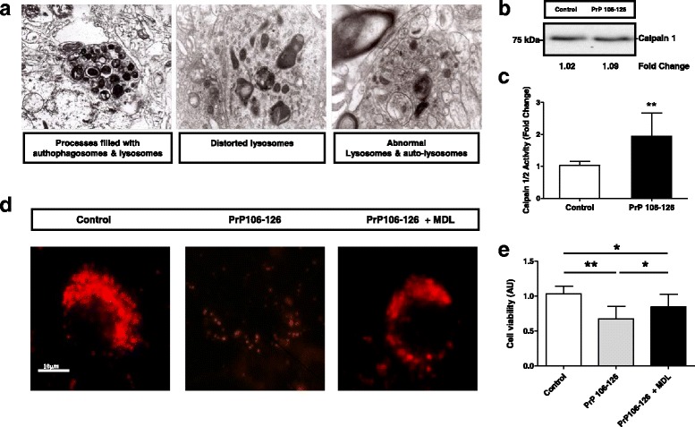
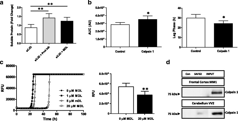
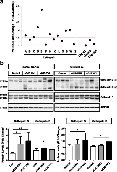
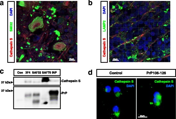
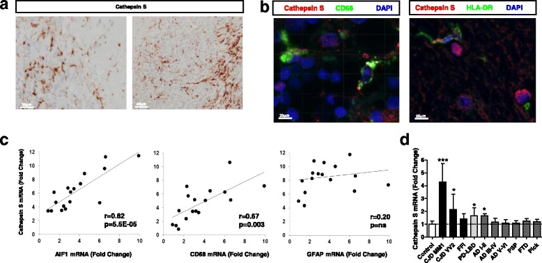
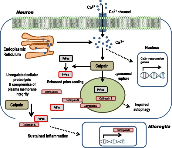
Sporadic Creutzfeldt-Jakob disease (sCJD) is the most prevalent form of human prion disease and it is characterized by the presence of neuronal loss, spongiform degeneration, chronic inflammation and the accumulation of misfolded and pathogenic prion protein (PrPSc). The molecular mechanisms underlying these alterations are largely unknown, but the presence of intracellular neuronal calcium (Ca2+) overload, a general feature in models of prion diseases, is suggested to play a key role in prion pathogenesis.Here we describe the presence of massive regulation of Ca2+ responsive genes in sCJD brain tissue, accompanied by two Ca2+-dependent processes: endoplasmic reticulum stress and the activation of the cysteine proteases Calpains 1/2. Pathogenic Calpain proteins activation in sCJD is linked to the cleavage of their cellular substrates, impaired autophagy and lysosomal damage, which is partially reversed by Calpain inhibition in a cellular prion model. Additionally, Calpain 1 treatment enhances seeding activity of PrPSc in a prion conversion assay. Neuronal lysosomal impairment caused by Calpain over activation leads to the release of the lysosomal protease Cathepsin S that in sCJD mainly localises in axons, although massive Cathepsin S overexpression is detected in microglial cells. Alterations in Ca2+ homeostasis and activation of Calpain-Cathepsin axis already occur at pre-clinical stages of the disease as detected in a humanized sCJD mouse model.Altogether our work indicates that unbalanced Calpain-Cathepsin activation is a relevant contributor to the pathogenesis of sCJD at multiple molecular levels and a potential target for therapeutic intervention.

Keywords: Ca2+; Calcium; Calpain; Cathepsin; Creutzfeldt-Jakob disease; Prion protein.


Figures

Fig. 1
Fig. 1
Altered Ca2+ homeostasis in sCJD. a Heat map analysis of regulated Ca2+ related genes in the cortical region of the tg340-PRNP129MM sCJD mice at 120 (pre-clinical) and 180 (clinical) days after inoculation with sCJD MM1 brain homogenates compared with control inoculated mice. Data were generated by RNA-sequencing analysis as indicated in material and Methods section. Regulated genes were considered those with log2FC superior or equal to 0.5 and p value <0.05. b qPCR validation of selected genes involved in Ca2+–dependent cellular responses at 120 dpi and 180 dpi in the sCJD infected tg340 mice. Four to five animals were analysed per time point and condition. c Western-blot (n = 14/group) and (d) immunohistochemistry validation in human sCJD MM1 cases from selected proteins regulated in the sCJD infected tg340 mice. Unpaired t-test (95% CI) was used for the comparisons of the two groups. *p < 0.05; **p < 0.01; ***p < 0.001
Fig. 2
Fig. 2
Regional and subtype-dependent expression and activation of Calpains in sCJD. a qPCR analysis of expression levels of Calpain 1 and Calpain 2 in the frontal cortex and cerebellum of control, sCJD MM1 and sCJD VV2 cases. b Western-blot and densitometry of Calpain 1 and Calpain 2 in the frontal cortex and cerebellum of control, sCJD MM1 and sCJD VV2 cases (n = 14/group). c Calpain activity by means of fluorometric assay based on the detection of cleavage of Calpain substrate Ac-LLY-AFC in the frontal cortex of control, sCJD MM1 and sCJD VV2 cases (n = 6/group). d Western-blot and densitometric analysis of Fodrin and N-terminal cleaved Calpain-1 in the frontal cortex of control, sCJD MM1 and sCJD VV2 cases (n = 6/group). ANOVA test followed by post-test Tukey’s Multiple Comparison Test was used to compare the values from different groups. P values for the comparisons of the three groups are indicated in the figure:*p < 0.05; **p < 0.01; ***p < 0.001
Fig. 3
Fig. 3
Neuronal localization of Calpains in sCJD. a Immunohistochemical staining of frontal cortex and cerebellum sCJD stained either with Calpain 1 (green) and Iba-1 or GFAP (red). Tissues were counterstained with DAPI (blue). b Immunohistochemical staining of cerebellum sCJD stained with Calpain 2 (green) and Cd68 (red). Tissues were counterstained with DAPI (blue). c Immunohistochemical staining of frontal cortex sCJD stained with Calpain 1 (green) and MAP2 (red). Calpains are mainly expressed in neurons as shown in the merged panels. d Western-blot analysis of solubility assays in control (n = 3) and sCJD MM1 cases (n = 3) by means of differential centrifugation in which cytoplasm fraction (S2), membrane fraction (S3) and insoluble fraction (S4) were obtained. Samples were developed for Calpain1 and PrP antibodies. . Unpaired t-test (95% CI) was used for the comparisons of the two groups. *p < 0.05; **p < 0.01; ***p < 0.001
Fig. 4
Fig. 4
Abnormal lysosomes in sCJD are dependent on Calpain over activation. a TEM images indicating the presence of processes with autophagosomes and lysosomes, distorted and abnormal lysosomes and auto-lysosomes in neurons of the frontal cortex of sCJD cases. b and c Increased Calpain 1/2 activity by fluorimetric Calpain activity (c) without alterations on Calpain 1 levels (b) in PCC treated with the prion protein peptide (106–126). Inhibition of Calpain activity by MDL28170 treatment partially reverses (d) decrease on Lysotracker signal and decrease on cell viability (e) caused by prion protein peptide treatment. Unpaired t-test (95% CI) was used for the comparisons of the two groups. Data from PCC are obtained from three independent experiments. ANOVA test followed by post-test Tukey’s Multiple Comparison Test was used to compare the values from different groups. P values for the comparisons of the three groups are indicated in the figure:*p < 0.05; **p < 0.01; ***p < 0.001
Fig. 5
Fig. 5
Calpain-dependent PrP solubility and prion conversion in sCJD. a Solubility assay in sCJD brain homogenates (n = 3) in presence of recombinant PrP (rPrP) and in the presence or absence of protein inhibitors and the Calpain inhibitor MDL28170. Quantification of soluble rPrP is shown. b RT-QuIC assay performed in the presence of sCJD brain homogenates (n = 4) as seeding material, previously treated or untreated with recombinant Calpain. Area under the curve (AUC) (left) and Lag Phase (hours) are shown. c RT-QuIC assay from tg340-PRNP129MM brain extracts (n = 3) inoculated with sCJD MM1 previously incubated with increased concentrations of MDL28170. Relative Fluorescence Units (RFU) are shown (d) Co-immunoprecipitation study of Calpain 1 and PrP in the frontal cortex of sCJD MM1 and in the cerebellum of sCJD VV2 cases. Calpain 1 antibody was used for western-blot immunodetection. Control indicates the use a non-specific antibody as immunoprecipitating antibody. Unpaired t-test (95% CI) was used for the comparisons of the two groups. ANOVA test followed by post-test Tukey’s Multiple Comparison Test was used to compare the values from different groups. P values for the comparisons of the three groups are indicated in the figure:*p < 0.05; **p < 0.01; ***p < 0.001
Fig. 6
Fig. 6
Altered Calpain levels in sCJD. a Expression of Cathepsin family, Calpain 1 (Capn1), Calpain 2 (Capn2) and Calpain 4 (Capns1) in the cortical region of the tg340-PRNP129MM mouse model at 180 (clinical) days after inoculation with sCJD MM1 brain homogenates. Data were generated by RNA-sequencing analysis. Fold Change line at 1.5 indicates the threshold of significant regulations in the expression levels for these genes between control and sCJD MM1 inoculated mice. b Western-blot and densitometry analysis of Cathepsin S and Cathepsin D expression in the frontal cortex and cerebellum of control (n = 9), sCJD MM1 (n = 9) and sCJD VV2 (n = 9) cases. ANOVA test followed by post-test Tukey’s Multiple Comparison Test was used to compare the values from different groups. P values for the comparisons of the three groups are indicated in the figure:*p < 0.05; **p < 0.01
Fig. 7
Fig. 7
Neuronal Cathepsin S in sCJD. Immunohistochemical staining of FC sCJD cases double-immunostained with Cathepsin S (red) and (a) SIM32 (green) or LAMP2 (b). Tissues were counterstained with DAPI (blue). c Co-Immunoprecipitation study of Cathepsin S and PrP in the frontal cortex of sCJD cases. Three different anti-PrP antibodies recognizing independent epitopes were used for Immunoprecipitation (3F4, SAF32 and SAF70). Western-blots were developed with a Cathepsin S antibody. Control indicates the use a non-specific antibody as immunoprecipitating antibody. d Immunohistochemistry images of Cathepsins S (green) in PCC treated or untreated with the prion peptide. Cells were counterstained with DAPI
Fig. 8
Fig. 8
Microglial overexpression of Cathepsin S in sCJD. a Immunohistochemical analysis of Cathepsin S expression in cerebellum of sCJD cases showing microglial localization. b Immunohistochemical staining of sCJD cases in the frontal cortex double-immunostained with Cathepsin S (red) and CD68 (green) left and Cathepsin S (red) and HLA-DR (green) right. Tissues were counterstained with DAPI (blue). c Correlations between the levels of Cathepsin S and glial markers (AIF1 and CD68 for microglia and GFAP for astroglia) in the frontal cortex of sCJD cases. R and p values (Pearson correlation) are indicated. d Expression levels of Cathepsin S in the frontal cortex of several neurodegenerative diseases with known cortical affection by means of qPCR analysis FFI: Fatal Familial Insomnia, PD-LBD: Parkinson Disease-Lewy Body Dementia, AD: Alzheimer’s Disease, Braak Stages I-II and III-IV, PSP: Progressive supranuclear palsy, FTD: Frontotemporal dementia, Pick: Pick’s disease. P values for the comparisons of the disease groups with control cases are indicated in the figure:*p < 0.05; **p < 0.01; ***p < 0.001
Fig. 9
Fig. 9
Activation of Calpain-Cathepsin in sCJD at pre-clinical stages of the disease. a qPCR analysis of expression levels of Calpain 1 and Calpain 2 in the cortex and cerebellum of the tg340-PRNP129MM mice inoculated with at sCJD MM1 at 120, 160, 180 and 210 dpi (10–1 inoculum dilution was used for animals sacrificed at 210 dpi). Fold Change represents the ratio between animals inoculated with sCJD-MM1 and control homogenates. b Western-blot and densitometry of Calpain 1, Calpain 2and Cathepsin S in the cortex and cerebellum of control (n = 3) and sCJD MM1 (n = 3) inoculated tg340-PRNP129MM mice at sCJD MM1 at 120, 160, 180 and 210 dpi (10–1 inoculum dilution was used for in animals sacrificed at 210 dpi). Fold Change represents the ratio between animals inoculated with sCJD-MM1and control homogenates. c Western-blot analysis of Calpain 1 N-ter, Cystatin C and Calpastatin in the cortex and cerebellum of control (n = 3) and sCJD MM1 (n = 3) inoculated tg340-PRNP129MM mice at pre-clinical (120dpi) and clinical (180dpi) stages. Fold change between sCJD MM1 and control inoculated animals is indicated. d qPCR analysis of expression levels of Cathepsin S in the cortex and cerebellum of the tg340-PRNP129MM mice inoculated with at sCJD MM1 at 120, 160, 180 and 210 dpi (10–1 inoculum dilution was used for animals sacrificed at 210 dpi). Fold Change represents the ratio between animals inoculated with sCJD-MM1 and control homogenates. e Western-blot analysis of PK-treated brain extracts from tg340-PRNP129MM mice inoculated with control and sCJD MM1 bran homogenates at 120 and 180 dpi. 180 dpi sample was diluted 1:5. ANOVA test followed by post-test Tukey’s Multiple Comparison Test was used to compare the values from different groups. P values for the comparisons of the three groups are indicated in the figure:*p < 0.05; **p < 0.01; ***p < 0.001
Fig. 10
Fig. 10
Proposed Calpain-Cathepsin S axis activation in sCJD. As a consequence of increased neuronal intracellular Ca2+ concentration in sCJD a broad range of pathologically related events occur such as i) direct or indirect alteration of gene expression patterns and ii) over activation of non-lysosomal cysteine proteases Calpains. On one side, pathological Calpain activity may cleave PrP, enhancing its misfolded conformation and enhancing prion seeding in new conversion cycles. On the other hand, Calpain compromise lysosomal membrane integrity, and as a consequence, Cathepsin proteases are liberated in the cytoplasm. Calpains and proteases with activity at neutral pH, such as Cathepsin S, unspecifically cleave cellular substrates and structures, interfering with physiological cellular functions. When plasma membrane is compromised, the cellular content is released into the extracellular space. Additionally, Cathepsin S expression is overexpressed in microglial cells as a consequence of chronic neuroinflammation

References

    1. Aguzzi A, Barres BA, Bennett ML. Microglia: scapegoat, saboteur, or something else? Science. 2013;339:156–161. doi: 10.1126/science.1227901. - DOI - PMC - PubMed
    1. Aguzzi A, Sigurdson C, Heikenwaelder M. Molecular mechanisms of Prion Pathogenesis. Annu Rev Pathol Mech Dis. 2008;3:11–40. doi: 10.1146/annurev.pathmechdis.3.121806.154326. - DOI - PubMed
    1. Anekonda TS, Quinn JF. Calcium channel blocking as a therapeutic strategy for Alzheimer’s disease: the case for isradipine. Biochim Biophys Acta. 2011;1812:1584–1590. doi: 10.1016/j.bbadis.2011.08.013. - DOI - PMC - PubMed
    1. Ansoleaga B, Garcia-Esparcia P, Llorens F, Hernandez-Ortega K, Carmona M, Del Rio JA, Zerr I, Ferrer I. Altered mitochondria, protein synthesis machinery, and Purine metabolism are molecular contributors to the pathogenesis of Creutzfeldt-Jakob disease. J Neuropathol Exp Neurol. 2016;75:755–769. doi: 10.1093/jnen/nlw048. - DOI - PubMed
    1. Atarashi R, Satoh K, Sano K, Fuse T, Yamaguchi N, Ishibashi D, Matsubara T, Nakagaki T, Yamanaka H, Shirabe S, Yamada M, Mizusawa H, Kitamoto T, Klug G, McGlade A, Collins SJ, Nishida N. Ultrasensitive human prion detection in cerebrospinal fluid by real-time quaking-induced conversion. Nat Med. 2011;17:175–8. doi: 10.1038/nm.2294. - DOI - PubMed

Publication types

MeSH terms

Supplementary concepts