. 2017 Apr 27;36(1):60.
doi: 10.1186/s13046-017-0533-1.

HIF-1α promoted vasculogenic mimicry formation in hepatocellular carcinoma through LOXL2 up-regulation in hypoxic tumor microenvironment

Affiliations

HIF-1α promoted vasculogenic mimicry formation in hepatocellular carcinoma through LOXL2 up-regulation in hypoxic tumor microenvironment

Meili Wang et al. J Exp Clin Cancer Res. .

Abstract

Background: The incidence and mortality rates of hepatocellular carcinoma (HCC) have steadily increased in recent years. A hypoxic microenvironment is one of the most important characteristics of solid tumors which has been shown to promote tumor metastasis, epithelial-mesenchymal transition and angiogenesis. Epithelial-mesenchymal transition and vasculogenic mimicry have been regarded as crucial contributing factors to cancer progression. HIF-1α functions as a master transcriptional regulator in the adaptive response to hypoxia. Lysyl oxidases like 2 (LOXL2) is a member of the lysyl oxidase family, which main function is to catalyze the covalent cross-linkages of collagen and elastin in the extracellular matrix. Recent work has demonstrated that HIF-1α promotes the expression of LOXL2, which is believed to amplify tumor aggressiveness. LOXL2 has shown to promote metastasis and is correlated with poor prognosis in hepatocellular carcinoma. The purpose of our study is to explore the role of HIF-1α in progression and metastasis of hepatocellular carcinoma by promoting the expression of LOXL2 as well as the potential regulatory mechanism.

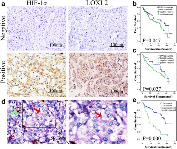
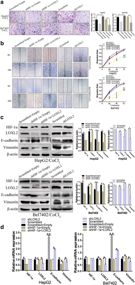
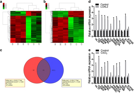
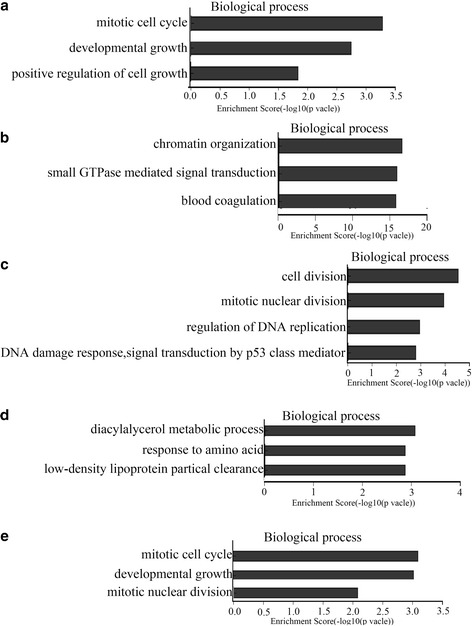
Methods: HIF-1α, LOXL2 expression and CD31/periodic acid-Schiff double staining in HCC patient samples were examined by immunohistochemical staining. shRNA plasmids against HIF-1α was used to determine whether LOXL2 been increased by HIF-1α. We monitored a series of rescue assays to demonstrate our hypothesis that LOXL2 is required and sufficient for HIF-1α induced EMT and VM formation, which mediates cellular transformation and takes effect in cellular invasion. Then we performed GeneChip® Human Transcriptome Array (HTA) 2.0 in HepG2 cells, HepG2 cells overexpressed LOXL2 and HepG2 cells treated with CoCl2.

Results: In clinical HCC tissues, it confirmed a positive relationship between HIF-1α and LOXL2 protein. Importantly, HIF-1α and LOXL2 high expression and the presence of vasculogenic mimicry were correlated to poor prognosis. HIF-1α was found to induce EMT, HCC cell migration, invasion and VM formation by regulating LOXL2. The results of microarray assays were analyzed.

Conclusion: HIF-1α plays an important role in the development of HCC by promoting HCC metastasis, EMT and VM through up-regulating LOXL2. This study highlights the potential therapeutic value of targeting LOXL2 for suppression of HCC metastasis and progression.

Keywords: EMT; HIF-1α; Hepatocellular carcinoma; Hypoxic tumor microenvironment; LOXL2; Tumor progression; Vasculogenic mimicry.


Figures

Fig. 1
Fig. 1
Expression of HIF-1α and LOXL2 correlates with vasculogenic mimicry (VM) and poor prognosis in HCC samples. a Hepatocellular carcinoma specimens were analyzed by immunohistochemistry. The first panel for negative expression and the second panel for positive expression of HIF-1α and LOXL2 (×200,bars 100um). b, c Overall survival of patients with HIF-1α-positive and HIF-1α-negative samples, LOXL2-positive and LOXL2-negative samples. Kaplan–Meier analysis showed that the patients with HIF-1α-positive and LOXL2-positive samples displayed poorer prognosis. d CD31/PAS double staining displayed VM channels in hepatocellular carcinoma specimens. The channels (red arrowhead) lined with tumor cells contained red blood cells and were CD31-negative and PAS-positive. The EDVs were CD31-positive (green arrowhead) (×400, bars 50um). e Prognostic significance of VM in HCC. Kaplan–Meier analysis of overall survival based on VM in 201 patients. Kaplan–Meier survival curves showed that the presence of VM was associated with poor overall survival
Fig. 2
Fig. 2
HIF-1α promotes LOXL2 expression was demonstrated by downregulation of HIF-1α with CoCl2 treatment in HepG2 and Bel7402 cells. a Western blot and b qRT-PCR results showed that knockdown of HIF-1α induced down-regulation of LOXL2 expression with CoCl2 treatment. c Immunofluorescence staining. Overexpression of HIF-1α increased the protein expression of LOXL2 and HIF-1α silencing expression inhibited LOXL2 expression (bar,50um). *P < 0.05
Fig. 3
Fig. 3
High HIF-1α expression promotes cell invasion, migration and EMT by regulating LOXL2 expression. a and b, The invasion and migration ability of HCC cells were decreased following HIF-1α knockdown and increased by LOXL2 overexpression; while shLOXL2 decreased invasion and migration ability, all groups treated with CoCl2 (a, ×100; bars, 100 μm) and (b,×40; bars, 100 μm). c and d, HepG2 and Bel7402 cells treated with CoCl2 were cotransfected with shHIF-1α and LOXL2 or the control vector and then western bolt and qRT-PCR assays were used to test the restoration of LOXL2 protein by LOXL2 plasmids in the presence of shHIF-1α. While HepG2 and Bel7402 cells were transfected with shLOXL2 plasmids or the control vector and then western bolt and qRT-PCR assays were used to test the restoration of LOXL2 protein by shLOXL2 in the presence of high HIF-1α expression. At the same time, expression of the epithelial protein E-cadherin and the mesenchymal protein vimentin in shHIF-1α transfected HepG2 and Bel7402 cells and LOXL2 stably transfected HepG2 and Bel7402 cells was detected by Western blot. qRT-PCR was used to detect mRNA expression. β-actin and GAPDH were used as loading controls. Error bars represent SD and *P < 0.05, **P < 0.01
Fig. 4
Fig. 4
HIF-1α promoted VM formation by regulating LOXL2 expression. a Effects of HIF-1α and LOXL2 on the tube formation abilities of the HCC cell lines. b and c The protein and mRNA levels of VE-cadherin in HepG2 and Bel7402 cells, as analyzed by Western blotting and qRT-PCR. β-actin and GAPDH were used as loading controls. Original magnification: 100×, bar100μm. Error bars represent SD and *P < 0.05
Fig. 5
Fig. 5
The results of the microarray among HepG2-LOXL2, HepG2-CoCl2, and HepG2-Control cells. a A cluster analysis showed 70 differentially expressed genes between LOXL2 and Control group, including 48 upregulated genes and 22 downregulated genes(O represents LOXL2 group and C represents Control group). b The gene expression analysis showed 1059 differentially expressed genes between CoCl2 and Control group, including 771 upregulated genes and 288 downregulated genes(D represents CoCl2 group and C represents Control group). (genes with a fold change ≥2 and a P-value (t-test) <0.05 were collected). c The results of venn analysis between LOXL2 vs Control and CoCl2 vs Control. d qRT-PCR identified changes in expression levels between LOXL2 and Control group. e qRT-PCR was used to to validate the microarray results between CoCl2 and Control group.*P < 0.05
Fig. 6
Fig. 6
Gene Ontology (GO) analysis of biological process of genes which were upregulated and downregulated and their enrichment score. a GO analysis of biological process of genes, which were upregulated, associated with our study between LOXL2 and Control group. b GO analysis of biological process of genes which were downregulated between LOXL2 and Control group. c GO analysis of biological process of genes, which were upregulated, associated with our study between CoCl2 and Control group. d GO analysis of biological process of genes which were downregulated between CoCl2 and Control group. e GO terms of biological process of genes which were upregulated associated with our present study between LOXL2 vs Control and CoCl2 vs Control

References

    1. Siegel RL, Miller KD, Jemal A. Cancer Statistics, 2017. CA Cancer J Clin. 2017;67:7–30. doi: 10.3322/caac.21387. - DOI - PubMed
    1. Marie-Egyptienne DT, Lohse I, Hill RP. Cancer stem cells, the epithelial to mesenchymal transition (EMT) and radioresistance: potential role of hypoxia. Cancer Lett. 2013;341:63–72. doi: 10.1016/j.canlet.2012.11.019. - DOI - PubMed
    1. Zhu P, Ning Y, Yao L, Chen M, Xu C. The proliferation, apoptosis, invasion of endothelial-like epithelial ovarian cancer cells induced by hypoxia. J Exp Clin Cancer Res. 2010;29:124. doi: 10.1186/1756-9966-29-124. - DOI - PMC - PubMed
    1. Soeda A, Park M, Lee D, Mintz A, Androutsellis-Theotokis A, McKay RD, Engh J, Iwama T, Kunisada T, Kassam AB, et al. Hypoxia promotes expansion of the CD133-positive glioma stem cells through activation of HIF-1alpha. Oncogene. 2009;28:3949–3959. doi: 10.1038/onc.2009.252. - DOI - PubMed
    1. Keith B, Simon MC. Hypoxia inducible factors, stem cells and cancer. Cell. 2007;129(3):465–472. doi: 10.1016/j.cell.2007.04.019. - DOI - PMC - PubMed

Publication types

MeSH terms

Substances

LinkOut - more resources