

Loss of WDFY3 ameliorates severity of serum transfer-induced arthritis independently of autophagy
- PMID: 28449847
- PMCID: PMC5515728
- DOI: 10.1016/j.cellimm.2017.04.001
Loss of WDFY3 ameliorates severity of serum transfer-induced arthritis independently of autophagy
Abstract
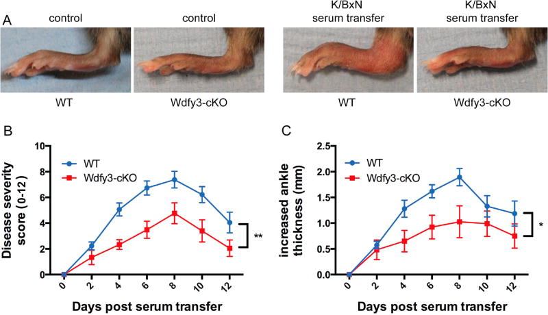
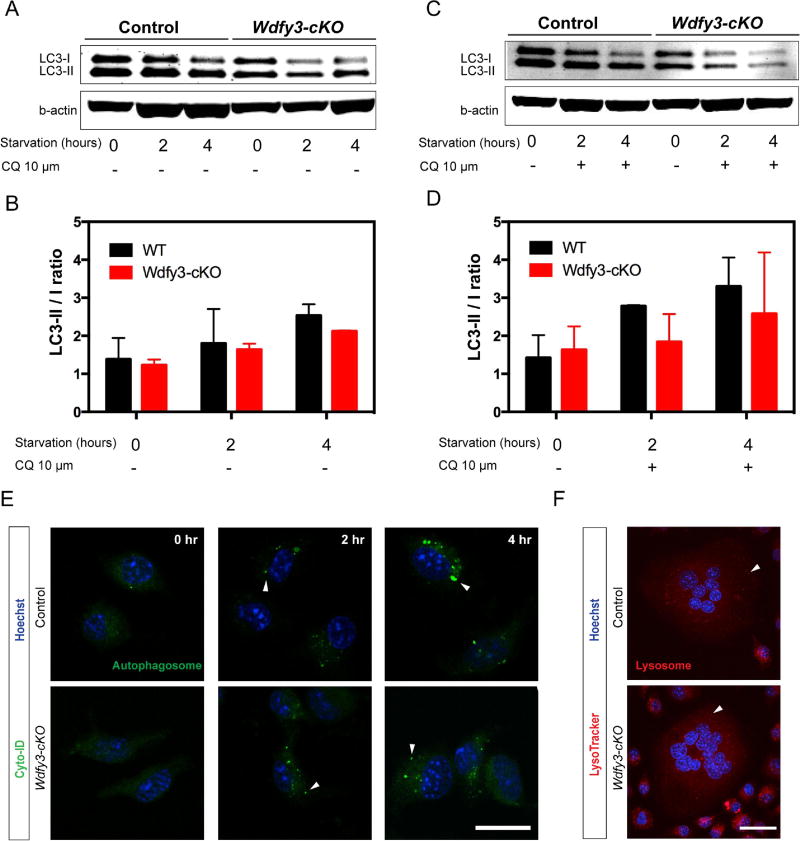
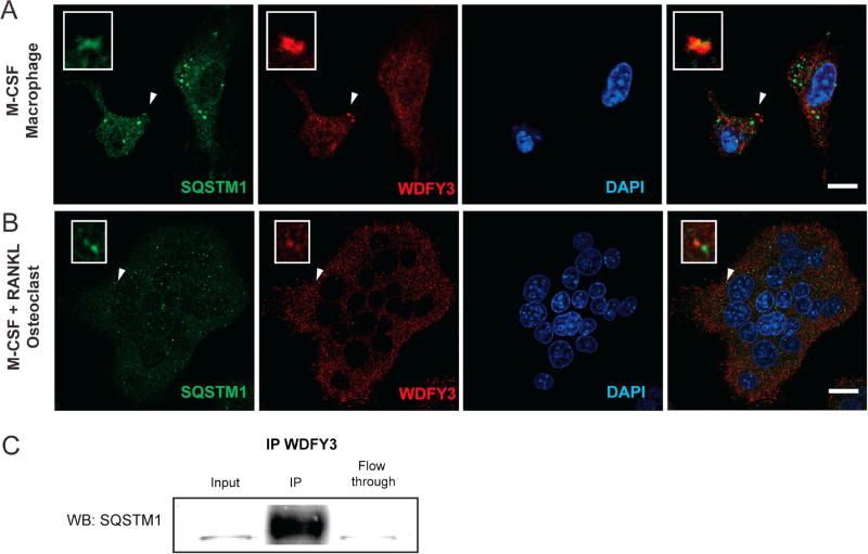
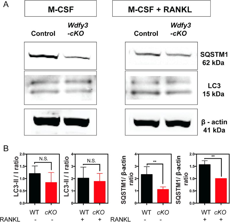
WDFY3 is a master regulator of selective autophagy that we recently showed to interact with TRAF6 and augment RANKL-induced osteoclastogenesis in vitro and in vivo via the NF-κB pathway. Since the NF-κB pathway plays a major role in inflammation herein, we investigate the role of WDFY3 in an arthritis animal model. Our data show that WDFY3 conditional knockout mice (Wdfy3loxP/loxP-LysM-Cre+) were protected in the K/BxN serum transfer-induced arthritis animal model. These effects were independent of alterations in starvation-induced autophagy as evidenced by Western blot analysis of the autophagy marker LC3, autophagosome formation in osteoclast precursors and lysosome formation in osteoclasts derived from WDFY3-cKO mice compared to controls. Moreover, we demonstrate by immunofluorescence and co-immunoprecipitation that WDFY3 interacts with SQSTM1 in macrophages and osteoclasts. Collectively, our data suggest that loss of WDFY3 in myeloid cells leads to reduced severity of inflammatory arthritis independently of WDFY3 function in starvation-induced autophagy.
Keywords: ALFY; Autophagy; Autophagy-linked FYVE containing protein; Musculoskeletal diseases; Osteoclast; WDFY3.
Copyright © 2017 Elsevier Inc. All rights reserved.
Conflict of interest statement
None of the authors has any potential financial conflict of interest related to this manuscript.
Figures
Similar articles
-
Autophagy-linked FYVE containing protein WDFY3 interacts with TRAF6 and modulates RANKL-induced osteoclastogenesis.J Autoimmun. 2016 Sep;73:73-84. doi: 10.1016/j.jaut.2016.06.004. Epub 2016 Jun 18. J Autoimmun. 2016. PMID: 27330028 Free PMC article.
-
Functional interaction between sequestosome-1/p62 and autophagy-linked FYVE-containing protein WDFY3 in human osteoclasts.Biochem Biophys Res Commun. 2010 Nov 19;402(3):543-8. doi: 10.1016/j.bbrc.2010.10.076. Epub 2010 Oct 29. Biochem Biophys Res Commun. 2010. PMID: 20971078
-
Beyond autophagy: a novel role for autism-linked Wdfy3 in brain mitophagy.Sci Rep. 2018 Jul 27;8(1):11348. doi: 10.1038/s41598-018-29421-7. Sci Rep. 2018. PMID: 30054502 Free PMC article.
-
Myeloid deletion of SIRT1 aggravates serum transfer arthritis in mice via nuclear factor-κB activation.PLoS One. 2014 Feb 3;9(2):e87733. doi: 10.1371/journal.pone.0087733. eCollection 2014. PLoS One. 2014. PMID: 24498364 Free PMC article.
-
Effects of autophagy on joint inflammation.Joint Bone Spine. 2017 Mar;84(2):129-132. doi: 10.1016/j.jbspin.2016.09.002. Epub 2016 Oct 21. Joint Bone Spine. 2017. PMID: 27777171 Review. No abstract available.
Cited by
-
Decaprenyl-phosphoryl-ribose 2'-epimerase (DprE1): challenging target for antitubercular drug discovery.Chem Cent J. 2018 Jun 23;12(1):72. doi: 10.1186/s13065-018-0441-2. Chem Cent J. 2018. PMID: 29936616 Free PMC article. Review.
References
-
- Gutierrez MG, Master SS, Singh SB, Taylor GA, Colombo MI, Deretic V. Autophagy is a defense mechanism inhibiting BCG and Mycobacterium tuberculosis survival in infected macrophages. Cell. 2004;119:753–766. - PubMed
MeSH terms
Substances
Grants and funding
LinkOut - more resources
Full Text Sources
Other Literature Sources
Medical
